
Scaphisoma hulai, Löbl, 2018
|
publication ID |
https://doi.org/ 10.2478/aemnp-2018-0013 |
|
publication LSID |
lsid:zoobank.org:pub:BF2CDC12-C555-4187-BFAB-274C9C674A5D |
|
DOI |
https://doi.org/10.5281/zenodo.3706175 |
|
persistent identifier |
https://treatment.plazi.org/id/03F94B1B-217A-FF8F-66B1-E7EA005D819C |
|
treatment provided by |
Tatiana |
|
scientific name |
Scaphisoma hulai |
| status |
sp. nov. |
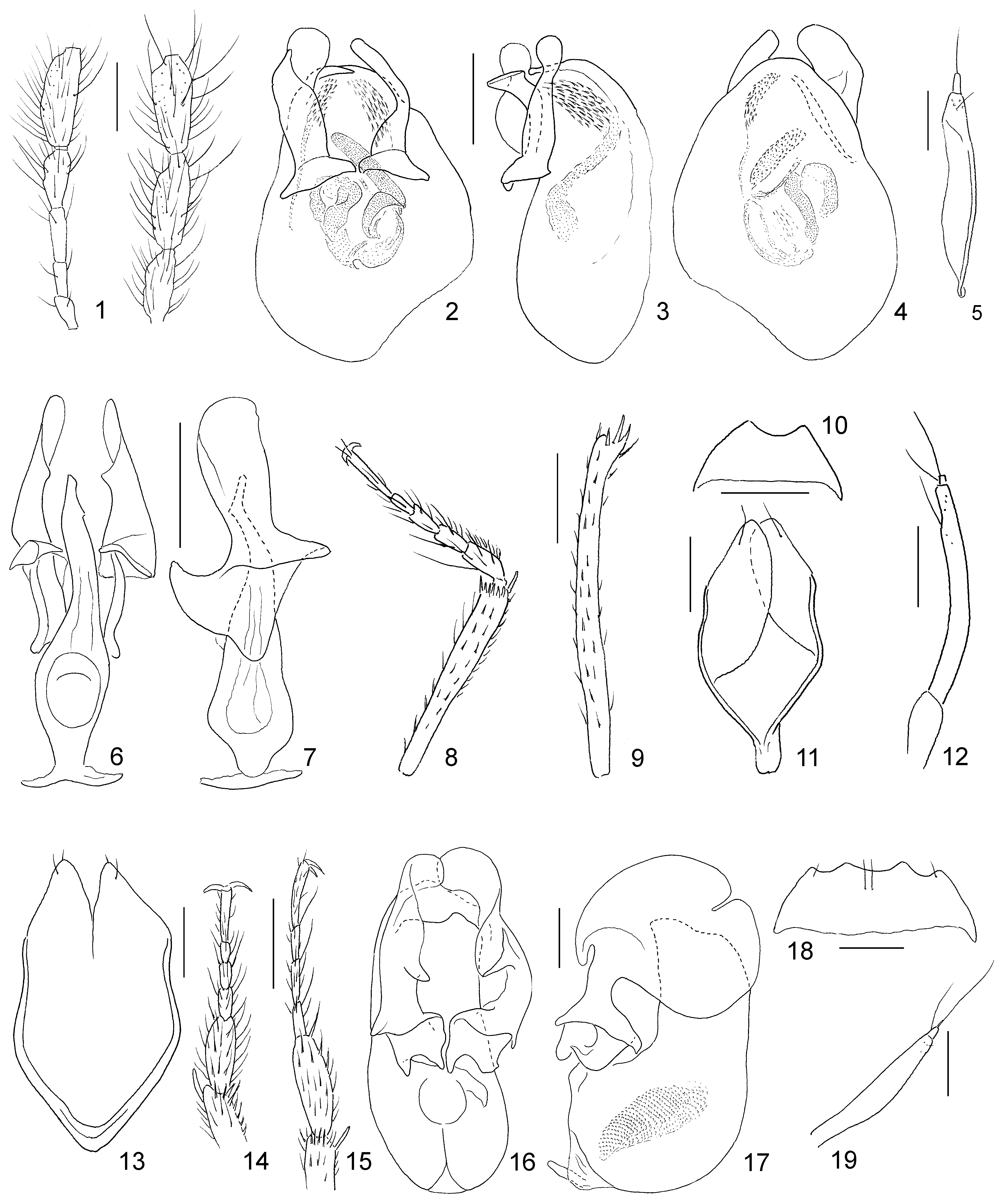
Scaphisoma hulai View in CoL sp. nov.
( Figs 13–19 View Figs 1–19. 1–5 )
Type locality. Indonesia, Central Sulawesi, Palu District, Taman Nasional Lore Lindu [National Park], environs of Wuasa, 1°24,445 ′ S, 120°18,801 ′ E.
Type material. HOLOTYPE: ( NMPC), SULAWESI, Palu dist. / Lore Lindu N.P. , Wuasa env. , / 1°24,445’S, 20°18,801’E [sic!], / 15.viii. 2013, V.Hula lgt. GoogleMaps PARATYPES: 2 3 ♀♀ ( NMPC, MHNG), with the same data as the holotype. GoogleMaps
Description. Length 1.90–2.24 mm, width 1.38–1.62 mm. Body uniformly light reddish-brown or ochreous, apex of abdomen, tibiae, tarsi and antennae ochreous, lighter than body. Antennae long, length/width ratios of antennomeres as: III 15/10: IV 47/10: V 63/10: VI 60/9: VII 62/14: VIII 45/10: IX 65/15: X 55/15: XI 58/15. Pronotum and elytra not microsculptured. Pronotum strongly narrowed anteriad, with regularly arcuate lateral margins, lateral margins carinae throughout visible in dorsal view; punctation on disc very fine, sparse, hardly visible at magnification 20×, consisting of sharply delimited punctures, puncture intervals much larger than puncture diameters, punctures along lateral carinae absent. Apical part of scutellum exposed. Elytra weakly narrowed apically, lateral margins almost regularly curved, lateral margin carinae exposed throughout in dorsal view, apical margin truncate, inner apical angles situated about in same level as outer angles, apical serration inconspicuous, sutural margin not raised, adsutural areas flat, narrow, each with single puncture row. Sutural striae almost parallel between level of scutellum up to apices, curved near pronotal lobe and extended laterad to form basal striae, extended laterally to outer halves or thirds of basal width. Punctures along lateral margins absent. Elytral disc with punctures very fine and shallow, somewhat larger than those on pronotal disc, not clearly delimited, puncture intervals mostly about three to four times as large as puncture diameters, punctures near apical margins denser, to part about as large as puncture intervals. Hind wings reduced. Exposed tergites impunctate, with punctulate microsculpture. Hypomera not microsculptured, smooth. Mesepimera somewhat longer than intervals to mesocoxae and about five times as long as wide. Metaventrite not microsculptured; punctation very fine and sparse on prevailing surface, antecoxal puncture rows absent; apical intercoxal margin straight. Submesocoxal lines convex, finely punctate; submesocoxal areas 0.04–0.05 mm, about as long as fourth of shortest intervals between them and metacoxae. Metanepisterna flat, slightly narrowed anteriad. Abdominal ventrite I not microsculptured, with punctation very fine and sparse, as that on lateral parts of metaventrite; submetacoxal lines convex, finely punctate; submetacoxal areas each 0.05 mm, about as long as fifth of intervals between them and apical margin of ventrite. Following ventrites with conspicuous punctulate microsculpture, lacking punctation.
Male characters. Pro- and mesotarsomeres I strongly enlarged, wider than apices of tibiae ( Figs 14, 15 View Figs 1–19. 1–5 ). Pro- and mesotarsomeres II and III slightly widened. Pro- and mesotibiae straight, metatibiae slightly curved and narrowed in apical halves. Mesal part of metaventrite with large, subtriangular impression and very densely punctate anterior metacoxal process, punctures to part about as large as puncture intervals. Apical margin of abdominal ventrite VI emarginate in middle ( Fig. 18 View Figs 1–19. 1–5 ). Tergite IX with two macrosetae on each plate, stalk widened basally, with lateral sections to part concave ( Fig. 13 View Figs 1–19. 1–5 ). Aedeagus ( Figs 16, 17 View Figs 1–19. 1–5 ) 1.10–1.20 mm long, asymmetrical. Median lobe with short, robust basal bulb bearing basoventral sclerotized apophysis.Articular process inconspicuous. Apical process stout, not bent. Parameres expanded by large, overlapping lobes. Inner sac compact, vesicular, bearing dense rows of scale-like structures; sclerotized pieces absent.
Female characters. Pro- and mesotarsi narrow. Tibiae straight, evenly thick. Metaventrite convex in middle, flattened between metacoxae, with dense punctation limited onto small areas near coxae. Gonocoxite slightly bent, widened in middle, with subapical microtrichiae ( Fig. 19 View Figs 1–19. 1–5 ).
Differential diagnosis. The new species resembles S. latitarse Löbl, 2012 , by its large-sized, finely punctate body, the presence of basal elytral striae, and the strongly enlarged basal protarsomeres. It may be readily distinguished from S. latitarse by its light body colour, the reduced hind wings, the long mesepimera and the abdominal ventrite I lacking microsculpture. The aedeagi of these two species are very distinctive. Several Moluccan congeners have aedeagi with basoventral processes, expanded parameres and bulbous internal sacs lacking sclerites (S. perdecorum Löbl, 2015, S. spatulatum Löbl, 2015, S. spinosum Löbl, 2015, S. permixtum Löbl, 2015). Scaphisoma hulai sp. nov. differs from all of them by its robust median lobe with wide, non-inflexed apical process, and by the much wider parameres.
Etymology. The species is named in honour of its collector, Vladimír Hula, Brno, Czech Republic; the name is a noun in the genitive case.
Distribution. Indonesia, Central Sulawesi.
No known copyright restrictions apply. See Agosti, D., Egloff, W., 2009. Taxonomic information exchange and copyright: the Plazi approach. BMC Research Notes 2009, 2:53 for further explanation.
|
Kingdom |
|
|
Phylum |
|
|
Class |
|
|
Order |
|
|
Family |
|
|
Genus |