
Bornella calcarata Mörch, 1863
|
publication ID |
https://doi.org/10.5281/zenodo.185130 |
|
DOI |
https://doi.org/10.5281/zenodo.5689393 |
|
persistent identifier |
https://treatment.plazi.org/id/03F98793-FF98-723F-2BCF-FDC2A312FE8D |
|
treatment provided by |
Plazi |
|
scientific name |
Bornella calcarata Mörch, 1863 |
| status |
|
Bornella calcarata Mörch, 1863 View in CoL
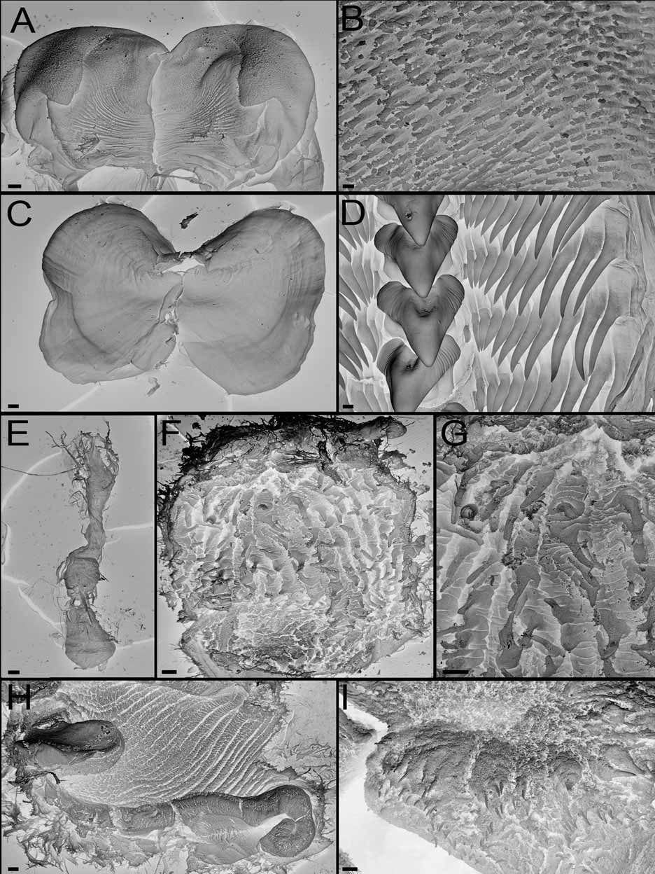
( Figures 1 View FIGURE 1 C–D, 4B, 5)
Bornella calcarata Mörch, 1863: 21 View in CoL ; Bergh, 1874: 289 –301, Pl. 36 figs 2–21, Pl. 37 figs 1–13, Pl. 38 figs 1–12; Er. Marcus, 1958: 28 –32, figs 46–54; Marcus & Marcus, 1967: 105 –106, Fig. 135; Thompson, 1980: 74 –75, figs 1,3, Pl. 2a; Bertsch, 1980: 33 –42.
Material examined: CASIZ 118665, Caribbean Sea, West Indies, Lesser Antilles, St. Lucia Island, S of P. 'Eux Fort', one specimen 20 mm preserved, dissected, 24-27 m depth, coll: J. Hamann. MZSP 84448, Brazil, BA, Marau. Barra Grande de Camamu. Recifes de Itaipus de fora, one specimen 50 mm preserved, 12 November 2006, dissected, 0.5 m depth, coll: C. Sampaio. MZSP 25294, Brazil, PE, Recife, one specimen 40 mm preserved, coll: E. Marcus. MZSP 39127, Brazil, ES, Guarapi, three specimens 50, 50 and 60 mm preserved, 30-50 m depth, coll: v/2000.
Geographic Distribution: Caribbean Sea: St. Thomas (Danish Antilles) ( Mörch 1863), Surinam (The Guianas) ( Nijssen-Meyer 1965; Valdés et al. 2006), Brazil ( Marcus 1958, 1977; Marcus & Marcus 1967; Valdés et al. 2006; present study), Colombia ( Marcus 1976; Valdés et al. 2006), Jamaica ( Thompson 1980; Valdés et al. 2006), Panama ( Marcus 1977), Honduras ( Wilk 2005), Mexico, Costa Rica, Cayman Island, Puerto Rico, Virgin Islands, Guadalupe, St. Lucia, Trinidad and Tobago, French Guyana ( Valdés et al. 2006), St. Vincent and the Grenadines ( Ianniello 2002; Valdés et al. 2006) and St. Lucia Island (present study).
External morphology: The general body shape is elongate and limaciform with the posterior end of the foot being long and tapering. The living adults reach 80 mm in total length ( Ianniello 2002). The background color is yellowish or whitish covered with a dense network of brilliant orange-red, forming a precise pattern on the main parts of the body ( Figs. 1 View FIGURE 1 C,D). On the dorsum the orange-red markings are arranged in a reticulate pattern with the transverse lines more dominant than the longitudinal lines. In some specimens the redorange pigmentation extends out from the lines to obscure much of the yellowish background color. This color pattern extends to the lobe-like oral tentacles, rhinophore sheaths and dorsolateral processes. On each side of the mouth, the oral tentacle is modified into a lobe, with 8 to 13 blunt papillae arranged in two rows. The red rhinophores are perfoliate with about 10 to 25 lamellae. The stalks of the rhinophore sheaths are tall, and there are usually four elongate and bluntly tipped papillae arranged around the upper edge. There is also a taller posterior crest inserted just below the upper edge of the stalk. This posterior crest has a central papilla and up to three secondary papillae, which have been interpreted previously as the fusing of the first dorsolateral processes with the rhinophore stalks ( Bergh 1874; Marcus 1958). Posterior to the rhinophores there are four pairs of well-developed dorsolateral processes, followed by one or two small pairs and two smaller unpaired processes in the dorsal midline. The first two dorsolateral processes have four long papillae, while the next processes have only two. There are up to four tripinnate secondary gills, associated with each dorsolateral process, and when fully extended they can be as large or larger than the papillae. The anus is small, located on the right side of the dorsum between the first and second pair of dorsolateral processes, closer to the second. The renal pore is situated very close to the anus. The reproductive opening is located on the right side, midway between the rhinophore sheath and the first dorsolateral process.
Alimentary Canal: The buccal bulb is relatively large. The labial cuticle is thick ( Fig. 5 View FIGURE 5 A), with a labial armature of cuticular rodlets. The rodlets are elongate and numerous, standing in irregular rows ( Fig. 5 View FIGURE 5 B). The jaws are roughly oval, without a distinct masticatory process ( Fig. 5 View FIGURE 5 C). The radular formulae of two specimens are 43 x 11.1.11 (CASIZ 118665, 20 mm preserved) and 35 x 13.1.13 (MZSP 84448, 50 mm preserved). The rachidian teeth are robust, slightly wider than higher. They lack well-developed denticles on each side of the cusp but have a number of irregular faint ridges at the posterior base of the strong cusp ( Fig. 5 View FIGURE 5 D). The blade-like laterals have a long basal portion and a pointed tip. In the specimens examined there are not vestigial outer laterals ( Fig. 5 View FIGURE 5 D). The lateral denticles increase in size from the innermost to the ninth then slightly decrease until the end. A long unpaired oral gland is found ventrally ( Fig. 5 View FIGURE 5 E). A pair of large and branched salivary glands is attached to the posterior side of the oesophagus. They open into the posterior buccal bulb on each side of the oesophageal opening. After passing through the nerve ring, the relatively long oesophagus runs back to the stomach. Opening into the dorsal side of the stomach are the anterior digestive glands, each of which forms a branch into the first pair of dorsolateral processes. There are no branches from the anterior digestive glands to the rhinophore sheaths. The posterior digestive gland opens into the lower left surface of the stomach. All dorsolateral processes, other than the anteriormost pair already mentioned, receive branches from the posterior portion of the digestive gland. The posterior chamber of the stomach is wide and thick walled. Its anterior half is armed with about 15 longitudinal rows of chitinous brown spines ( Fig. 5 View FIGURE 5 F) which are spatuliform at their tips ( Fig. 5 View FIGURE 5 G). From the stomach the intestine descends ventrally and runs out to the anus.
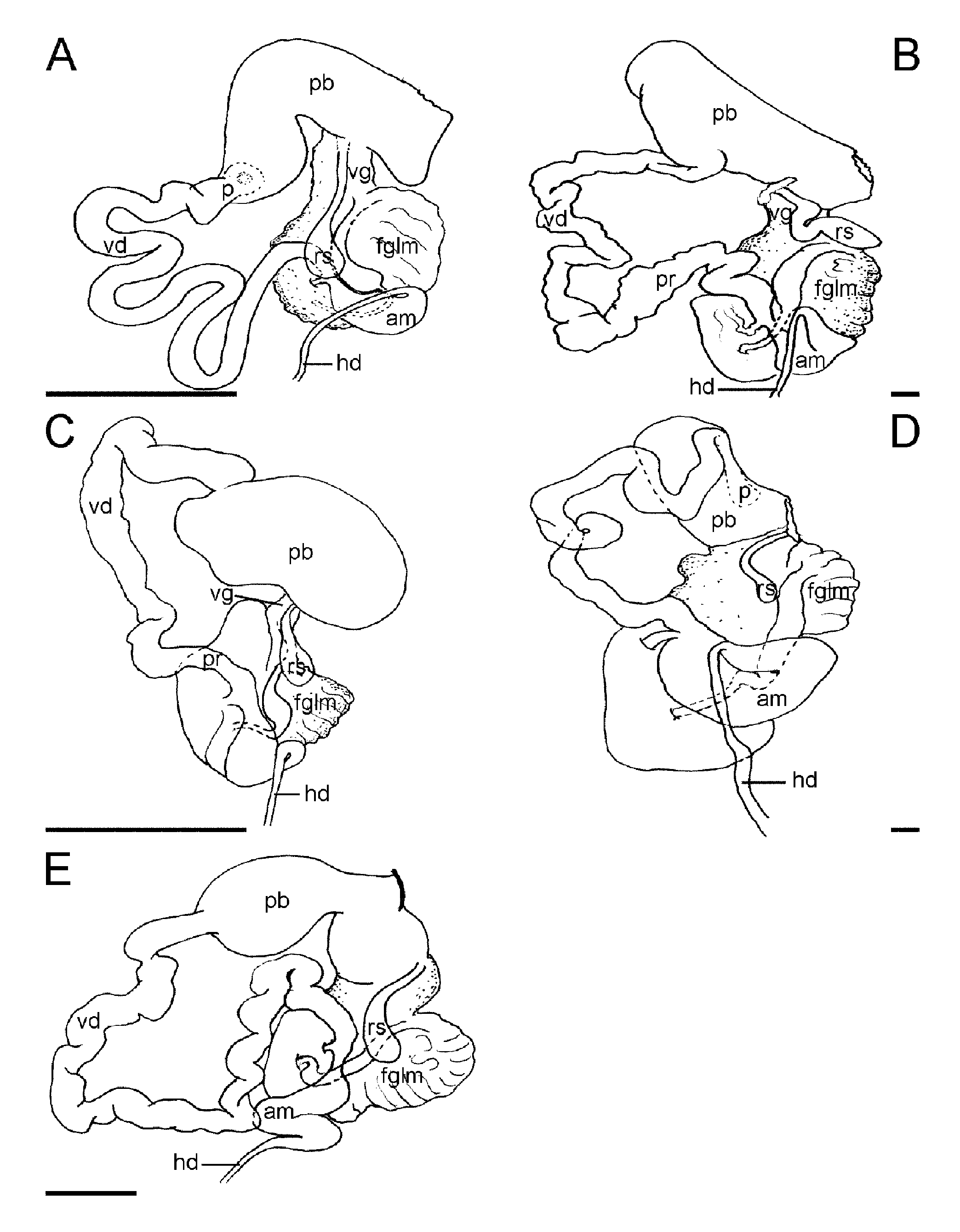
Reproductive system: ( Fig. 4B View FIGURE 4. A – E ): The ovotestis consists of six rounded follicles lying in the middle third of the body cavity above the posterior digestive gland. From each follicle, a thin-walled duct passes forwards to the hermaphroditic duct which passes around the stomach on the left side to join the ampulla on the dorsal side of the female gland mass. The inflated and folded ampulla narrows, before dividing into two ducts, the vas deferens and the oviduct. The vas deferens is long and folded with a soft glandular consistency suggesting that there is a prostatic layer along its whole length. The vas deferens runs to the large elongate penial bulb, which contains an elongate fleshy ridge, lobed at each end. Along the crest of the ridge is a band of three to five irregular rows of chitinous hooked spines ( Figs. 5 View FIGURE 5 H,I). This ridge was described by Bergh (1874) and Marcus (1958) illustrates how it forms a raised ring of spines at the tip of the everted penis. The oviduct, from the ampulla to the female gland mass is short. A medium-sized distal pyriform allosperm receptacle opens directly to the genital aperture through a long duct.
Remarks: Bornella calcarata is the only known species of the genus from the Caribbean. Mörch (1863) recorded B. calcarata from Virgin Islands (Danish Antilles). The author described the external morphology but he did not mention any coloration or internal features. Mörch’s specimen was later dissected and reported on by Bergh (1874). No further information was available on the species until Marcus (1958) published an anatomical description of a specimen from Recife, Brazil. There are some differences between these two specimens and the present examined specimens, for example, in the number of dorsolateral processes. Bergh stated that the holotype had five pairs of dorsolateral processes with gills, while Marcus described six pairs followed by two unpaired medial processes. Both descriptions match our specimen since only the five anterior pairs of dorsolateral processes bear gills. The description of Nijssen-Meyer (1965) of a specimen from Surinam also matches our description. The number of papillae of the oral tentacles slightly varies from one specimen to another. Also the number of rhinophore lamellae may vary. Bergh and Marcus described about 25 to 30 lamellae. Thompson (1980) described tiny lamellate rhinophores in a specimen from Jamaica, giving also a description of the general color of the animal. In our specimens some have about 25 lamellae while some of them have only ten. Regarding the internal anatomy, it must be clarified that the rachidian teeth are not smooth as most of the previous mentioned authors stated (as well as Odhner 1936 and Marcus and Marcus 1967), if not with a number of irregular faint denticles at the posterior base of the median cusp. Thompson (1980) also noticed this denticulation. Another species that shares similar denticulation on either side of the median cusp is B. sarape . The reproductive system also permits differentiation between species. In B. calcarata the penis is armed, with at least 3 to 5 irregular rows of penial spines along a ribbon. The description of the shape and arrangement of the penis was well detailed by Bergh (1874). This kind of penis is also found in the new species from South Africa described in this paper.

| MZSP |
Sao Paulo, Museu de Zoologia da Universidade de Sao Paulo |
No known copyright restrictions apply. See Agosti, D., Egloff, W., 2009. Taxonomic information exchange and copyright: the Plazi approach. BMC Research Notes 2009, 2:53 for further explanation.
|
Kingdom |
|
|
Phylum |
|
|
Class |
|
|
Order |
|
|
Family |
|
|
Genus |
Bornella calcarata Mörch, 1863
| Pola, Marta, Rudman, William B. & Gosliner, Terrence M. 2009 |
Bornella calcarata Mörch, 1863 : 21
| Thompson 1980: 74 |
| Bertsch 1980: 33 |
| Marcus 1967: 105 |
| Er 1958: 28 |
| Bergh 1874: 289 |
| Morch 1863: 21 |