
Bornella valdae, Pola, Marta, Rudman, William B. & Gosliner, Terrence M., 2009
|
publication ID |
https://doi.org/10.5281/zenodo.185130 |
|
DOI |
https://doi.org/10.5281/zenodo.5689411 |
|
persistent identifier |
https://treatment.plazi.org/id/03F98793-FFB0-7216-2BCF-FDFFA502FF7D |
|
treatment provided by |
Plazi |
|
scientific name |
Bornella valdae |
| status |
sp. nov. |
Bornella valdae View in CoL sp. nov.
( Figures 16 View FIGURE 16 C–D, 15C, 18)
Bornella View in CoL sp. 1.: Fraser, 1999a, b; Warren, 2002 Bornella View in CoL sp. 2.: Debelius & Kuiter 2007
Material Examined: Holotype: AM C. 212720, South Africa, Natal, Oslo Beach, 30°47’S, 30°25’E, adult specimen, 40 mm preserved, 37 m depth, 27 December 1999, coll: V. Fraser (only buccal mass extracted). Paratypes: AM C. 379068, South Africa, Natal, Oslo Beach, 30°47’S, 30°25’E, adult specimen, 35 mm preserved, 37 m depth, 27 December 1999, coll: V. Fraser, completely dissected. SAM A55952 View Materials , South Africa, KwaZulu-Natal, off Pumula, one adult specimen, 40 mm preserved, 42 m depth, 10 March 2008, coll: V. and M. Fraser. CASIZ 176832, South Africa, KwaZulu-Natal, off Pumula, one adult specimen, 40 mm preserved, 42 m depth, 10 March 2008, coll: V. and M. Fraser.
Etymology: This species is named after Valda Fraser, who kindly collected the specimens and provided excellent pictures of it.
Distribution: At present this species is known only from the coast of KwaZulu-Natal, South Africa ( Fraser 1999a, b; Warren 2002; Debelius & Kuiter 2007).
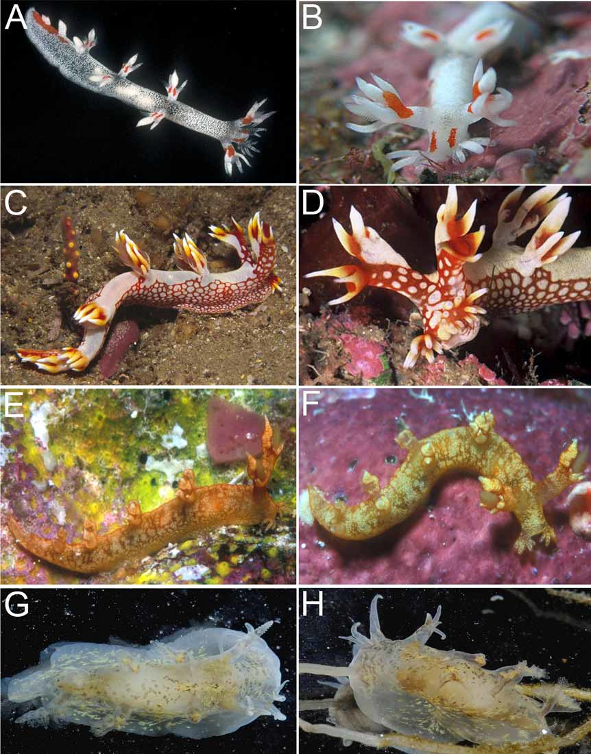
External morphology: The general body shape is elongate and limaciform with the posterior end of the foot being long and tapering ( Fig. 16 View FIGURE 16 C). The living adults are large, up to 70 mm in length ( Fraser 1999a, b; Warren 2002). The notum is smooth but when the body is slightly retracted there are traces of transverse wrinkling. The background color of the body is orange to orange-red with scattered opaque white spots, which are arranged in different densities in different parts of the body ( Figs. 16 View FIGURE 16 C,D). Over the whole of the dorsal surface the white spots have fused to form an opaque white band from just behind the rhinophores to the posterior tip of the foot. In most cases, the dorsolateral processes are opaque white with the inner bases of the papillae being orange-red. In one photo, the trunks of the dorsolateral papillae are not a uniform white but have a broken network of orange, suggesting that this white region developed from a close aggregation of white spots. The head is orange, with scattered white spots, including a group of large irregular spots, which are arranged along the dorsal midline, back to the rhinophores. The papillae along the edge of the lobe-like oral tentacles are white with an orange basal region. Often a yellowish zone can be seen between the orange and the white. The trunk of the rhinophore sheaths is orange with scattered white spots except for an opaque white band up the postero-dorsal side. The rhinophoral papillae are all opaque white, except for the inner bases, which are orange-red. Wherever the white pigmentation meets the orange-red, on both the rhinophore sheaths and the dorsolateral processes, there is a yellowish band. The actual rhinophores are orange-red. The sides of the body are orange-red with white spots. Towards the posterior end of the body the spots become smaller and more closely packed, reducing the orange background color to a reticulate pattern.
On either side of the mouth is a lobe-like oral tentacle, with approximately 11 finger-like papillae of unequal length arranged in at least two different rows. Each rhinophore sheath has a tall stalk from which the rhinophore protrudes. The rhinophores have between 28 and 38 lamellae. Surrounding the upper edge of the rhinophore sheath, are three long, narrow anterior and anterolateral papillae and a taller, wide posterior sail ( Fig. 16 View FIGURE 16 D). The posterior sail has three large, similar-sized, sharply pointed branches. Posterior to the rhinophores are five pairs of dorsolateral processes, followed by one single process in the dorsal midline ( Fig. 16 View FIGURE 16 C). The processes in the first and second pair have four papillae, two larger inner ones and two smaller outer ones, while the processes in the third and fourth pair have only the two inner papillae. The processes in the fifth pair are simple. The papillae are rounded, with a pointed tip. Except for the fifth dorsal pair, the dorsolateral processes bear large tri- or quadripinnate gills. The first and second pair has three gills; one large and highly branched inner gill at the base of the two inner papillae and two smaller outer gills at the base of each outer papilla. On the third and fourth processes only the large inner gill is present. The anus is small, located on the right side of the dorsum between the first and second pair of dorsolateral processes, closer to the second. The reproductive opening is located on the right side, midway between the rhinophore sheath and the first dorsolateral process.
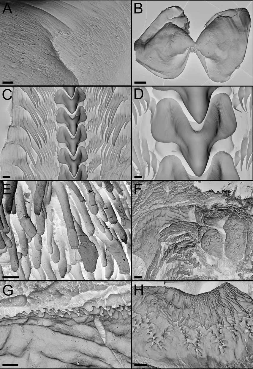
Alimentary Canal: The buccal bulb is large. The labial cuticle is thick, with a labial armature of cuticular rodlets surrounding the opening to the buccal bulb ( Fig. 18A View FIGURE 18. A – H ). The rodlets are elongate and numerous. The jaws are roughly oval in shape, without a distinct masticatory process ( Fig. 18B View FIGURE 18. A – H ). The radular formula of the holotype is 30 x 14.1.14. The paratype’s formula is 29 x 13.1.13. The rachidian teeth are somewhat quadrangular, without denticles on either side of a strong central cusp ( Figs. 18C,D View FIGURE 18. A – H ). The lateral denticles are bladelike, with a long basal portion and a pointed tip. They increase in size from the inner lateral to the eleventh. The twelfth is a little bit shorter and then the antepenultimate is very short but still with a small cusp. The last denticle is vestigial and without any cusp ( Fig. 18C View FIGURE 18. A – H ). A long unpaired oral gland is found ventrally. The oesophagus is almost completely covered by the large salivary glands, which open anteriorly into the posterior buccal bulb. The two large anterior digestive glands (left bigger than right) open into the upper wall of the stomach. Each anterior digestive gland has a single branch which enters the first dorsolateral process on its side of the body. The posterior portion of the digestive gland opens into the stomach on the lower left surface. The posterior chamber of the stomach is large and thick walled and its anterior half is armed with about 20 to 25 longitudinal rows of chitinous brown spines. These spines are straight and spatuliform ( Fig. 18E View FIGURE 18. A – H ). From the stomach the intestine descends to the ventral side and then bends dorsally to the anus.
Reproductive system: ( Fig. 15C View FIGURE 15. A – E ). The ovotestis consists of 16 closely packed, rounded or somewhat pyriform follicles lying over the posterior digestive gland. The hermaphrodite duct runs around the stomach on the left side and expands into the ampulla on the dorsal side of the female gland mass. From the ampulla, the hermaphrodite duct divides into the vas deferens and the oviduct, which descends into the female gland mass. The vas deferens, when compared with the other species examined, is extraordinarily long and folded. It is of similar width along its entire length and appears to be lined with a layer of prostate gland, however as the first half of the vas deferens was white in dissections, and the second half orange, it is possible that only the first half is prostatic. The penial bulb is large and muscular penial and the penial apparatus is similar to that described for B. calcarata . There are three to five irregular rows of chitinous hooked spines, which are arranged in zig-zag rows along a ribbon on a fleshy ridge or crest ( Figs. 18 View FIGURE 18. A – H F-H). A medium-sized distal pyriform allosperm receptacle opens directly to the genital aperture through a long duct.
Remarks: Two other species of Bornella have also been recorded from the Indian Ocean coast of South Africa: Bornella stellifer ( Adams & Reeve 1848) and Bornella anguilla ( Johnson 1984; Gosliner 1987a). Bornella valdae is clearly distinguishable from these species by its external coloration and external features. The mosaic color pattern of B. anguilla is completely different from that of B. valdae , while B. stellifer shares with Bornella valdae the orange-red reticulation pattern on parts of the body. However the dorsolateral processes and the papillae on the posterior rhinophore sheaths of B. stellifer and B. anguilla are simple, while they are branched in B. valdae . Internally, important differences are found in the radula, and the penial morphology. The rachidian teeth of Bornella valdae have smooth edges on both sides of the strong median cusp while B. stellifer and B. anguilla have very well developed denticles. Regarding the penis, Bornella valdae has at least three to five irregular rows of penial spines arranged along a ridge, similar to those of B. calcarata , while B. stellifer and B. anguilla have a single circular or semicircular (respectively) row of penial spines.

| SAM |
South African Museum |
No known copyright restrictions apply. See Agosti, D., Egloff, W., 2009. Taxonomic information exchange and copyright: the Plazi approach. BMC Research Notes 2009, 2:53 for further explanation.
|
Kingdom |
|
|
Phylum |
|
|
Class |
|
|
Order |
|
|
Family |
|
|
Genus |