
Bornella pele, Pola, Marta, Rudman, William B. & Gosliner, Terrence M., 2009
|
publication ID |
https://doi.org/10.5281/zenodo.185130 |
|
DOI |
https://doi.org/10.5281/zenodo.5689409 |
|
persistent identifier |
https://treatment.plazi.org/id/03F98793-FFB4-7214-2BCF-F8C5A4C2FE6D |
|
treatment provided by |
Plazi |
|
scientific name |
Bornella pele |
| status |
sp. nov. |
Bornella pele View in CoL sp. nov.
( Figures 16 View FIGURE 16 A–B, 15B, 17)
Material Examined: Hawaii: Holotype: CASIZ 101139, Hawaii, Molokini Island, floating about 30 m off the wall, adult specimen, 19 mm alive, 13 m depth, 12 September 1994, coll: P. Fiene. Paratypes: CASIZ 0 86356, Hawaii, Kahoolawe Island, Halona Point, on surge-washed arches, six specimens (three dissected), 12–15 mm alive, 21 and 23 January 1992, coll: P. Fiene, slide. Australia: AM C. 140380, Australia, Coral Sea, Wreck Reef, Porpoise Cay, 22° 13 S, 155° 18 E under rock, very exposed intertidal reef crest, 8 mm preserved, dissected, 29 October 1983, coll: I. Loch, slides. AM C. 137536, Australia, Queensland, Great Barrier Reef, 12° 5 S, 143° 58 E, near corner of Wreck Bay Reef, on rubble in deep hole in very exposed reef, 17 mm preserved, 12 December 1982, coll: I. Loch.
Etymology: This species is named after the Hawaiian goddess “ Pele ”, goddess of fire, lightning, dance, volcanoes and violence. This species was first collected in Hawaii and the red markings of the animal remind the fire and violence of the Hawaiian goddess.
Geographic distribution: Thus far this species is known from Hawaii, eastern Australia (present study), and Japan (present study and Nakano 2004: 211 in part).
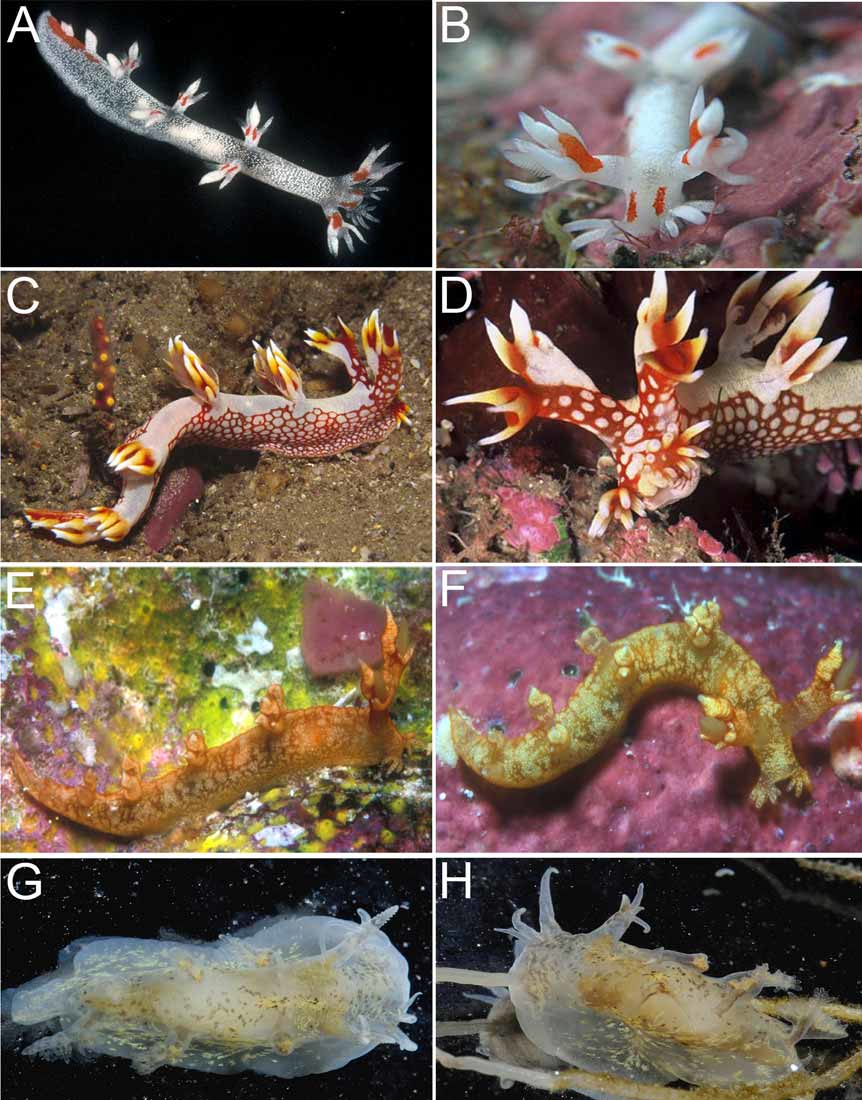
External morphology: The general body shape is elongate and limaciform with the posterior end of the foot being long and tapering. The living adults are small, up to 20 mm in length. The general color pattern consists of a translucent whitish body with scattered subepithelial opaque white granules ( Fig. 16 View FIGURE 16 A). There are two parallel red marks on the inner side of each dorsolateral process and of the rhinophore sheaths. There is also a pair of red marks on the head running between the oral tentacles and the base of the rhinophore sheaths. Another irregular, elongate red patch runs along the dorsal midline, from behind the last pair of dorsolateral processes to the posterior tip of the foot ( Figs. 16 View FIGURE 16 A,B). On either side of the mouth is a lobe-like oral tentacle, each bearing 9 to 13 finger-like papillae of unequal length arranged in three different rows. Usually the outer row bears seven papillae. The oral tentacles do not have any red pigmentation. Each rhinophore sheath has a tall stalk from which the rhinophore protrudes. The translucent whitish rhinophores are perfoliated with about 15 to 19 lamellae. Surrounding the upper edge of the rhinophore sheath, are three elongate anterior and anterolateral papillae and a taller, posterior papilla, which is distinctly bifid ( Fig. 16 View FIGURE 16 B). Posterior to the rhinophores, there are three pairs of dorsolateral processes, followed by two single processes along the dorsal midline ( Fig. 16 View FIGURE 16 A). The number of processes was consistent in all specimens examined. Each dorsolateral process has a stout base topped with three papillae, all acutely pointed at the tip. The central papilla is larger and more elongate than the other two. There are two bipinnate translucent gills on each of the paired dorsolateral processes. They are located on the inner surface, at the base of the lateral papillae. The bases of the most posterior pair of dorsolateral processes are joined. The two posterior, single, dorsal processes are simple and decrease in size towards the posterior end of the foot. The anus is small, located on the right side of the dorsum between the first and second pair of dorsolateral processes, closer to the second. The reproductive opening is located on the right side, midway between the rhinophore sheath and the first dorsolateral process.
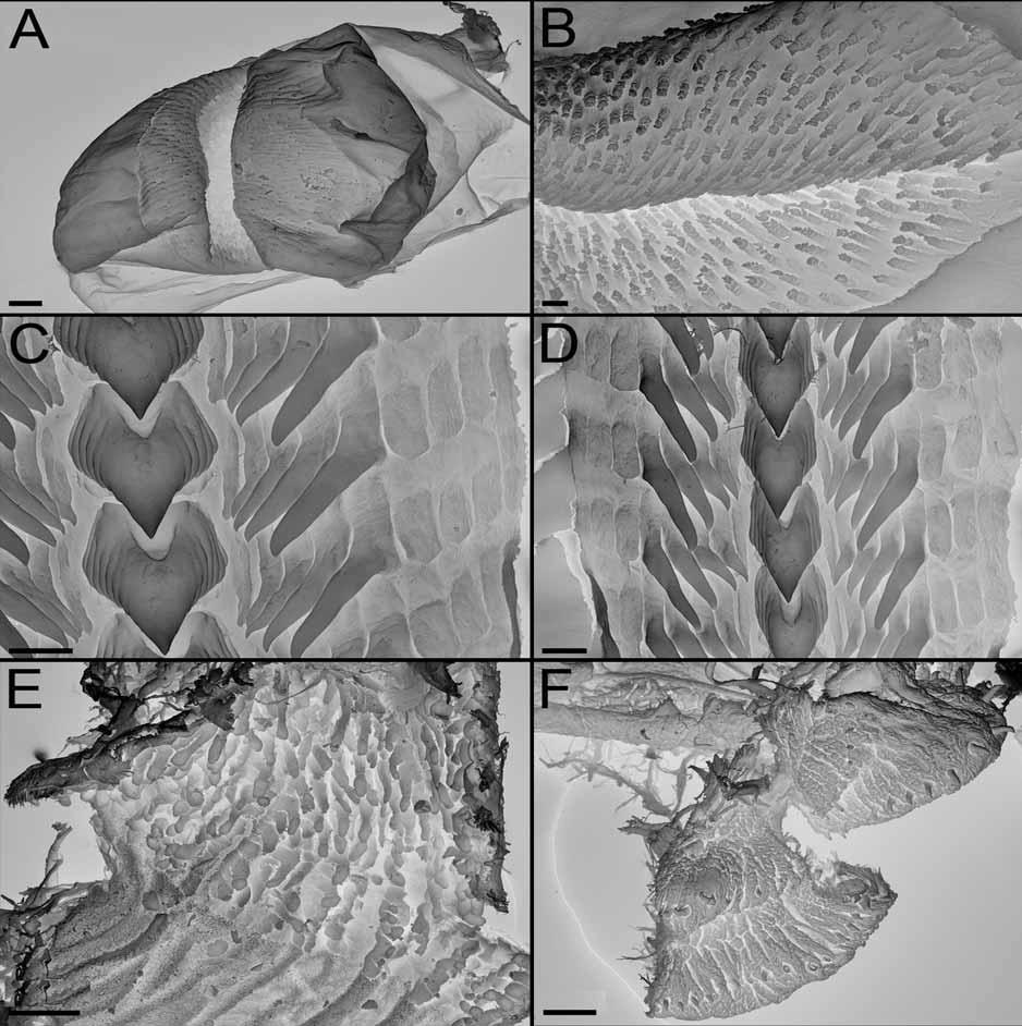
Alimentary Canal: The buccal bulb is relatively small. The labial cuticle is thin, consisting of small overlapping scales, arranged in fairly regular rows ( Fig. 17A View FIGURE 17. A – F ). The rodlets are elongate but not as dense or numerous as in other Bornella species ( Fig. 17B View FIGURE 17. A – F ). The jaws are roughly oval in shape, without a distinct masticatory process. The radular formulae are: 23– 24 x 9–10.1.9– 10 in the three specimens dissected (CASIZ 0 86356, 12– 15 mm preserved) and 21 x 4 –6.1.4–6 (AM C. 140380, 8 mm preserved). The rachidian teeth are quite elongate, with seven denticles on either side of a strong central cusp ( Figs. 17C,D View FIGURE 17. A – F ). The denticles increase in size away from the cusp. The innermost five lateral denticles have a long basal portion and then go straight or with slightly smooth-edge hook, which terminate in a fine point. They increase in size to the outer side. After these blade-like laterals there are three or four outer plate-like denticles, which lack any cusp ( Figs. 17C,D View FIGURE 17. A – F ). A long unpaired oral gland is found ventrally. It extends back from the ventral side of the mouth to the region of the reproductive system. A pair of compact salivary glands is attached to the posterior side of the oesophagus. They open into the posterior buccal bulb on each side of the oesophageal opening. The oesophagus is relatively short and wide, running back to the thin walled stomach. The two bilobed anterior digestive glands are of similar size and open on the upper surface of the stomach. The larger lower lobes run forward to the first dorsolateral processes, but unlike some other species, the smaller upper lobes do not enter the rhinophore sheaths. The posterior digestive gland opens on the ventral side of the stomach. The posterior chamber of the stomach is relatively smaller than in the other species of the genus, and its anterior half is lined with about 20 longitudinal rows of chitinous brown spines. These spines are straight, cylindrical and spatuliform at tip ( Fig. 17E View FIGURE 17. A – F ). From the stomach the intestine descends to the ventral side and then bends dorsally to the anus.
Reproductive system: ( Fig. 15B View FIGURE 15. A – E ) The ovotestis consists of between three-five closely packed, rounded or somewhat pyriform follicles lying over the posterior digestive gland. From each of these lobes a thin-walled duct joins to the hermaphrodite duct which runs around the stomach on the left side before enlarging to form the ampulla lying on the dorsal side of the female gland mass. On leaving the ampulla the hermaphrodite duct branches to give rise to the oviduct and the vas deferens. The visible oviduct is very short before it descends into the female gland mass. The vas deferens is relatively short and folded and along its length it is enclosed in a uniform layer of prostate gland. The penis is similar to that of B. hermanni , with a fleshy lobed structure, armed with a single circular row of chitinous hooked spines ( Fig. 17F View FIGURE 17. A – F ). A medium-size distal pyriform allosperm receptacle opens at the end of the vagina.
Remarks: Bornella pele is a relatively small species with a very characteristic color pattern. The translucent white body with its characteristic red markings clearly distinguishes this species from all others in the genus. Internally, the rachidian teeth are elongate with denticles on either side of the strong median cusp but a good character to distinguish this species is the presence of at least three conspicuous plate-like outermost lateral teeth. In other species of the genus, these plate-like outer laterals, if present, are really difficult to see but in B. pele they are strong developed. Bornella dotoides resembles B. pele in several internal features such as their compact salivary glands, small buccal bulb, less numerous rodlets on the labial cuticle and single circular row of penial spines. Nevertheless, the upper branches of the anterior digestive glands run into the rhinophore sheaths in Bornella dotoides , while they do not reach the rhinophore in B. pele . Bornella pele has been collected in Hawaii and the Coral Sea and it has also been reported here from Japan ( Fig 16 View FIGURE 16 B).

No known copyright restrictions apply. See Agosti, D., Egloff, W., 2009. Taxonomic information exchange and copyright: the Plazi approach. BMC Research Notes 2009, 2:53 for further explanation.
|
Kingdom |
|
|
Phylum |
|
|
Class |
|
|
Order |
|
|
Family |
|
|
Genus |