
Pseudobornella orientalis Baba, 1932
|
publication ID |
https://doi.org/10.5281/zenodo.185130 |
|
DOI |
https://doi.org/10.5281/zenodo.5689415 |
|
persistent identifier |
https://treatment.plazi.org/id/03F98793-FFBD-721A-2BCF-FA52A0E9F81D |
|
treatment provided by |
Plazi |
|
scientific name |
Pseudobornella orientalis Baba, 1932 |
| status |
|
Pseudobornella orientalis Baba, 1932 View in CoL
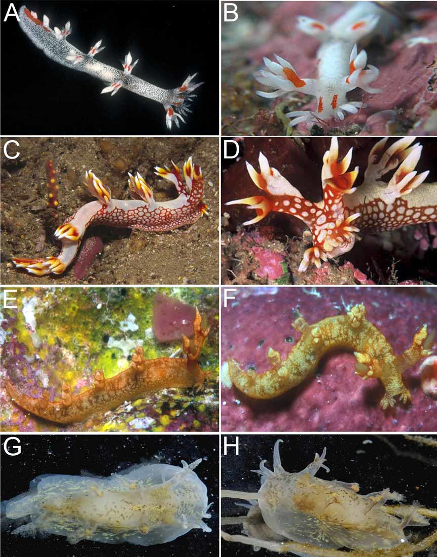
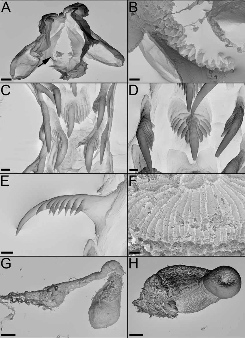
( Figures 16 View FIGURE 16 G–H, 15E, 20)
Pseudobornella orientalis Baba, 1932: 369 View in CoL , figs. 1–7; Baba, 1933: 278 –279, fig. 5.
Material examined: CASIZ 174988, China, Luoyuan Bay (E119°82’, N26°41’), 1 to 2.5 m depth on hydroids, two adult specimen 7 mm preserved, dissected, 15 March 2007, coll: S. Xikum. CASIZ 174989, China, Daisong Bay (E117°57’, N24°12’), 1 to 2.5 m depth on hydroids, 47 specimens 6-10 mm alive, 5 dissected, 1 April 2007, coll: S. Xikum.
Geographic Distribution: This species has been recorded from Japan ( Baba 1932; Okutani 2000; Masayoshi 2002) and China (Song 2006; present study).
External morphology: The general body shape is relatively elongate but the foot is very wide and posteriorly it tapers to a pointed tip. The living adults are small, the largest reported animal being less than 10 mm in length. The general background color is translucent whitish with numerous scattered brown spots and patches, and some small yellow diagonal streaks ( Figs. 16 View FIGURE 16 G-H). On either side of the vertical mouth are three smooth tapering oral tentacles, which increase in size outward from the mouth. From photographs of living animal, the outermost tentacle on each side is approximately half the body length; the middle tentacle is half that length, and the inner tentacle one quarter. The rhinophore sheath is very elongate, able to extend to a length similar to that of the outer oral tentacles. On the upper edge of each rhinophore sheath there are two lateral elongate papillae and one posterior one which is extremely slender and elongate, extending in the living animal, according to Baba (1933), to five times the body length. The rhinophores have approximately 10 lamellae. Posterior to the rhinophores there are four pairs of elongate dorsolateral processes, decreasing in size towards the posterior end of the foot, the first being almost as long as the rhinophore stalks. The dorsolateral processes are unbranched and taper to a rounded tip. There are numerous branched and unbranched gills attached along the inner side of each dorsolateral papilla. The anus is small, located on the right side of the dorsum between the first and second pair of dorsolateral processes. The reproductive opening is located on the right side, midway between the rhinophore sheath and the first dorsolateral process.
Alimentary Canal: The general external anatomy and the alimentary canal were described by Baba (1932). The buccal bulb is relatively large and the labial cuticle is thin, without armature. The jaws are elongate, with a distinct long masticatory process consisting of about 7 to 10 rows of small rodlets ( Figs. 20A,B View FIGURE 20. A – H ). A ventral oral gland is absent. The radular formulae of three specimens dissected are: 12 x 2.1.2 (CASIZ 174988), 10-11 x 2.1.2 (CASIZ 174989). The rachidian teeth are stout, elongate with a very strong and prominent cusp and with about six to seven very strong denticles on both sides of the cusp ( Figs. 20C,D View FIGURE 20. A – H ). These denticles appear to be about the same size, a little bit shorter close to the cusp. There are only two lateral denticles ( Fig. 20C View FIGURE 20. A – H ). The inner lateral is larger, with a very strong elongate, sharp cusp and eight to 9 denticles on both side of the cusp. These denticles are all about the same size, elongate and sharp ( Fig. 20E View FIGURE 20. A – H ). The outer lateral is shorter than the inner lateral, elongate and smooth ( Figs. 20C View FIGURE 20. A – H ). A pair of elongate salivary glands, opening into the posterior buccal bulb, is attached to the posterior side of the oesophagus. The oesophagus is short and wide opening into the large stomach. The stomach has a dorsal opening for each of the two anterior digestive glands, and an opening on the floor of the stomach for the posterior portion of the digestive gland. The anterior digestive glands each have a single elongate anterior lobe but it does not enter the most anterior dorsolateral process on its side of the body. There are also no branches of the posterior digestive gland to the other dorsolateral processes. There is no evidence of a posterior chamber in the stomach, with or without spines ( Fig. 20F View FIGURE 20. A – H ). From the stomach the intestine runs directly to the anus.
Reproductive system: The reproductive system is shown in Figure 15E View FIGURE 15. A – E . The ovotestis is a single mass not identifiable as separate follicles and lies dorsolaterally on the right side of the posterior digestive gland. From the ovotestis, the relatively short hermaphrodite duct opens into the large elongate ampulla. From the ampulla, the hermaphrodite duct continues for a short distance before branching into the oviduct and the vas deferens. The relatively unfolded vas deferens appears to be lined with a layer of prostate gland along its whole length. The penial bulb is relatively small and unarmed ( Fig. 20H View FIGURE 20. A – H ). A very large distal pyriform allosperm receptacle opens directly to the genital aperture via a long duct ( Fig. 20G View FIGURE 20. A – H ).
Remarks: Described from two 7 mm long preserved specimens, a good illustration of a living animal was published by Baba (1933). Since then little published information has been available, until a photo in Okutani (2000), and number of photos in the Sea Slug Forum ( Masayoshi 2002; Kurihara 2002; Hayashi 2002). The most distinctive external feature is the development on the rhinophoral sheath of a long slender tentacle, which can extend to at least five times the length of the body. The specimens studied in this paper perfectly match the original description of the species. The reproductive system is described here for the first time. Baba (1932) reported that it was found feeding on the hydroid Tubularia mesembryanthemum attached to Zostera . Three other records on the Sea Slug Forum ( Kinoshita 2002; Kurihara 2004 - both from Japan, and Kitagawa 2004 - from the Philippines) differ slightly in having longer lateral papillae on the rhinophore sheath. Until their anatomy is studied we cannot say if they are P. orientalis or a different species within this genus.

No known copyright restrictions apply. See Agosti, D., Egloff, W., 2009. Taxonomic information exchange and copyright: the Plazi approach. BMC Research Notes 2009, 2:53 for further explanation.
|
Kingdom |
|
|
Phylum |
|
|
Class |
|
|
Order |
|
|
Family |
|
|
Genus |
Pseudobornella orientalis Baba, 1932
| Pola, Marta, Rudman, William B. & Gosliner, Terrence M. 2009 |
Pseudobornella orientalis
| Baba 1933: 278 |
| Baba 1932: 369 |