
Mahurubia clava, Willems & Artois & Vermin & Backeljau & Schockaert, 2005
|
publication ID |
https://doi.org/ 10.1080/00222930400014239 |
|
persistent identifier |
https://treatment.plazi.org/id/03FA2A32-4811-FFB6-2AC1-E391FC6EFEC8 |
|
treatment provided by |
Felipe |
|
scientific name |
Mahurubia clava |
| status |
gen. nov. |
Mahurubia clava View in CoL gen. nov. sp. nov.
mahurubia-clava sp. nov.
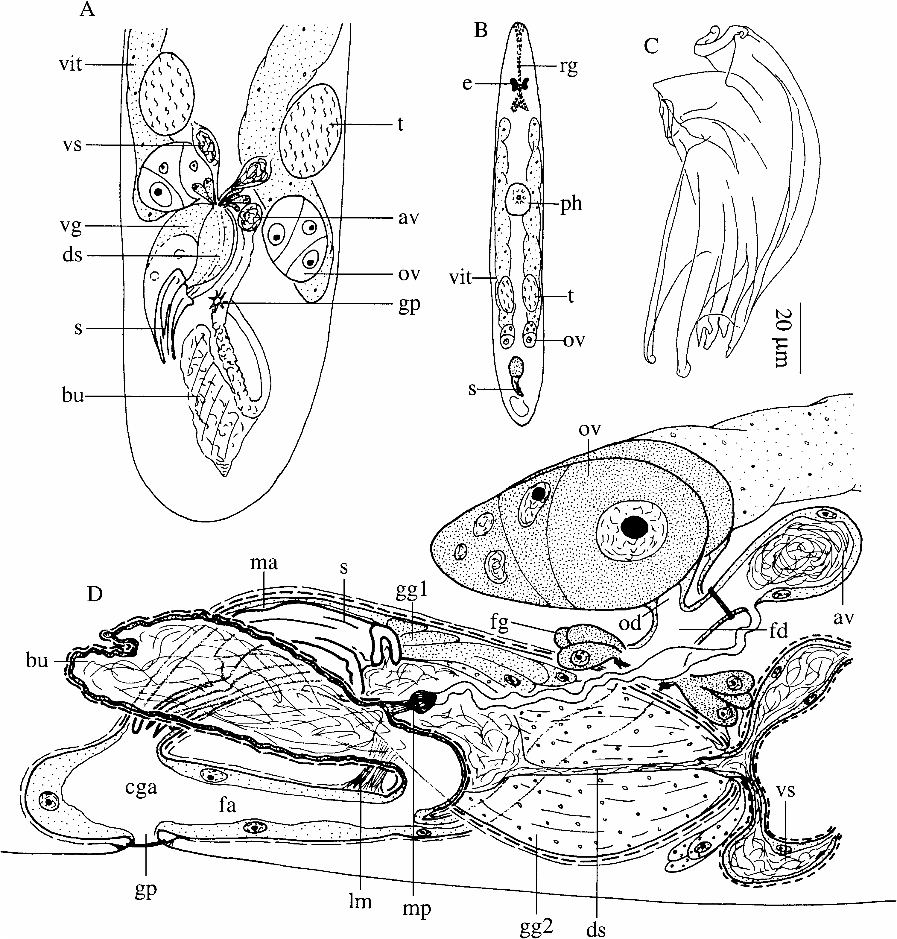
( Figure 2 View Figure 2 )
Diagnosis
Mahurubia gen. nov. Trigonostominae with the pharynx in the middle of the body. Testes and seminal vesicles paired, far behind the pharynx. Prostate stylet consisting of two parts, one of which is gutter-shaped and receives the prostate secretion, the other one a folded plate-like structure, which carries several spines distally. Additional vesicle (with sperm) at the proximal part of the female (efferent) duct. Afferent system consisting of tortuous duct and large, thick-walled bursa. Mouthpiece of bursa club-shaped. Type species: Mahurubia clava .
Mahurubia clava sp. nov. Provisionally with the same diagnosis as the genus. Stylet 87 Mm long.
Locality
Zanzibar, Mahurubi Palace Ruins : open beach with fine sand in front of the mangroves, at low tide (5 August 1995) (type locality); same locality: sandflat with crab holes (17 August 1995) .
Material
Three individuals studied alive and mounted: one designated holotype ( LUC no. 275), another one paratype ( LUC no. 276). Three serially sectioned animals (paratypes; LUC no. 277–279) .
Etymology
The genus name/praenomen refers to the type locality, Mahurubi ( Zanzibar, Tanzania); gender: feminine. The species epithet refers to the shape of the mouthpiece; clava (Latin) : club .
Description
The animals are ¡ 0.6–0.9 mm long (measured on whole mounts), with two eyes. The cellular epidermis is about 2 Mm thick, with cilia 1.5 Mm long. Large rhabdites occur throughout the epidermis, but are slightly more concentrated at the rostral and caudal ends. Rostrally there are two long strands of rhabdite glands ( Figure 2B View Figure 2 : rg). Caudal glands are also well developed.
The pharynx is situated at about 40%. There are basophilic and eosinophilic pharyngeal glands. The nucleated epithelium of the pharyngeal lumen is low. There are 24 internal longitudinal and 24 radial muscles in horizontal sections.
The common genital pore lies at ¡90%. The common genital atrium is rather wide, lined with a high, nucleated epithelium, and surrounded by longitudinal muscles only. It extends rostrally to form the female atrium and communicates dorsally with the male atrium.
Paired testes are situated far behind the pharynx, just in front of the ovaries ( Figure 2A, B View Figure 2 ). Seminal vesicles are paired, lined with low, nucleated epithelium and surrounded by spiral muscles. Both vesicles narrow and subsequently fuse to form the seminal duct ( Figure 2D View Figure 2 : ds), just before entering the prostate vesicle. The seminal duct continues through the prostate vesicle as a very narrow duct and widens just before entering the stylet. The prostate vesicle ( Figure 2A View Figure 2 : vg) is very large and elongated. It is surrounded by inner circular and outer longitudinal muscles. Two kinds of prostate glands are present: eosinophilic ones ( Figure 2D View Figure 2 : gg2) with extracapsular nucleated parts, and basophilic glands, which are completely intracapsular ( Figure 2D View Figure 2 : gg1). The very complex stylet ( Figure 2C View Figure 2 ) is 81–90 Mm long (mean587 Mm; n 53) and consists of two parts: a bent, gutter-shaped one (maybe a closed tube) and a folded plate-like part at the concave side, which is attached to the gutter-shaped part over most of its length. Sperm is probably discharged in between these two parts, while the gutter-shaped one receives the prostate secretion. The male genital atrium is lined with a membranous epithelium and surrounded by inner circular and outer longitudinal muscles. Distally, the circular layer ends where the male atrium enters the common genital atrium. A short, but strong bundle of longitudinal muscle fibres ( Figure 2D View Figure 2 : lm) connects the proximal part of the male atrium with the common genital atrium.
The paired, ovoid ovaries are situated on both sides of the prostate vesicle. They form the most caudal part of the elongated ovovitellaria ( Figure 2B View Figure 2 : ov+vit), which extend from just behind the eyes to the prostate vesicle. There is a large variation in size of the ovaries between different individuals, possibly due to differences in developmental stage. The female duct (5efferent duct; Figure 2D View Figure 2 : fd) enters the female atrium ( Figure 2D View Figure 2 : fa) anteriorly. The common genital atrium, the female atrium and the female duct are all lined with a high, nucleated epithelium and surrounded by longitudinal muscles. Distally from the junction of the oviducts a large bundle of eosinophilic glands ( Figure 2D View Figure 2 : fg) opens into the female duct, which is guarded by a sphincter. Proximally from the junction, the female duct ends in a small sperm-containing vesicle ( Figure 2D View Figure 2 : av), lined with a low, nucleated epithelium and filled with motile sperm in the living animal. This additional vesicle can be closed by a weak sphincter. At the transition between the female duct and the vesicle a tortuous duct (5part of the afferent duct) connects a large bursa ( Figure 2D View Figure 2 : bu) with the ovaries. Where the duct leaves the bursa, a sclerotized, club-shaped mouthpiece ( Figure 2D View Figure 2 : mp; not visible on whole mounts) is present. The bursa proper is thickwalled, surrounded by strong spirally running, almost longitudinal muscles, and enters the female atrium dorsally, just above the female duct. A uterus is lacking.
No known copyright restrictions apply. See Agosti, D., Egloff, W., 2009. Taxonomic information exchange and copyright: the Plazi approach. BMC Research Notes 2009, 2:53 for further explanation.