
Triconia komo, Cho & Böttger-Schnack & Kim & Lee, 2019
|
publication ID |
https://doi.org/ 10.5252/zoosystema2019v41a28 |
|
publication LSID |
urn:lsid:zoobank.org:pub:EB5117B3-49C3-4F42-B8A7-65C9F3841A5A |
|
DOI |
https://doi.org/10.5281/zenodo.4450186 |
|
persistent identifier |
https://treatment.plazi.org/id/CA4A0457-8738-4141-BE3C-1A3F55CBD619 |
|
taxon LSID |
lsid:zoobank.org:act:CA4A0457-8738-4141-BE3C-1A3F55CBD619 |
|
treatment provided by |
Felipe |
|
scientific name |
Triconia komo |
| status |
sp. nov. |
Triconia komo n. sp.
( Figs 2-5 View FIG View FIG View FIG View FIG ; 13A View FIG ; Tables 2-4)
urn:lsid:zoobank.org:act:CA4A0457-8738-4141-BE3C-1A3F55CBD619
TYPE LOCALITY. — Northeastern equatorial Pacific Ocean (10°30’N, 131°20’W, 0-100 m).
TYPE MATERIAL. — Holotype. 1 ♀; NIBRIV0000838000 ; 10°30’N, 131°20’W; 0-100 m; dissected and mounted on 10 slides, collected from the type locality on 21.VIII.2009 by D. J. Ham. GoogleMaps
Paratypes. 5 ♀; NIBRIV0000838001-005; 10°30’N, 131°20’W; each dissected and mounted on each of 5, 8 or 10 slides (lost a slide of urosome with P5 of the third paratype) . — 2 ♂; NI-BRIV 0000838006-007; each dissected and mounted on 8 or 9 slides, respectively. — 4 ♀, 1 ♂ kept in one vial in alcohol, MNHN-IU-2019-2282 . All specimens are from the type locality.
ETYMOLOGY. — The species is named after the station name KOMO [KODOS (the Korea Deep Ocean Study) Long-term Monitoring Station’ in the Korean mining area for manganese nodules in the central part of the Clarion-Clipperton Fracture Zone], which is the type locality of the new species in the northeastern equatorial Pacific Ocean.
DESCRIPTION
Female
Body length. 613-658 Μm, based on six specimens.
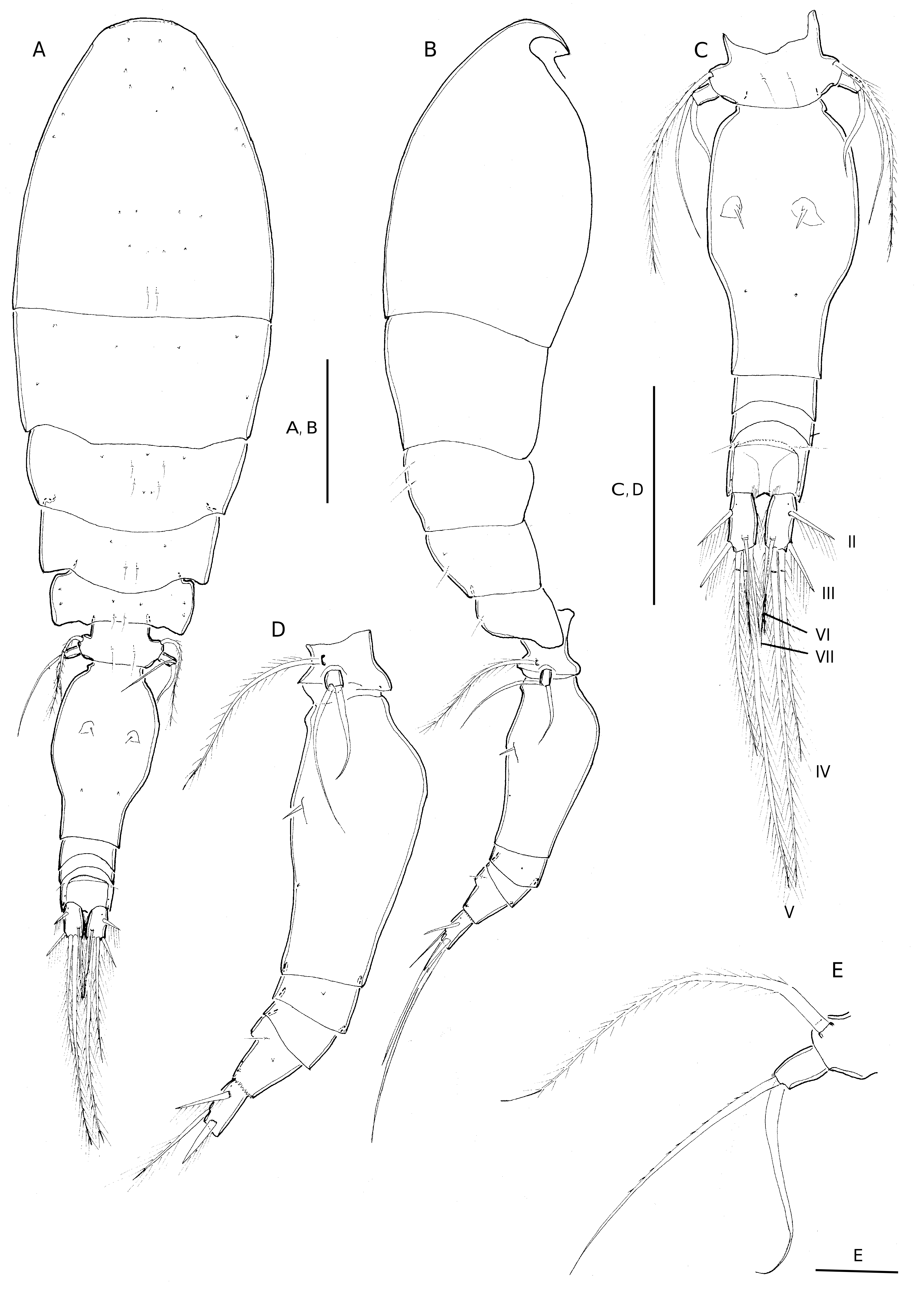
Exoskeleton weakly chitinized. Prosome 2.0 times length of urosome, excluding caudal rami, 1.8 times urosome length including caudal rami. P2-bearing somite without conspicuous dorsoposterior projection in lateral aspect ( Fig. 2B View FIG ). Integumental pores as figured in Fig. 2A, B View FIG . Pleural areas of P4-bearing somite with rounded posterolateral corners in lateral aspect. One pair of secretory pores laterally discernible on first postgenital somite ( Fig. 2D View FIG ).
Genital double-somite. 1.7 times as long as maximum width (measured in dorsal aspect) and about 2.1 times as long as postgenital somites combined; bottle-shaped with largest width measured at anterior two thirds and with moderately rounded lateral margins, posterior part tapering gradually. Paired genital apertures located at about 2/5 distance from anterior margin of genital double-somite; armature represented by 1 long spine and two minute spinous processes ( Fig. 13A View FIG ). Pore pattern on dorsal surface as in Fig. 2C View FIG .
Anal somite. Slightly wider than long; slightly longer than caudal rami ( Fig. 2C View FIG ). Secretory pore discernible on either side of anal opening and additional pair of pores anterolaterally on anal somite. Anterior margin of anal opening with transverse row of minute denticles. Posterior margin of somite finely serrate ventrally and laterally ( Fig. 2D View FIG ).
Caudal ramus ( Fig. 2C View FIG ). 1.8 times longer than wide. Dorsal seta (VII) about half the length of seta IV, about as long as seta VI. Inner margin of somite with few long, fine spinules. Dorsal anterior surface ( Fig. 2C View FIG ) with secretory pore near insertion of seta II.
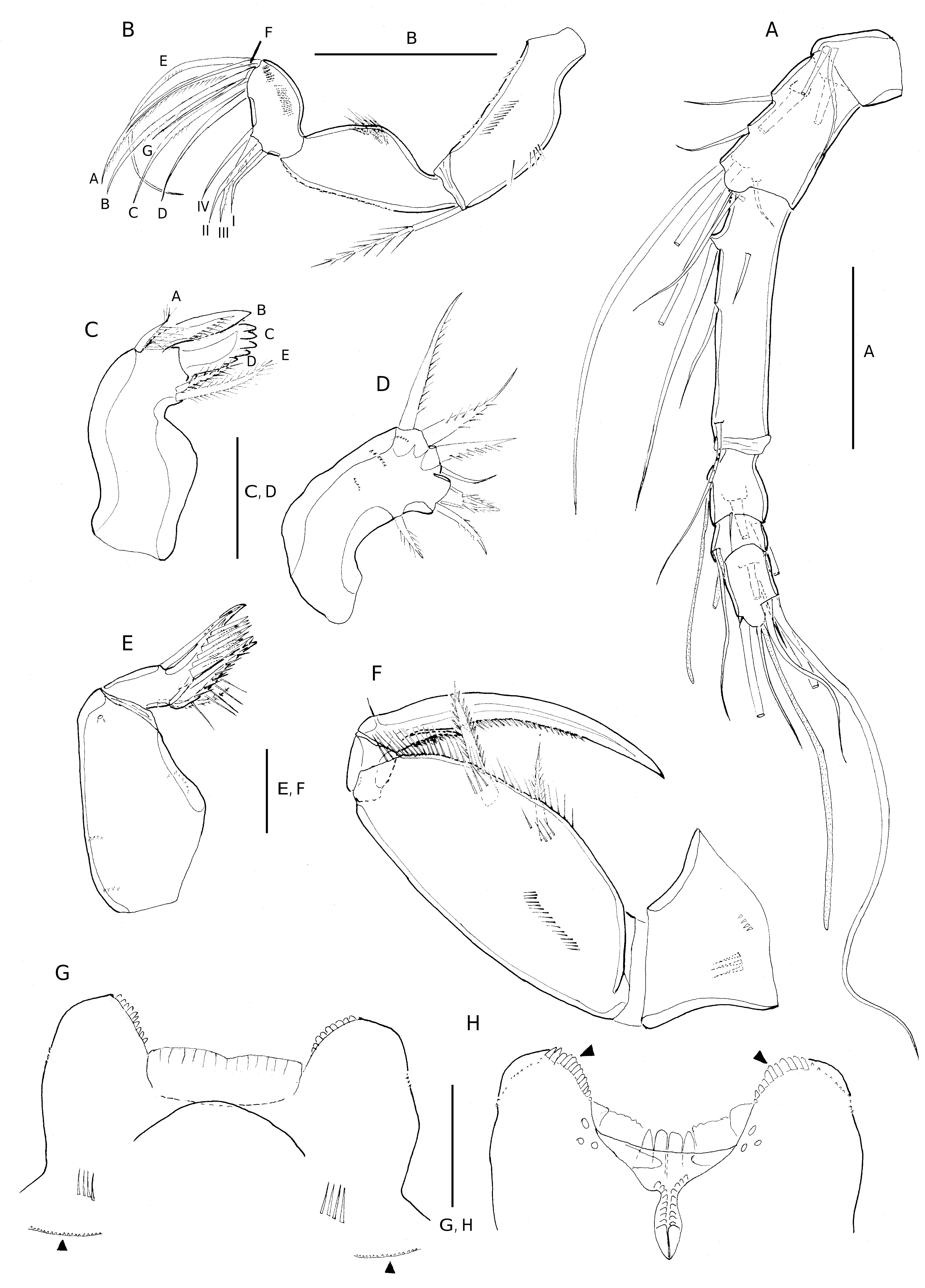
Antennule ( Fig. 3A View FIG ). 6-segmented. Armature formula: 1-[3], 2-[8], 3-[5], 4-[3+ae], 5-[2+ae], 6-[6+(1+ae)].
Antenna ( Fig. 3B View FIG ). 3-segmented, distinctly reflexed.Coxobasis armed with bipinnate seta at inner distal corner; surface of coxobasis with row of long, fine spinules along outer and inner margins and with few minute denticles along outer margin. Endopod segments unequal in length; proximal endopod segment subtriangular forming outer lobate outgrowth bearing spinular patch, with row of denticles along posterior inner margin. Distal endopod segment distinctly shorter than proximal endopod segment and articulating with narrow cylindrical base; with two patches of short spinules along outer margin; lateral armature consisting of one pinnate seta (III) and three naked setae (I, II, IV), seta II + III longest; distal armature consisting of one long curved seta (E), four curved setae (A-D) of graduated length, ornamented with pinnules along entire inner margin (A) or at middle part (B, C), and two slender bare setae (F and G), seta G shorter than seta D and slightly shorter than F.
Labrum ( Fig. 3G, H View FIG ). Distinctly bilobed. Each lobe with row of minute denticles around outer ventral margin and strong dentiform processes converging and slightly decreasing in size medially (arrowed in Fig. 3H View FIG ). Lobes separated by medial concavity covered anteriorly by single hyaline lamella ( Fig. 3G View FIG ). Posterior part of medial concavity ornamented with four long sclerotized, dentiform processes (“teeth”), posterior surface with group of three secretory pores located on proximal part of each lobe ( Fig. 3H View FIG ). Anterior surface ( Fig. 3G View FIG ) with paired row of long setules either side of median swelling, and paired integumental pockets latero-posteriorly, which are difficult to illustrate under a light microscope because of their three-dimensional nature, but have been shown in detail for other oncaeid species by a scanning electron microscope (cf. Böttger-Schnack 2001: fig. 9A, C, D), free margin of pockets ornamented with minute denticles (arrowed in Fig. 3G View FIG ).
Mandible ( Fig. 3C View FIG ). With surface of coxa unornamented; gnathobase with five elements, numbered using capital letters in Fig. 3C View FIG : element A (ventral element) much shorter than ventral blade B, with long setules; ventral blade B strong and broad, with row of setules on posterior surface; dorsal blade C strong and broad, with six or seven dentiform processes around distal margin and along distal half of dorsal margin; dorsal elements setiform, shorter spinulose (D), longer bipinnate (E).
Maxillule ( Fig. 3D View FIG ). Weakly bilobed, surface ornamented with few spinules. Inner lobe with three elements: outermost one spiniform and swollen at base, with three strong spinules at midregion and spinulose at distal part, middle element setiform and indistinctly bipinnate, innermost element bipinnate, located at some distance from others. Outer lobe with four elements: innermost element setiform and naked, element next to outermost spiniform and strong, with row of short spinules, two outermost elements setiform and pectinate, outermost element longest.
Maxilla ( Fig. 3E View FIG ). 2-segmented. Syncoxa unarmed, surface ornamented with few spinular rows and one large secretory pore; allobasis produced distally into slightly curved claw bearing two rows of strong spinules along inner margin; outer margin with stout naked seta almost extending to tip of allobasal claw; inner margin with slender spinulose seta and strong basally swollen spine ornamented with two spinular rows along medial margin and few long spinules at outer margin.
Maxilliped ( Fig. 3F View FIG ). 4-segmented, comprising syncoxa, basis and 2-segmented endopod. Syncoxa unarmed, posterior surface ornamented with few spinules. Basis robust, inner margin with two spiniform bipinnate elements unequal in length, proximal one more slender and slightly shorter than distal one; fringe of long pinnules along inner margin between distal spine and articulation with endopod and row of long spinules between distal and proximal spine; few short transverse rows of long setules on anterior surface and additional longitudinal row as illustrated in Fig. 3F View FIG . Distal endopod segment drawn out into long stout claw, with row of pinnules on proximal 2/3 of concave margin; accessory armature consisting of minute, naked seta on outer proximal margin and unipectinate spine basally fused to inner proximal corner of claw.
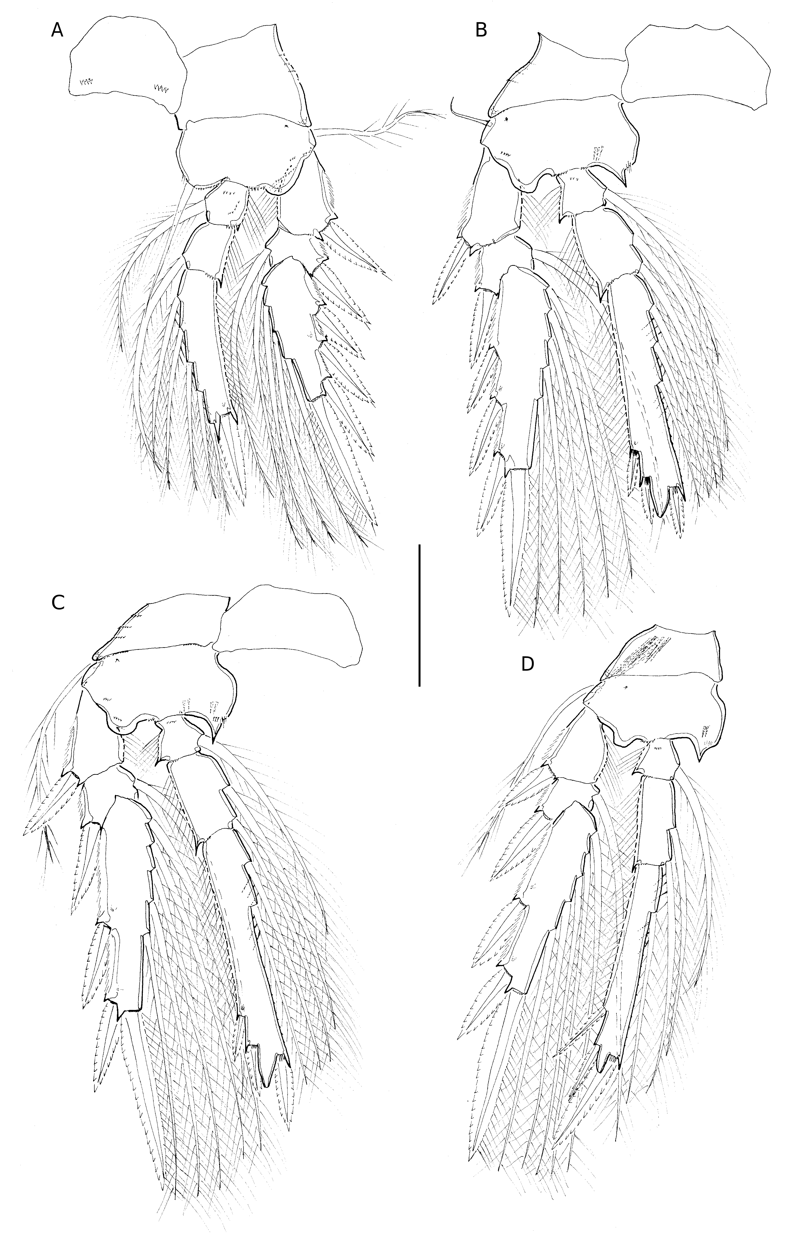
Swimming legs 1-4. Biramous ( Fig. 4 View FIG A-D). With three-segmented rami. Intercoxal sclerites well developed, ornamented with paired row of few denticles distal corner in P1. Coxae and bases of P1-P4 with surface ornamentation as in Fig. 4 View FIG A-D. Coxae of P1-P4 with raised secretory pore on posterior face near outer distal corner. Bases with short naked (P2) or long plumose (P1, P3, P4) outer seta; with anterior secretory pore near outer proximal corner. Coxa of P4 with tuft of very long fine setules posteriorly at outer proximal corner. Inner basal seta on P1 long and spiniform.
Leg armature formula
See Table 1.
Exopods. Outer margin of exopod segments with well-developed serrated hyaline lamella; inner margin of proximal exopod segments with long setules. Secretory pore located on posterior surface of distal segments; hyaline lamellae on outer spines well developed; outer and distal spines of P1 with subapical tubular extension; this extension lacking on proximalmost spine of exp-3. Distal spine equal in length to (P1) or shorter than (P2-P4) distal exopod segment. Length ranges of outer spine on exp-1 relative to outer spine on exp-2 of P3 and P4 are given in Table 3.
Endopods. Distal endopod segments with (P1-P3) or without (P4) single secretory pore on posterior surface. Distal margin of P2-P4 produced into conical process with apical pore, cone smaller in size and more slender on P4 ( Fig. 4 View FIG B-D). Length data of endopodal spines of holotype and five paratype females as shown in Table 2; length ranges of outer subdistal spine (OSDS) and outer distal spine (ODS) relative to distal spine (DS) are given in Table 3.
P5 ( Fig. 2E View FIG ). Comprising free exopod segment with few denticles on distal margin; very long and plumose outer basal seta. Exopod about 1.5 times longer than wide, bearing strong, curved inner seta and very long, slender outer seta, outer seta about 1.3 times longer than inner seta, reaching about to middle of genital double-somite.
P6 ( Figs 2C View FIG ; 13A View FIG ). Represented by operculum closing off each genital aperture; armed with long spine and two minute spinules (only one of which is discernible under a light microscope).
Male
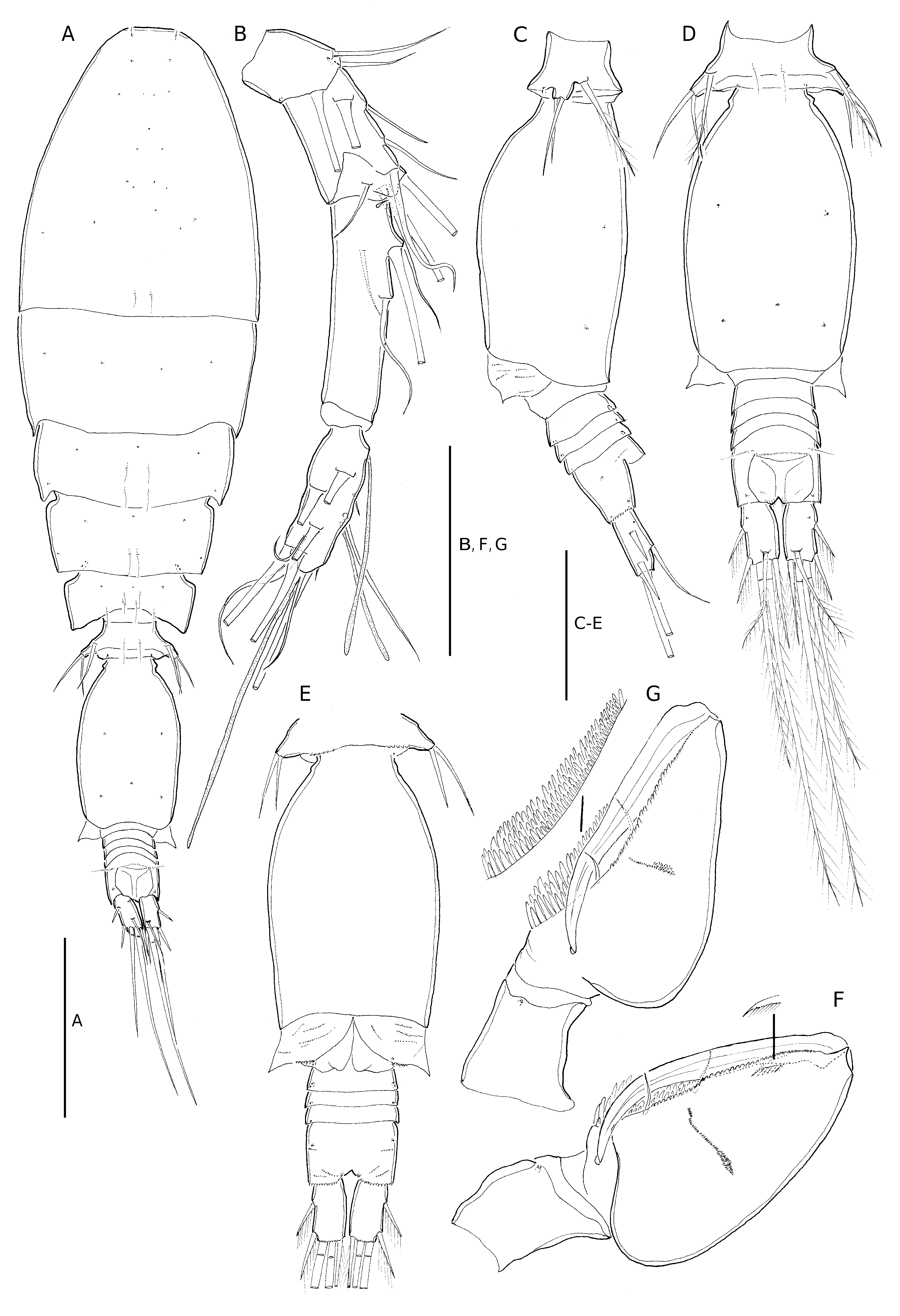
Body length. 503-507 Μm, based on two specimens. Sexual dimorphism in antennule, maxilliped, P5-P6 and in genital segmentation. Posterior margin of P5-bearing somite with paired row of minute denticles or spinules ventrally ( Fig. 5E View FIG ).
Prosome. 2.1 times the length of urosome, excluding caudal rami, about 1.9 times urosome length, including caudal rami ( Fig. 5A View FIG ). Integumental pores on prosome and urosome as figured ( Fig. 5A View FIG ).
Caudal rami. 1.7 times longer than wide, slightly shorter than in female; with length to width ratio and proportional lengths of caudal setae as in female. Dorsal surface of genital somite with five secretory pores as indicated in Fig. 5D View FIG . Surface of genital flaps ornamented with several rows of small spinules ( Fig. 5E View FIG ). Anal somite slightly wider than long as in female; with few rows of minute spinules on ventral surface.
Antennule ( Fig. 5B View FIG ) 4-segmented with distal segment corresponding to fused segments 4-6 of female. Armature formula 1-[3], 2-[8], 3-[4], 4-[11+2ae+(1+ae)].
Maxilliped ( Fig. 5F, G View FIG ). 3-segmented, comprising syncoxa, basis and 1-segmented endopod. Syncoxa without surface ornamentation except for single secretory pore, unarmed. Basis robust, particularly inflated in proximal half forming bulbous swelling; anterior surface with 1-2 transverse spinular rows, and with small flat spinules along inner margin between proximal spine and articulation with endopod ( Fig. 5F, G View FIG ); posterior surface with three rows of spatulate spinules of graduated length along palmar margin ( Fig. 5G View FIG ); with two small naked setae of equal length inserted within longitudinal cleft. Endopod drawn out into long curved claw, concave margin unornamented: accessory armature consisting of short, unipectinate spine basally fused to inner proximal corner of claw, figured separately in Fig. 5F View FIG ; claw with minute hyaline apex.
Swimming legs. With armature and ornamentation as in female; length data of endopodal spines of two males as shown in Table 2; length ranges of outer subdistal spine (OSDS) and outer distal spine (ODS) relative to distal spine are given in Table 4. Length ranges of outer spine on exp-1 relative to outer spine on exp-2 of P3 and P4 are given in Table 4.
P5 ( Fig. 5C, D View FIG ). Exopod not delimited from somite, shorter than that of female, armature as in female, but setal lengths much shorter than in female, with outer exopodal seta slightly longer than inner seta; outer basal seta bipinnate, much shorter than in female.
P6 ( Fig. 5E View FIG ). Represented by posterolateral flap closing off genital aperture on either side; covered by pattern of spinules as shown in Fig. 5E View FIG ; posterolateral corners protruding laterally and visible in dorsal aspect ( Fig. 5A, D View FIG ).
REMARKS
Triconia komo n. sp. belongs to the 1st group within the similis - subgroup of Triconia , based on the proportional length of the distal spine on P2 enp-3, which is longer than the conical process in all species of this group (cf. Böttger-Schnack & Machida 2011, see also introduction of the present paper). Among the four described species of this group, the females of T. komo n. sp. from the northeastern equatorial Pacific Ocean are most similar to T. hawii in the shape of the genital double-somite in dorsal view and the proportional lengths of the exopodal setae on P5. However, the females of the two species differ in: 1) body length, which is larger in T. komo n. sp. (613-658 Μm) than in T. hawii (490-560 Μm); 2) the relative length of outer basal seta on P5, which is longer than the outer exopodal seta (1.36:1) in T. komo n. sp. reaching beyond the genital apertures in dorsal view ( Fig. 2E View FIG ), while the outer basal seta is shorter than the outer exopodal seta (0.84:1) in T. hawii , not reaching as far as the genital apertures (cf. Böttger-Schnack 1999, fig. 21B); 3) the length to width ratio of the caudal rami is slightly larger (about 1.8:1) than in T. hawii (1.6:1); 4) the relative length of caudal seta VI, which is about half the length of seta IV in T. komo n. sp., while seta VI is about 1/3 the length of seta IV in T. hawii ; and 5) in the form and length to width ratio of the genital double-somite, which is somewhat more elongate (1.7:1) than in T. hawii (1.5:1), with the narrower posterior part being longer (1/3 the length of genital double-somite) than in T. hawii (1/4-1/5 the length of genital double-somite).
In addition to the differences in morphological characters stated above morphometric differences are indicated in the proportional lengths of the exopodal spines on P3: the exopodal spine of exp-1 relative to the exopodal spine of exp-2 on P3, tends to be higher in T. komo n. sp. (0.77-0.87:1) than in T. hawii (0.69-0.78:1).
Another morphometric difference between T. komo n. sp. and T. hawii was assumed to exist in the endopodal spine lengths on P2 and P3 enp-3: the outer distal spine is reaching only slightly beyond the tip of the conical process in T. komo n. sp. ( Fig. 4B, C View FIG ), while this spine is reaching far beyond the tip of the cone in T. hawii (according to Böttger-Schnack & Boxshall 1990: fig. 4C, D). However, a detailed re-examination of the swimming legs of T. hawii from the type locality in the Red Sea, based on the copepod material from the personal collection of R. Böttger-Schnack [cf. Böttger-Schnack 1999, p. 85 (c)], showed that the lengths of outer distal endopodal spines had not been figured entirely correctly in the original description by Böttger-Schnack & Boxshall (1990: fig. 4C, D): the outer distal spine on P2 and P3 enp-3 is somewhat shorter than figured in that account and is not reaching far beyond the tip of the cone, but only slightly beyond the cone. In the redescription of the species from the Red Sea by Böttger-Schnack (1999) these morphometric characters had not be re-examined in detail and so the error was not noted. A detailed revision of the endopodal spine lengths on P2-P4 of the genuine T. hawii from the Red Sea including new figures is in progress (Böttger-Schnack, pers. obs.). In the present paper, the updated measurements of outer distal spines on the endopods of P2-P4 of T. hawii were used to recalculate the length ratio of the outer distal to the distal spine on these legs, which, however, did not change significantly so that the given values are still valid for comparison with other species of the similis -subgroup ( Table 3).
Further minor differences between T. komo n. sp. and T. hawii are found in several ornamentation details, such as of seta I on the second endopod segment of the antenna, of the innermost element on the outer lobe of maxillule and of the seta on the outer allobasal margin of the maxilla, all of which are naked in T. komo n. sp., but minutely ornamented in T. hawii .
Table 4 presents a comparison of morphological features for the described males of the similis -subgroup. The male of T. komo n. sp. differs from previously described four males of the similis -subgroup (including the male of T. onnuri n. sp. described below) by the following morphological characters: a smaller size (503-507 Μm) than males of T. similis and T. onnuri n. sp., which are longer than 570 Μm; the pore pattern on the dorsal surface of the genital somite, with 5 pores compared to 3 in T. similis , T. onnuri n. sp. and T. recta ; the relative length of the inner seta on P5, which is shorter than the outer seta in the male of T. komo n. sp., but slightly longer in T. hawii ; the length ratio of seta VI to seta VII on the caudal ramus, which is about 1 in the male of T. komo n. sp., while it is less than 1 in males of T. hawii and T. recta . Additional small differences between the four species are found in the few minute denticles or spinules on the ventral face of the P5-bearing somite in T. komo n. sp., which are absent in the other males except T. onnuri n. sp., and in the length to width ratio of the caudal ramus (cf. Table 4).
The male of T. denticula , representing another species of the similis -subgroup, is still unknown. Females of T. denticula were found to co-occur with T. komo n. sp. in the study area during the present study, but were very rare, while T. komo n. sp. was common. No mating pairs of either species were found, which would have provided clear evidence of the identity of the male; however, the male of T. komo n. sp. was tentatively identified on the basis of the relative lengths of caudal seta VI, being similar to seta VII as in female of T. komo n. sp., while in female of T. denticula , seta VI is much shorter than seta VII. The proportional spine lengths on the endopods of P2-P4, which were examined in detail for both sexes in the present study, are not helpful for a distinction, as they are similar between females of the two species ( Table 3); however, the ratio of the outer distal spine to distal spine on P4 enp- 3 in the male ( Table 4) appears to be more similar to T. komo n. sp. than to T. denticula . Also, the proportional spine lengths on the exopods of P3 and P4, which are proposed as new diagnostic characters for the identification of oncaeid males, appear to be more similar to T. komo n. sp. than to T. denticula ( Tables 3, 4). However, in view of the limited number of male individuals examined for these morphometric characters, their variability is still insufficiently known and support for a positive identification can only be given in further studies on a larger number of specimens in order to more clearly define the indicative value of these characters.
No known copyright restrictions apply. See Agosti, D., Egloff, W., 2009. Taxonomic information exchange and copyright: the Plazi approach. BMC Research Notes 2009, 2:53 for further explanation.
|
Kingdom |
|
|
Phylum |
|
|
Class |
|
|
Order |
|
|
Family |
|
|
Genus |