
Utivarachna dusun Deeleman-Reinhold, 2001
|
publication ID |
https://doi.org/ 10.11646/zootaxa.5343.1.3 |
|
publication LSID |
lsid:zoobank.org:pub:20BF9AB4-F86D-42EA-AEAA-7C03EBA44EB6 |
|
DOI |
https://doi.org/10.5281/zenodo.8324637 |
|
persistent identifier |
https://treatment.plazi.org/id/03FA87E7-FFB4-FFDD-FF7D-3BBEFE5F8957 |
|
treatment provided by |
Plazi |
|
scientific name |
Utivarachna dusun Deeleman-Reinhold, 2001 |
| status |
|
Utivarachna dusun Deeleman-Reinhold, 2001 View in CoL
Figs 4–7 View FIGURE 4 View FIGURE 5 View FIGURE 6 View FIGURE 7
Utivarachna dusun Deeleman-Reinhold, 2001: 389 View in CoL , figs 610–619.
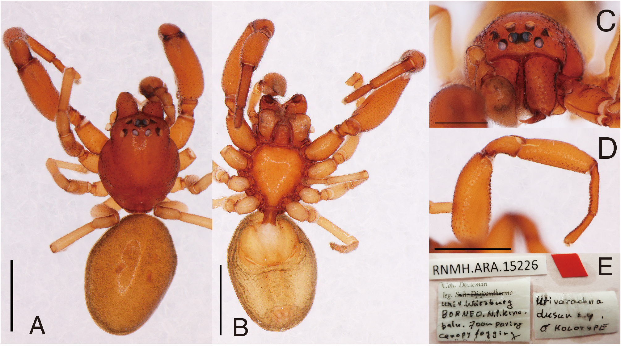
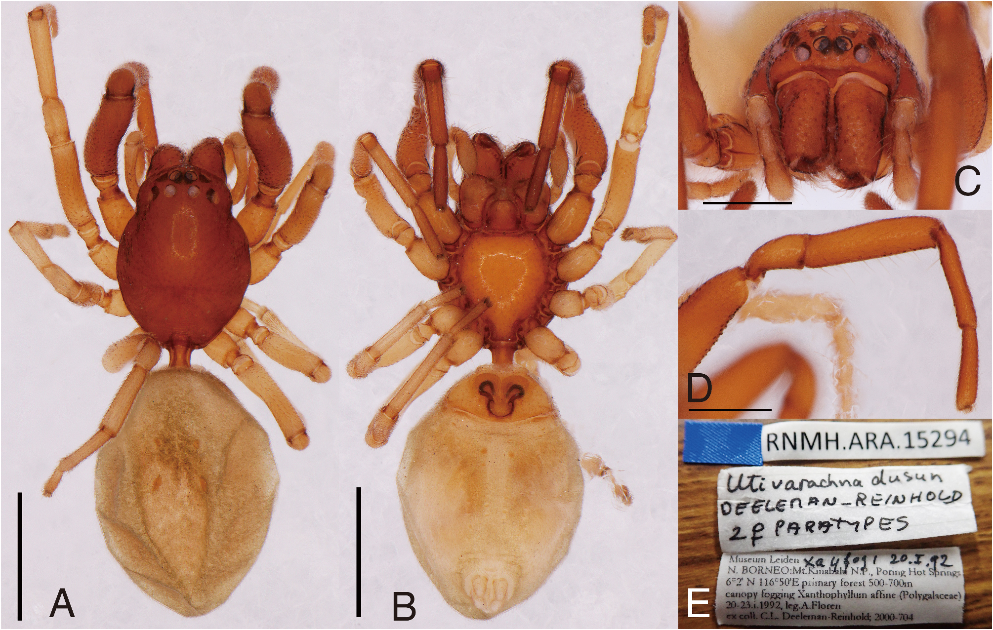
Type material examined. Holotype ♁ ( RNMH.ARA.15226; label data shown in Fig. 4E View FIGURE 4 ), Canopy fogging, [alt.] 700 m, Poring [Poring Hot Spring], N. P. Kinabalu, Borneo, leg. Univ. Würzburg, Coll. Deeleman. Paratypes: 2 ♀ ( RMNH. ARA. 15294; label data shown in Fig. 6E View FIGURE 6 ), canopy fogging, Xanthophyllum affine (Polygalaceae) , primary forest (6º2’N 116º50’E), 500–700 m, Poring Hot Springs, Mt. Kinabalu N. P., N. Borneo, 20.I.1992, leg. A. Floren, ex coll. C.L. Deeleman-Reinhold; 2000-704.
Diagnosis. Males of U. dusun are distinguishable from those of U. rubra by the shape of the RTA. Apical third of the RTA is curved in U. dusun , whereas apical half is curved in U. rubra . Regarding females, U. dusun is distinguishable from U. rubra by the size of the copulatory atrium and the shape of bursae. In females of U. dusun , the copulatory atrium is smaller than the area occupied by the internal ducts, and the bursae are club-shaped and swollen distally. In females of U. rubra , the atrium is large, and the bursae are slender than those of U. dusun .
Measurements (holotype ♁/ paratype ♀). Carapace length 1.65/1.52; cephalic width 0.86/0.78; thoracic width 1.21/1.10. Diameter of eyes: AME 0.10/0.08; ALE 0.12/0.12; PME 0.10/0.11; PLE 0.11/0.10. Interdistances between eyes: AME–AME 0.03/0.03; AME–ALE 0.01/0.01; ALE–ALE 0.27/0.25; PME–PME 0.08/0.08; PME– PLE 0.10/0.08; PLE–PLE 0.50/0.46; ALE–PLE 0.08/0.06; AME–PME 0.08/0.03. MOA: length 0.27/0.21; anterior width 0.23/0.22; posterior width 0.31/0.30. Clypeus height: 0.27/0.21. Abdomen length 1.82/2.07; width 1.32/1.41. Legs: I 4.07 (1.35, 0.50, 1.01, 0.70, 0.51)/3.45 (1.08, 0.37, 0.83, 0.65, 0.52); II n/a (0.98, 0.38, 0.75, lost, lost)/3.03 (0.92, 0.31, 0.71, 0.66, 0.43); III 2.57 (0.76, 0.35, 0.52, 0.62, 0.32)/2.42 (0.71, 0.32, 0.50, 0.56, 0.33); IV 3.16 (0.87, 0.35, 0.72, 0.86, 0.36)/3.21 (0.91, 0.32, 0.78, 0.83, 0.37).
Male ( Fig. 4A–D View FIGURE 4 ). Carapace oval and superficially granulated with truncated posterior end. Thoracic lateral margins slightly undulating. AER slightly recurved. PER longer than AER, almost straight. Fovea shallow, indistinct. Ventral margin of clypeus with small projection. Chelicerae superficially granulated, with three teeth on each prolateral and retrolateral margin. Pedicel short. Abdomen oval; entire dorsum weakly sclerotized. Legs superficially granulated; leg I enlarged, with bearing cusps ventrally.
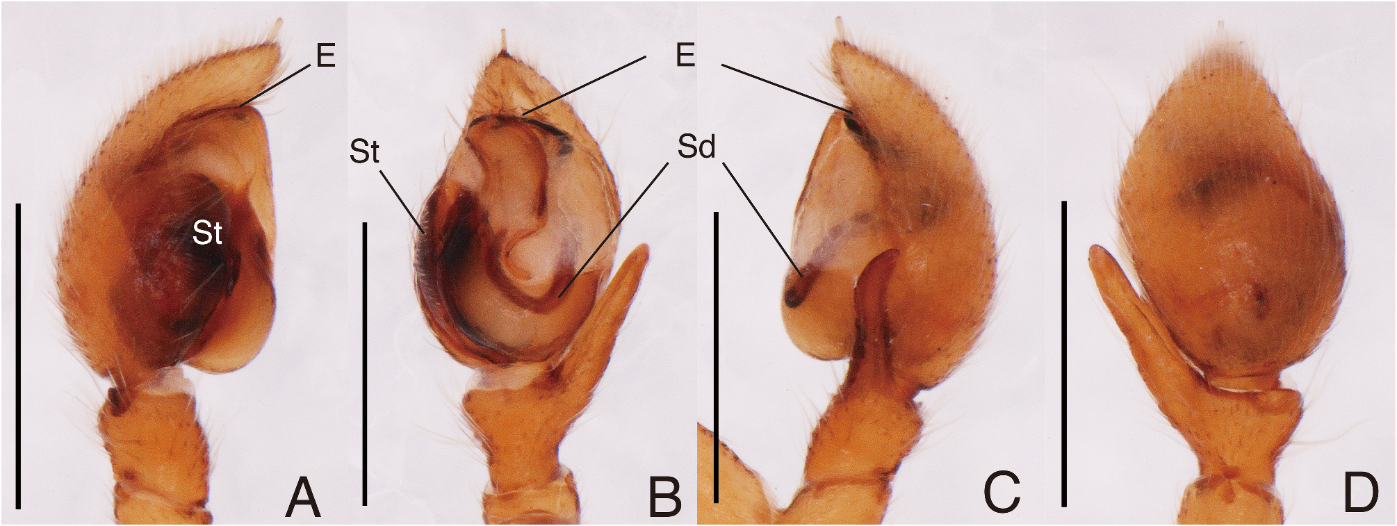
Palp ( Fig. 5A–D View FIGURE 5 ). Cymbium oval. Bulb oval. Sperm duct U-shaped, visible through median tegulum. Embolus coiled at horizontal plane; embolus coil plane inclining dorsally, covered by cymbium; basal embolus part accompanied by sclerotized plate originated in middle of tegulum. RTA long; apical third of RTA curved dorsally.
Coloration and setation ( Fig. 4A–D View FIGURE 4 ). Carapace light brown. Chelicerae light brown. Abdomen covered with fine setae; dorsum weakly sclerotized and light brown; lateral and ventral surfaces cream, tinged with grey. Legs covered with fine setae; legs I and II light brown, legs III and IV cream.
Female ( Fig. 6A–D View FIGURE 6 ). Carapace almost same as in male. Chelicerae with three prolateral and two retrolateral teeth. Abdomen oval, without sclerotized surface. Leg I robust but weaker than that of male; ventral side sparsely covered with cusps; ventral depression at proximal end of metatarsus present but not clear compared with that of male.
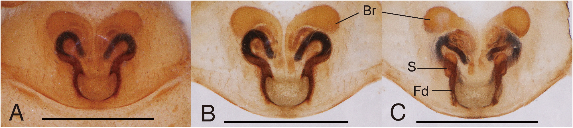
Copulatory organs ( Fig. 7A–C View FIGURE 7 ). Copulatory atrium wider than long, with sclerotized lateral margins. Copulatory ducts opening at anterior corner of copulatory atrium, extending anteriorly with strong curve and then looping several times. Bursae club-shaped, with spherical terminals. Spermathecae bean-shaped, with long narrow fertilization ducts extending posteriorly.
Coloration and setation ( Fig. 6A–D View FIGURE 6 ). Almost same as in male, except sclerotized dorsum of abdomen absent.
Distribution. Sabah.
| RMNH |
National Museum of Natural History, Naturalis |
No known copyright restrictions apply. See Agosti, D., Egloff, W., 2009. Taxonomic information exchange and copyright: the Plazi approach. BMC Research Notes 2009, 2:53 for further explanation.
|
Kingdom |
|
|
Phylum |
|
|
Class |
|
|
Order |
|
|
Family |
|
|
Genus |
Utivarachna dusun Deeleman-Reinhold, 2001
| Yamasaki, Takeshi, Hashimoto, Yoshiaki, Endo, Tomoji, Hyodo, Fujio, Itioka, Takao, Mohamed, Maryati & Meleng, Paulus 2023 |
Utivarachna dusun
| Deeleman-Reinhold, C. L. 2001: 389 |