
Petrarca, Fowler, 1889
|
publication ID |
https://doi.org/ 10.1093/zoolinnean/zlad009 |
|
publication LSID |
lsid:zoobank.org:pub:A2863AB5-855B-4549-89EC-A57C46140CD9 |
|
DOI |
https://doi.org/10.5281/zenodo.8152390 |
|
persistent identifier |
https://treatment.plazi.org/id/03FA8E1E-AB4D-FF98-473C-F927FA7AFBBA |
|
treatment provided by |
Plazi |
|
scientific name |
Petrarca |
| status |
SP. NOV. |
PETRARCA View in CoL NOZAWAI SP. NOV.
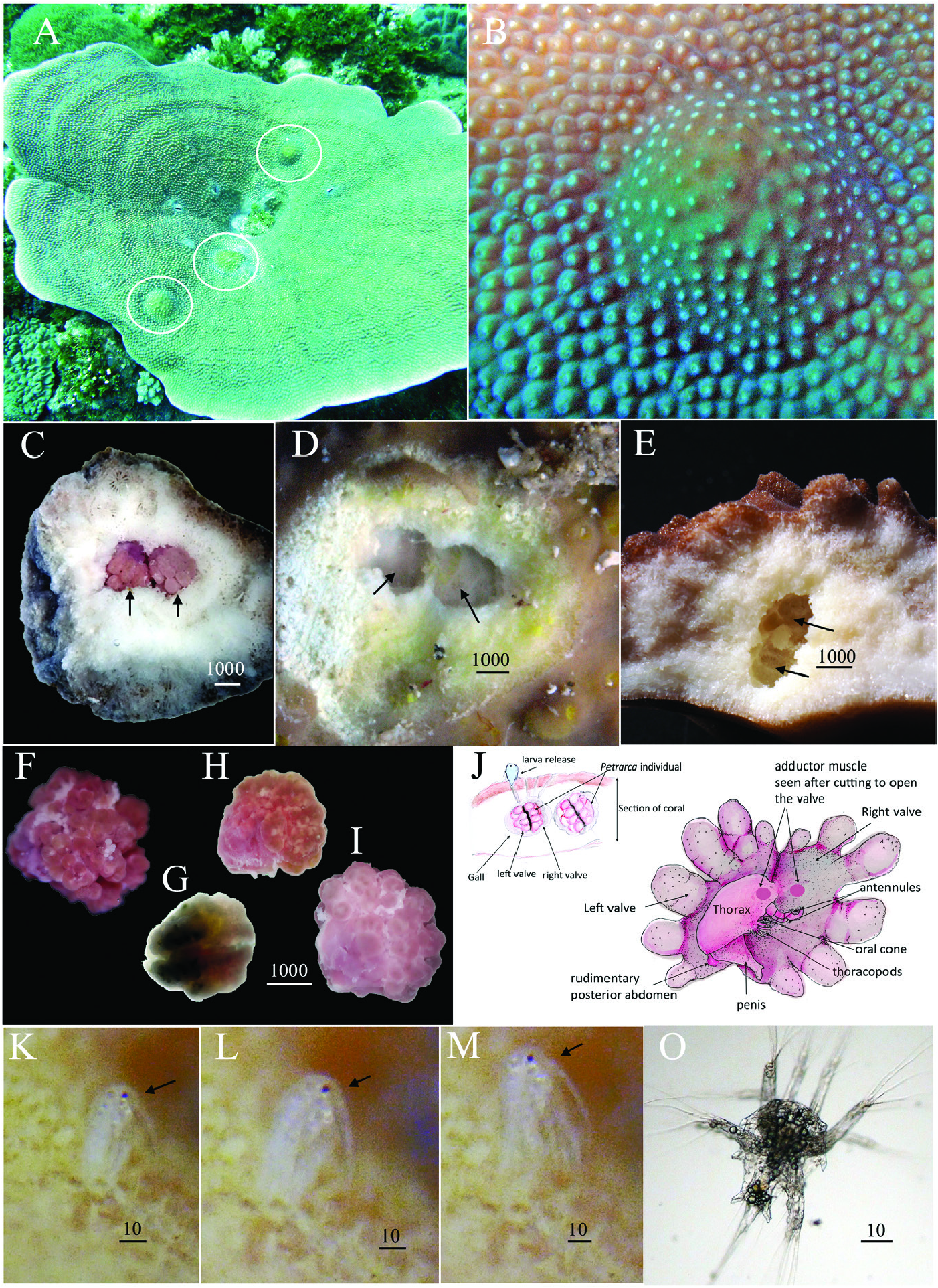
( FIGS 1H View Figure 1 , 14–18 View Figure 14 View Figure 15 View Figure 16 View Figure 17 View Figure 18 )
Probably as P. morula in Grygier & Nojima, 1995: 93–96, fig. 18.
Z o o b a n k R e g i s t r a t i o n: u r n: l s i d: z o o b a n k. o r g: a c t: A E C 7 C 3 E F - C 4 0 5 - 4 4 B 0 - B 0 6 9 - 364D18F6B6A3
Material examined: Two specimens (including holotype) in Turbinaria mesenterina , 22°40 ′ 39.2 ″ N, 121°28 ′ 57.2 ″ E, Green Island (L ǜ d ǎ o), Taiwan, 24.04.2015, at 4–20 m depth. GoogleMaps Six specimens in Turbinaria sp. , 25°7 ʹ 49 ″ N, 121°51 ʹ 26 ″ E, Keelung, Taiwan, 17.08.2015, at 4–10 m depth. GoogleMaps Glycerin slide of the holotype (no. Mg 1248) with the carapace valves, the dissected mouth parts, antennules and rest of the body proper with thoracopods and penis, and SEM stub of paratype (no. Mg 1250) with the carapace valves and the body proper are deposited in the Zoological Museum of Moscow State University in Moscow, Russian Federation.
Diagnosis: Carapace roughly ovoid, valves with eight to ten coalesced lumpy inflations, without distinct radial ridges; carapace margin uneven but not crenulated; ventral side of carapace with numerous, large conical papillae, lateral surface with small papillae. Mandible with 15 sharp, simple teeth; maxillules with 13–15 teeth; six pairs of thoracopods, first thoracopod setiform; penis with small, rounded rami.
Etymology: The species is named after Dr Yoko Nozawa, our colleague and friend, in appreciation of his help with identification of coral hosts for our Ascothoracida studies.
Description: Living specimens pink ( Fig. 1H View Figure 1 ). Adult (mature) specimens 3.70–4.22 mm long, 3.52–3.60 mm high and 3.2–3.3 mm wide ( Figs 14 View Figure 14 , 17A, G View Figure 17 ). Carapace ( Fig. 14 View Figure 14 , 17A, G View Figure 17 ) roughly ovoid, narrowing towards anterior end; each valve with eight to ten coalesced irregular lumpy inflations but without distinct radial ridges; dorsal and posterior margins of carapace uneven but not crenulated; anterioventral margins slightly curved or straight, with numerous, large conical papillae ( Figs 14C, E View Figure 14 , 17A, D View Figure 17 ); lateral surfaces and posterioventral margins with small papillae ( Fig. 17B, E View Figure 17 ). Carapace papillae volcano-shaped, terminating at central microscopic pore ( Fig. 17C, F View Figure 17 ). Cuticle of carapace with dense, polygonal, small swellings or bumps ( Fig. 17B, E, D View Figure 17 ).
Body inflated, curved, enclosed between carapace valves, tip of penis close to oral cone ( Figs 14F View Figure 14 , 17H View Figure 17 ). Cephalon with large adductor muscle lying above big oral cone flanked by five-segmented antennules. Thorax with sinusoid arched dorsal margin, with hump in posterior part but without distinct segmentation, with cluster of rudimentary uniramous thoracopods ( Figs 14F View Figure 14 , 17H View Figure 17 ). Abdomen with massive first segment bearing long penis and vestigial rear part ( Figs 14E, F View Figure 14 , 17H View Figure 17 ).
Antennules somewhat W-shaped and prehensile, with little armament of external sculpture on two distal cuticle with tiny pores on postaxial (ventral) margin of fifth antennular segment. H, distal parts of claw and claw guard, setae of claw guard and aesthetasc indicated by arrowheads. I, oral cone, lateroventral view. J, ctenoid scales on lateral surface of labrum. Abbreviations: ae, aesthetasc; cl, claw; clg, claw guard; lb, labrum; mo, mouth opening; mx2, maxillae; oc, oral cone; pe, penis; rab, rudimentary abdomen; thp, thoracopods. Scale bars in µm.
segments ( Figs 14F View Figure 14 , 15A–D View Figure 15 ). First segment irregularly rectangular, narrowing somewhat distally; second segment trapezoidal; third segment almost triangular, narrowing toward lower/ventral margin; fourth segment longer than wide, with curved ventral margin, short distal seta inserted at anteriodorsal corner ( Fig. 15A, B View Figure 15 ). Fifth segment rectangular, narrowing towards distal end, shorter and narrower than fourth and armed with sensory and grasping structures, ventral/postaxial margin almost straight, dorsal/preaxial margin concave ( Fig. 15C, D View Figure 15 ). Short, massive curved claw with smooth concave margin arising from distal end of segment. Three rudimentary setae at base of claw and to each side ( Fig. 15C, D View Figure 15 ); tiny pores (three to five) on inner and outer lateral sides ( Fig. 15C, D View Figure 15 ). Claw sheathed by large, hood-shaped, oval claw guard on posteriodistal corner; claw guard with three vestigial, distal setae; developed subterminal aesthetasc half as long as claw guard, terminates with two outgrowths ( Fig. 15C, D View Figure 15 ).
Oral cone prominent ( Figs 17H View Figure 17 , 18A View Figure 18 ). Massive prow-shaped labrum with short posteriolateral extensions, leaving maxillae largely exposed, dense ctenoid scales on the exterior ( Fig. 18A, B View Figure 18 ). Mandibles ( Fig. 15E View Figure 15 ) elongated, cutting edge straight, with 15 sharp, simple teeth. Maxillules ( Fig. 15F, G View Figure 15 ) with sclerotized, triangular distal parts, inner margin with 13–16 teeth, teeth in upper half with blunt tips, while those in lower half smaller and spiniform. Fused maxillae ( Figs 15H View Figure 15 , 18A, B View Figure 18 ) with dense ctenoid scales on lateral surfaces, distal ends with rounded zones of sclerotized, thick and wrinkled cuticle without denticles, pores or setules.
Six pairs of thoracopods; thoracopods 2–6 uniramous, unsegmented, grouped in cluster and arranged in an unorderly sequence in lateral view ( Figs 16B View Figure 16 , 17H View Figure 17 ). Thoracopod 1 setiform ( Fig. 16A View Figure 16 ); thoracopods 2–4 conical, longer and wider than thoracopods 5 and 6. Batteries of ampuliform seminal receptacles ( Fig. 16B View Figure 16 ) associated with thoracopods 2–5 (~14, 11–12, 9–10 and 9 receptacles, respectively). Cuticle of thoracopods 2–6 with dense and long ctenoid scales ( Fig. 18D View Figure 18 ).
Long (~ 1.2 mm) and massive, terminally bifid penis originates from large first abdominal segment ( Fig. 17H View Figure 17 ). Shaft of penis supports two small, flat and rounded rami about 100 µm long; distal part of penis, including rami, bearing numerous short but wide conical setae ( Figs 16D View Figure 16 , 18E View Figure 18 ). Rest of abdomen vestigial, consisting of two indistinct segments; posterior end of abdomen with shallow cleft ( Figs 16C View Figure 16 , 18C View Figure 18 ).
Remarks: The new species Petrarca nozaaeai sp. nov. is similar to the other two congeners, P. morula and P. goanna in utilizing Turbinaria corals as hosts. These species are characterized by the large lumpy inflations of the carapace, which are absent in other species of Petrarca . Petrarca nozaaeai differs from P. morula in having ovoid but not a spherical carapace and larger teeth on the inner margin of maxillules (small denticles in P. morula ). Petrarca nozaaeai is distinguished from P. goanna : (1) by the absence of radial ribs and the crenulated margin of the carapace; (2) by numerous large, long conical papillae on the anterioventral part of the carapace; and (3) by simple and fewer teeth on the mandible (simple and bifid teeth present in P. goanna ).


No known copyright restrictions apply. See Agosti, D., Egloff, W., 2009. Taxonomic information exchange and copyright: the Plazi approach. BMC Research Notes 2009, 2:53 for further explanation.