
Draconyx loureiroi, MATEUS & ANTUNES, 2001
|
publication ID |
https://doi.org/ 10.1093/zoolinnean/zlab113 |
|
DOI |
https://doi.org/10.5281/zenodo.6536743 |
|
persistent identifier |
https://treatment.plazi.org/id/03FB87A0-EB6E-6411-FF4F-FCB6B4D2FDCF |
|
treatment provided by |
Plazi |
|
scientific name |
Draconyx loureiroi |
| status |
|
DRACONYX LOUREIROI MATEUS & ANTUNES, 2001
( FIGS 2–7 View Figure 2 View Figure 3 View Figure 4 View Figure 5 View Figure 6 View Figure 7 )
Type material: The holotype specimen (newly reported material marked with *), ML 357 (subnumbers from 1 to 31) includes two maxillary teeth, carpal bones, two metacarpal distal ends*, three left carpal phalanges, one right carpal phalanx*, two left unguals, three right unguals* and one right leg including proximal femoral epiphysis, proximal and distal epiphysis of the tibia, astragalus, calcaneum, four metatarsals, five phalanges and two unguals.
Referred specimen: An isolated left femur, ML 434 from 1 km south of the type locality previously referred to Draconyx loureiroi by Mateus & Antunes (2001), now referred to Ankylopollexia indet.
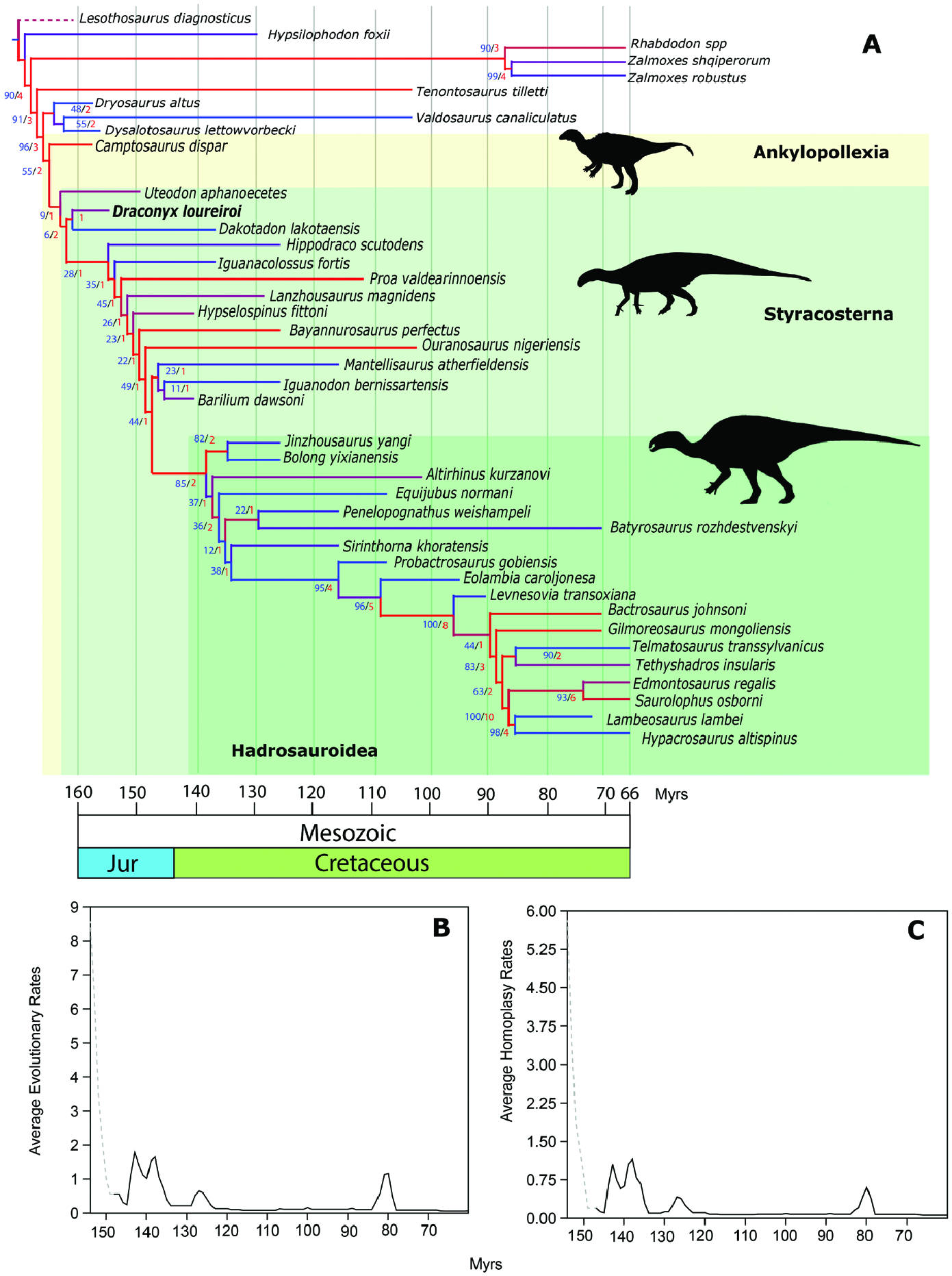
Type locality, horizon and age: Vale de Frades , Lourinhã Municipality , Portugal. Praia Azul Member of Lourinhã Formation , lower Tithonian, 151–152 Mya.
Emended diagnosis: Styracosternan iguanodontian distinguished from other basal iguanodontians by the following combination of characters: unfused and noninterlocked carpus; absence of a sharp crest running from the medial condyle of the femur towards of the lesser trochanter, fully open U-shaped extensor groove on distal epiphysis of the femur; fully open V-shaped flexor groove without overhangs on distal epiphysis of the femur; concave medial margin of proximal epiphysis of the tibia; caudally pointing fibular condyle of the tibia; and a splint-like metatarsal I.
Description and comparisons
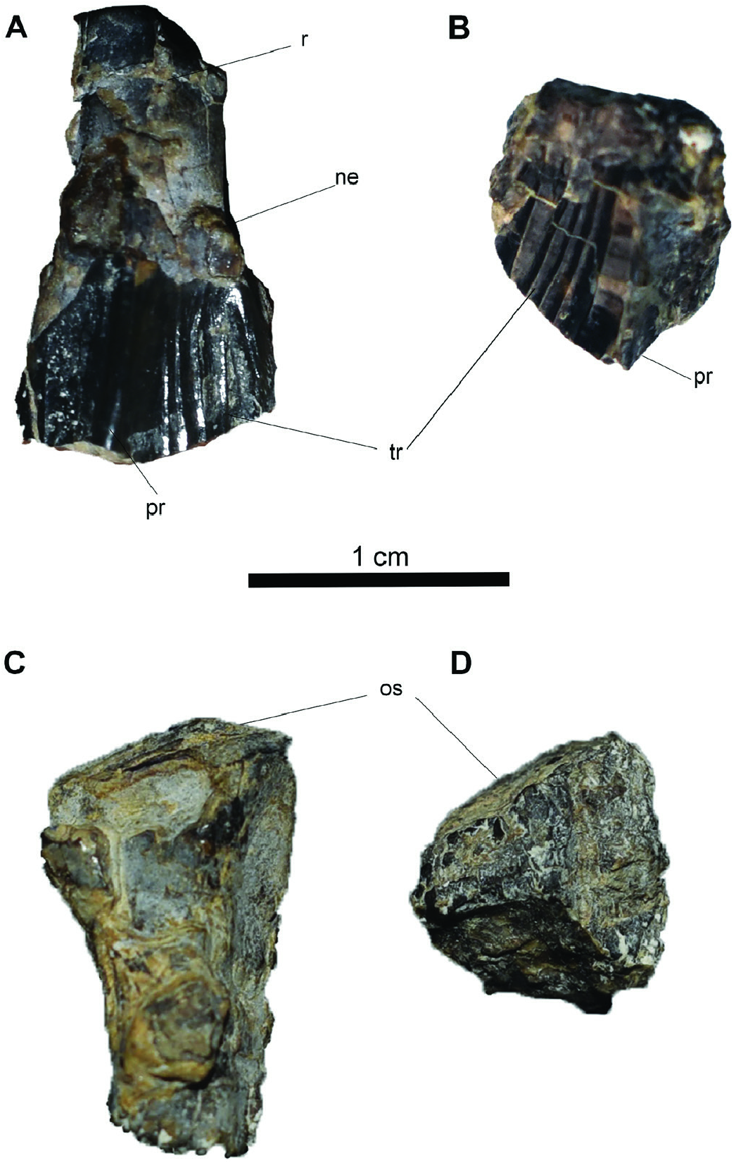
Cranial material: Maxillary teeth (ML 357–31) ( Fig. 2A, B View Figure 2 ): One of the maxillary teeth preserves part of the root and its crown is complete ( Fig. 2A View Figure 2 ). The other tooth is just an isolated crown ( Fig. 2B View Figure 2 ). The specimens appear to have suffered some erosion and post-mortem breakage. The root is slightly labiolingually curved and tapers smoothly into the crown ( Fig. 2A View Figure 2 ). There is no cingulum at the junction between the root and the crown. Overall, the crown is leaf-shaped and the veneer of enamel is thicker on the labial side ( Fig. 2A View Figure 2 ). Labially a thick primary ridge is distally offset and five accessory ridges are present on the mesial surface ( Fig. 2A, B View Figure 2 ). Non-mammillated hook-like denticles are coarsely present on the mesial crown margin, while distally they appear to have been obliterated by either erosion or occlusal wear. An extensive occlusal surface develops on the apex of the crown and is inclined labiolingually approximately around 30° ( Fig. 2A, B View Figure 2 ).
Remarks: The tapering root, slightly labiolingually recurved and the leaf-shaped crown are common characteristics of Dryomorpha ( Galton, 1983, 2006; Norman, 1986, 2004). The maxillary crowns possess a distally offset primary ridge resembling the condition of ankylopollexians but differing from dryosaurids in which the primary ridge is located towards the centre of the crown ( Galton, 2006). In the original description, Mateus & Antunes (2001) indicated the presence of five accessory ridges in the distal half of the crown as an autapomorphy of Draconyx loureiroi . However, the number of accessory ridges is variable throughout the dental series ( Galton, 2006). Therefore, we regard it as a non-informative character if the tooth position is not articulated.
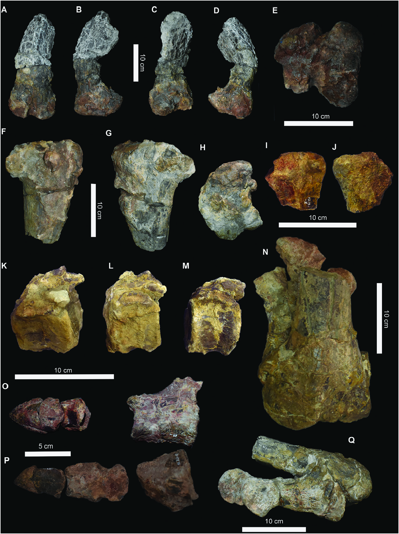
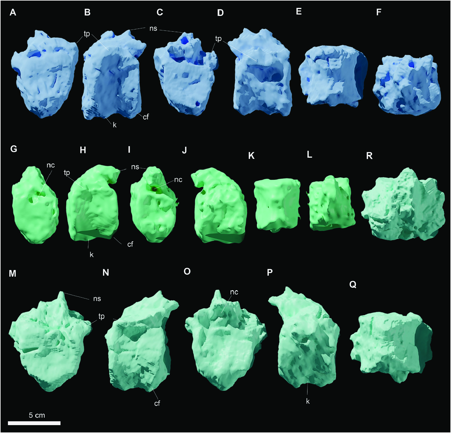
Axial skeleton: Caudal vertebrae (ML 357–9–11) ( Figs 3K–M View Figure 3 , 4A–F View Figure 4 ): Three proximal caudal vertebrae are preserved and, as for the rest of the skeleton, they have suffered breakage and erosion. The centra are stout and craniocaudally compressed. The largest centrum is ML 357–9, while ML 357–10 and ML 357–11 are slightly smaller. The cranial and caudal surfaces are slightly amphicoelous, having a subelliptical, rounded shape ( Fig. 4 A, C, G, I, M, O View Figure 4 ). Dorsally, the neural arch preserves the ventralmost portion of the neural spine but the neural suture is not visible. Immediately caudal to the spine, in the most complete specimens ML 357-9 and ML 357 - 10, ( Fig. 4C, O View Figure 4 ) two small, rounded and steeply inclined post-zygapophyses diverge laterally. On the lateral surface of the neural arch, the articulation for the transverse processes is overlain by a broken surface. The presence of this surface indicates that these vertebrae represent the cranialmost portion of the caudal series. In ventral view, the centra have an hourglass-shaped outline ( Fig. 4E, K, Q View Figure 4 ). The ventral surfaces of the centra possess a narrow keel and the margin is highly concave ( Fig. 4B, P View Figure 4 ). Caudally, broad facets for chevrons are present.
Remarks: The three caudal vertebrae are stout and cylindrical, similar in overall shape and proportions to those of other basal iguanodontians, such as: Camptosaurus dispar, Cumnoria prestwichii , Dryosaurus altus , Dysalotosaurus lettowvorbecki , Mantellisaurus atherfieldensis Mantell, 1825 and Uteodon aphanoecetes ( Galton & Powell, 1980; Galton, 1981; Norman, 1986; Carpenter & Wilson, 2008; Carpenter & Galton, 2018). They do differ remarkably from those of Iguanodon bernissartensis Van Beneden, 1881 and Iguanacolossus fortis McDonald et al., 2010b in being less discoidal in shape, and from Barilium dawsoni Lydekker, 1888a by being less compressed dorsoventrally ( Norman, 1980, 2011; McDonald et al., 2010b).
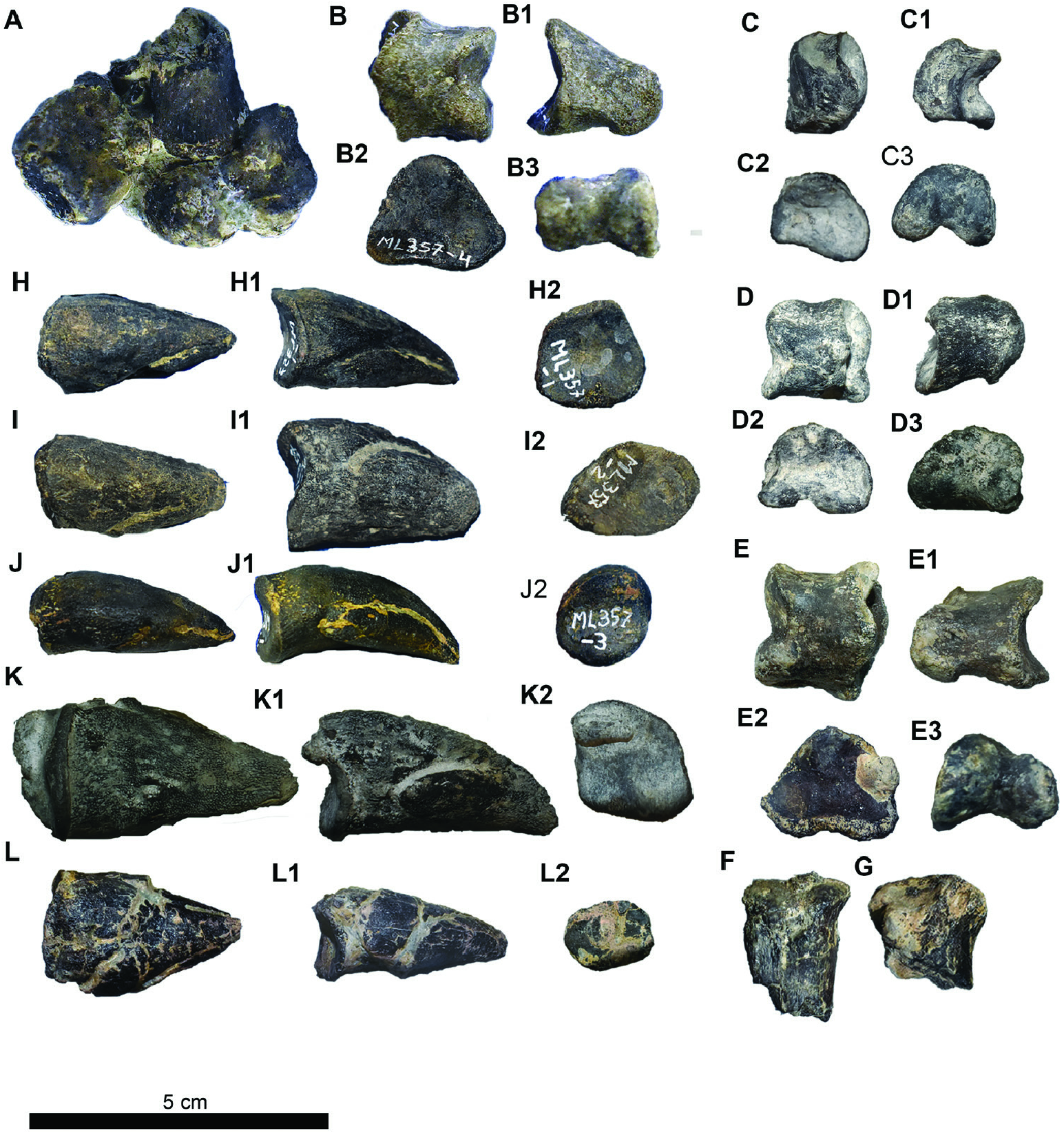
Manus (ML 357–1–5, 20–26) ( Fig. 5 View Figure 5 ): The partial carpus (ML 357–5) is composed of the proximal part of a metacarpal, two distal carpals and a proximal carpal ( Fig. 5A View Figure 5 ). Two isolated distal epiphyses of metacarpals (ML 357–25, 26) are associated with the semiarticulated carpus, being both consistent in size and their state of preservation ( Fig. 5F, G View Figure 5 ). Four manual phalanges and five ungual phalanges are associated with the carpus ( Fig. 5B–E View Figure 5 ). Three manual phalanges (phalanx n.2 ML 357–22, phalanx n.3 ML 357–21 and phalanx n.4 ML 357–24) and two unguals (ungual n.1 ML 357–1 and ungual n.4 ML 357–23) are likely to belong to the left manus ( Fig. 4 View Figure 4 ), while the remaining three unguals (ungual n.2 ML 357–2, ungual n.3 ML 357–3 and ungual n.5 ML 357–20) and manual phalanx n.1 ML 357–4 are attributed to the right manus. Given the fragmentary condition and the weathering of the specimen, it is not possible to identify the bones of each individual carpal. The two distal carpals are cuboid in shape and are stout elements, while the proximal carpal is more lightly built and slightly arched ( Fig. 5A View Figure 5 ). The proximal end of the metacarpal preserves a concave proximal margin. Despite the poor preservation, it is possible to determine that the carpus does not include fused elements, as all of the contacts between all the identifiable carpals are clearly visible ( Fig. 5A View Figure 5 ).
The two isolated distal metacarpals fragments are similar in shape: they are compressed along their extensor–palmar axis, broadening mediolaterally towards their distal ends. The medial and lateral ginglymi are subequal in size and preserved on their collateral ligament pits. The manual phalanges, ML 357–22 and ML 357–21, articulate with each other. Furthermore, phalanx ML 357–22 articulates with the ungual ML 357–23. Therefore, these latter manual elements compose a complete finger ( Fig. 5C, D, K View Figure 5 ). In general, all of the phalanges have a distal triangular section, moderately arched ventral surface and strongly inclined and robust ginglymi. The lateral ginglymus is smaller with respect to the medial one, with the exception of phalanx n.2 where they are subequal ( Fig. 5B–E View Figure 5 ). The unguals are generally elongated and clawlike, slightly arched along the extensor–palmar axis and have a subtriangular articular facet ( Fig. 5H–L View Figure 5 ). The only exception, ungual n.5 ( Fig. 5L View Figure 5 ), appears instead to be compressed along its extensor–palmar axis. A fracture develops mediolaterally and is slightly inclined craniocaudally. Both parts are separated by this fracture and have suffered mediolateral displacement.
R e m a r k s: D e s p i t e t h e p o o r p r e s e r v a t i o n o f the carpus of ML 3 5 7, certain features allow comparisons with other taxa. The small metacarpals are not significantly different from those of other iguanodontians (Norman, 2004). However, the carpus is constituted by isolated blocky elements, resembling the condition in dryosaurids and Tenontosaurus spp. ( Dodson, 1980; Galton, 1981; Forster, 1990; Winkler et al., 1997). The distal carpals of ML 357 differ from the ones of Camptosaurus dispar , which are arranged in two co-ossified and highly interlocked blocks. Furthermore, the carpus of ML 357 does not exhibit the total ossified condition present in Iguanodon bernissartensis, Magnamanus soriaensis Vidarte et al., 2016, Mantellisaurus atherfieldensis and other styracosternans ( Fig. 12A–J View Figure 12 ) ( Dodson, 1980; Norman, 1980, 1986, 2004, 2011, 2015, Vidarte et al., 2016). The carpals and the manual unguals are similar to the ones present in Camptosaurus dispar, Cumnoria prestwichii and Uteodon aphanoecetes , differing from the ones of more derived styracosternans (i.e. Iguanodon bernissartensis, Magnamanus soriaensis, Mantellisaurus atherfieldensis and all other hadrosauriformes) in being, respectively, more arched and more claw-like instead of extremely compressed and hoof-like ( Fig. 12A–J View Figure 12 ) ( Galton & Powell, 1980; Norman, 1980, 1986, 2004; Carpenter & Wilson, 2008, Vidarte et al., 2016).
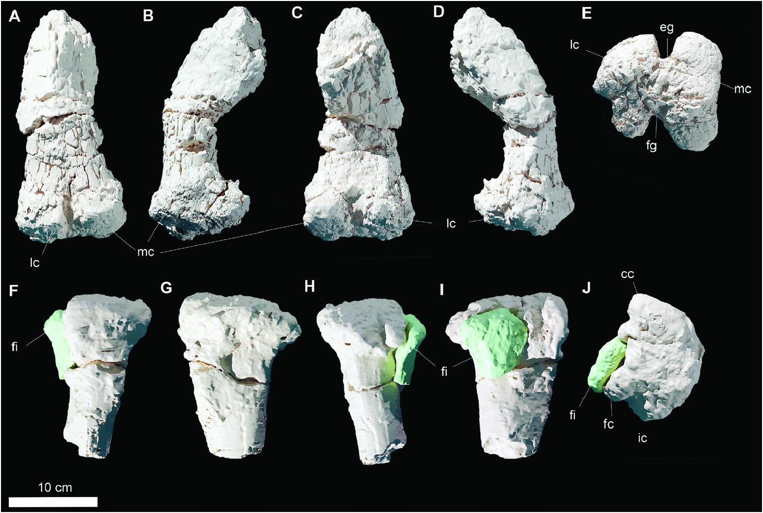
Femur ML 357–6 ( Figs 3A–E View Figure 3 , 6A–E View Figure 6 ): The preserved right femur consists of the heavily eroded distalmost part of the shaft and the distal epiphysis. The proximalmost part of the preserved shaft is mediolaterally crushed and compressed, due to taphonomic processes. The femur is strongly bowed craniocaudally; the section of the shaft was subcircular but is heavily distorted proximally by the compression and damaged distally ( Fig. 6B, D View Figure 6 ). The distal epiphysis is subrectangular in shape in distal view, extending slightly more mediolaterally than craniocaudally. The two distal condyles are preserved, appearing subequal in size with the lateral condyle slightly larger than the medial one ( Fig. 6E View Figure 6 ). A deep, extensive, fully open and U-shaped extensor groove separates the two condyles cranially ( Fig. 6E View Figure 6 ). The cranial process of the lateral condyle is rounded and deflected caudally. The caudal fingerlike process of the lateral condyle (condylid, according to Bertozzo et al., 2017) is not preserved, being broken at its base, but the crest for the muscular insertion is distinguishable. The medial condyle appears stout and rectangular, although its cranial and caudal processs have been eroded. The flexor groove is fully open and its margin, consisting of the caudal process of the lateral condyle and the condylid of the medial condyle, are V-shaped in outline.
Remarks: The preserved femoral shaft is strongly bowed craniocaudally, as in dryosaurids, most elasmarians, Camptosaurus dispar and differently from Tenontosaurus sp. and other styracosternans ( Norman, 1980, 1986, 2004; Carpenter & Wilson, 2008; Carpenter & Galton, 2018; Herne et al., 2019; Rozadilla et al., 2019, 2020). On the cranial surface there is no crest developing from the medial condyle, extending proximally towards the lesser trochanter, as seen in other ankylopollexians, such as Camptosaurus dispar , Iguanodon sp. , Mantellisaurus atherfieldensis and Uteodon aphanoecetes ( Gilmore, 1909; Norman, 1980, 1986, 2004; Carpenter & Wilson, 2008; Carpenter & Galton, 2018). The lateral condyle of the distal epiphysis of the femur in Draconyx loureiroi is concave in outline and it extends more craniocaudally than the ones of Camptosaurus dispar, Cumnoria prestwichii and Uteodon aphanoecetes ( Fig. 7 View Figure 7 ; Galton & Powell, 1980; Carpenter & Wilson, 2008). The inflection point of the curvature is located more cranially in Draconyx loureiroi than in Camptosaurus dispar and Uteodon aphanoecetes ( Carpenter & Wilson, 2008; Carpenter & Galton, 2018), while Cumnoria prestwichii exhibits a smoother outline without abrupt changes in curvature. The lateral condyle of Early Cretaceous species, such as Barilium dawsoni and Mantellisaurus atherfieldensis , are larger in proportions with respect to the total size of the epiphysis and extend more caudally than the ones of the above-mentioned taxa, including D. loureiroi ( Norman, 1986; 2011). The medial condyle of Draconyx loureiroi is subrectangular and its medial margin is straight, as is the one of Mantellisaurus atherfieldensis and Barilium dawsoni ( Norman, 1986; 2011). However, in Jurassic taxa such as Uteodon aphanoecetes and Camptosaurus dispar , the medial margin of the medial condyle is rounded ( Carpenter & Wilson, 2008; Carpenter & Galton, 2018). The flexor and extensor grooves are fully open as in many basal and cursorial iguanodontians, contrasting with more derived forms (Norman, 2004). Extensor grooves of the Jurassic taxa Camptosaurus dispar and Uteodon aphanoecetes are fully open, but shallower compared to the ones of Barilium dawsoni, Cumnoria prestwichii , Draconyx loureiroi , Iguanodon sp. and Mantellisaurus atherfieldensis ( Norman, 1980, 1986, 2004, 2011, Carpenter & Wilson, 2008; Carpenter & Galton, 2018; Verdú et al., 2018). Moreover, the flexor groove walls of Cumnoria prestwichii and Uteodon aphanoecetes are slightly divergent from one another ( Fig. 7 View Figure 7 ). The extensor groove of the Early Cretaceous iguanodontians Barilium dawsoni , Mantellisaurus atherfieldensis and other styracosternans are partially enclosed by overhangs of medial and lateral condyles. Draconyx loureiroi does not exhibit an overhang of the medial condyle on the flexor groove, unlike the condition present in Camptosaurus dispar, Cumnoria prestwichii , Mantellisaurus atherfieldensis , Ouranosaurus nigeriensis Taquet, 1976 and Uteodon aphanoecetes ( Norman, 1980, 1986, 2004, 2011; Carpenter & Wilson, 2008; Bertozzo et al., 2017; Carpenter & Galton, 2018, Verdú et al., 2018). This condition more closely resembles the plesiomorphic condition within Ornithopoda ( Norman et al., 2004).
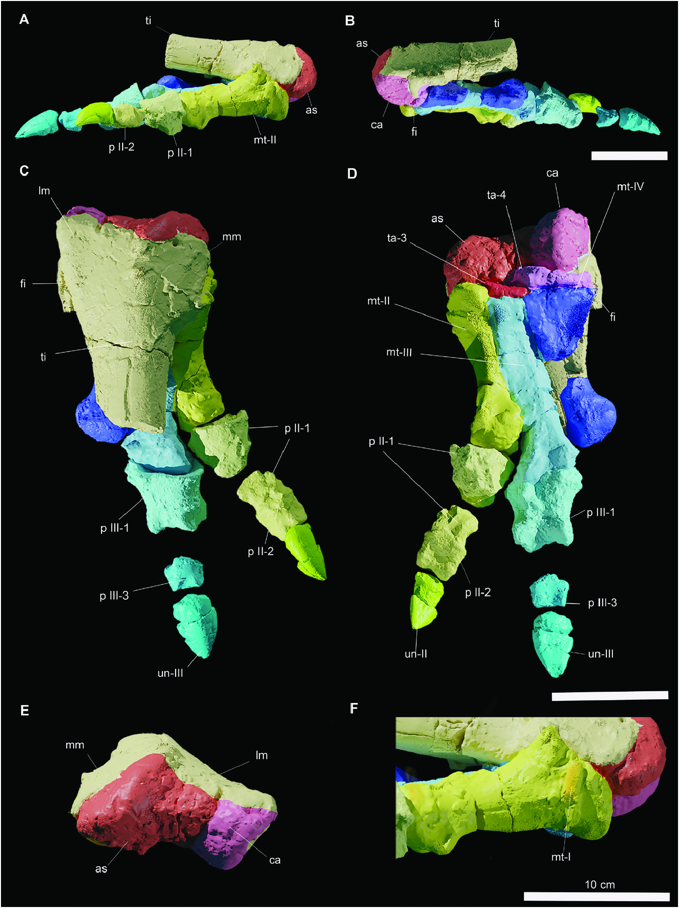
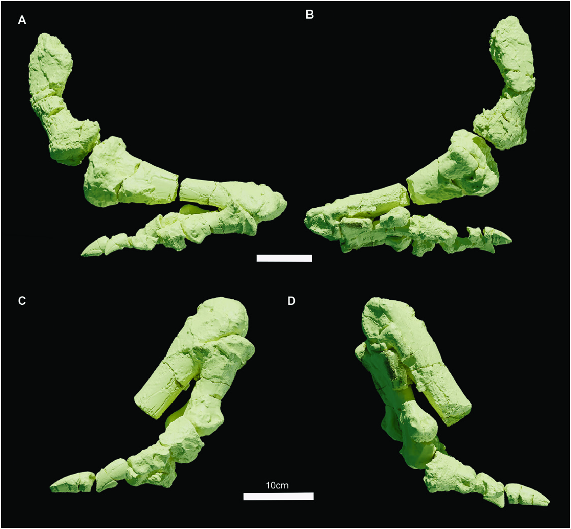
Tibia (ML 357–7, 12) ( Figs 3F, G View Figure 3 , 6F–J View Figure 6 , 8A–D View Figure 8 ): The proximal ( Fig. 6F–J View Figure 6 ) and distal ( Fig. 8A–E View Figure 8 ) epiphyses of the tibia are preserved, but the tibial shaft is missing. Both extremities are heavily eroded and covered by matrix and adhesives, but relevant characters are still distinguishable. The proximal epiphysis preserves a conspicuous cnemial crest, a robust fibular condyle and the internal condyle. The cnemial crest tapers dorsally forming a smooth edge; in dorsal view its margins are laterally concave and medially convex ( Fig. 6G, I View Figure 6 ). Laterally, it is divided by the fibular condyle to form a deep and extensive scar (sulcus tibialis). The fibular condyle is a stout process that deflects strongly caudally, its articular facet is rounded and it blends smoothly with the anterior and posterior surfaces of the condyle. The internal condyle is a blunt eminence directed caudally and projecting gently laterally. Like the cnemial crest, its lateral margin is concave, while the medial margin is strongly convex ( Fig. 6E View Figure 6 ). This convexity forms a deep sulcus that separates the internal condyle from the fibular condyle. Immediately ventral to the proximal epiphysis, the proximal part of the diaphyseal shaft is preserved and it is teardrop-shaped in cross-section. The distal part of the tibia is articulated with the astragalus, calcaneum and the metatarsals ( Fig. 8A–E View Figure 8 ). The distal epiphysis flares mediolaterally, the medial malleolus is rounded and slightly more expanded mediolaterally than the lateral one, which is narrow and elongate, expanding proximodistally. They articulate between each other forming an angle of approximately 45°. The intermuscular line is located on the caudal surface of the distal epiphysis, separating the surface of the two malleoli and reaching the astragalus, forming with its apex a continuous concave surface.
Remarks: The medial margin of the proximal epiphysis of the tibia is convex, as is typical of many iguandontians (Norman, 2004), except for Talenkauen santacrucensis Novas et al., 2004 and Eousdryosaurus nanohallucis Escaso et al., 2014 ( Escaso et al., 2014; Rozadilla et al., 2019; Dieudonné et al., 2021). As noted by Dieudonné et al. (2021), the cnemial crest apex is directed strongly craniolaterally, a characteristic common to Laurasian dryomorphans and Dysalotosaurus Pompeckj, 1920 , differing from the condition exhibited by Elasmaria and the Portuguese ornithopod Eousdryosaurus nanohallucis ( Escaso et al., 2014) . As in many styracosternans, the cnemial crest is well developed (Norman, 2004). The fibular condyle differs from dryosaurids (with the exception of Valdosaurus canaliculatus ), Camptosaurus dispar and Uteodon aphanoecetes in being partially posterolaterally deflected, similar to the condition seen in Cumnoria prestwichii , Talenkauen santacrucensis and more derived styracosternanans ( Galton & Powell, 1980; Norman, 1980, 1986, 2011; Norman et al., 2004; Carpenter & Wilson, 2008; Barrett et al., 2011; Rozadilla et al., 2019). The distal epiphysis, found in articulation with the rest of the pes, does not differ significantly from that of other iguanodontians (Norman, 2004).
Fibula (ML 357–8, 12) ( Figs 3I, J View Figure 3 , 6F–J View Figure 6 , 8A–D View Figure 8 ): The distal and proximal epiphyses of the fibula are preserved, whereas the diaphysis is completely absent. The proximal end is a flattened subtriangular element. Dorsally, it appears to be slightly medially convex. The cranial margin deflects abruptly dorsally into the cranial process, which is slightly eroded ( Fig. 6I View Figure 6 ). In contrast, the caudal margin deflects less abruptly dorsally. A deep fossa is present close to the caudal margin on the lateral surface.
The distal epiphysis is in articulation with the rest of the pes, located on the lateral surface of the tibia and contacts the calcaneum ( Fig. 7A–D View Figure 7 ).
Remarks: The proximal epiphysis has cranial and caudal margins that diverge smoothly, as in Dryosaurus altus , Dysalotosaurus lettowvorbecki , Eousdryosaurus nanohallucis , Talenkauen santacrucensis and Valdosaurus canaliculatus , but differs from Iguanodon bernissartensis , Mantellisaurus atherfieldensis and Ouranosaurus nigeriensis ( Norman, 1980, 1986; Galton, 1981; Barrett et al., 2011; Escaso et al., 2014; Barrett, 2016; Bertozzo et al., 2017; Rozadilla et al., 2019). As in Sektensaurus sanjuanboscoi Ibiricu et al., 2019 and Talenkauen santacrucensis , the caudal margin of the proximal part of the fibula is almost vertical ( Ibiricu et al., 2019; Rozadilla et al., 2019).
Pes (ML 357–12–19) ( Figs 3N, Q View Figure 3 , 8A–F View Figure 8 , 9C, D View Figure 9 ): The pes is articulated with the distal tibia, distal fibula, astragalus and calcaneum and proximal metatarsals. The diagenetic permineralization between the tibia and the rest of the pes prevented a reliable segmentation by CT techniques. This also hampered views of the internal anatomy of the astragalus and calcaneum. The articulated elements share the same subnumber (ML 357–12), while the isolated elements have their own numbers. Therefore, the same element may have two subnumbers, depending on whether part of it belongs to the articulated block.
Calcaneum
The calcaneum is preserved in its entirety and does not show signs of significant breakage, erosion or distortion. In its general appearance, the calcaneum i s a c o m p a c t a n d n a r r o w e l e m e n t, e x p a n d i n g craniocaudally more than mediolaterally ( Fig. 8B, E View Figure 8 ). Distally, its surface is subrectangular in outline and extends dorsally, appearing as a small rhomboidal element in caudal view ( Fig. 8B, E View Figure 8 ). Caudally, the medial margin of the calcaneum forms a smooth concavity that accommodates the lateralmost margin of the astragalus. In lateral view, the calcaneum appears triangular with a caudal apex and is slightly deflected proximally. The proximal and distal margins progressively flare cranially, with the latter forming a scoop-like concavity.
Astragalus
Like the calcaneum, the astragalus is complete and not significantly distorted or eroded. In distal view, the bone has a subtrapezoidal shape ( Fig. 8A, E View Figure 8 ). Laterally, it contacts the medial malleolus of the tibia via a deep concavity, while medially the astragalus/calcaneum contact is straight ( Fig. 8A, E View Figure 8 ). In caudal view, the astragalus is triangular, with a high ascending process representing the apex, which protrudes extensively dorsally. Immediately ventral to the ascending process, a stout steeply inclined caudomedial process forms a well-distinguishable relief.
Remarks: The astragalus of ML 357 is subtriangular and its caudal surface is moderately high, as in dryosaurids, Camptosaurus dispar, Cumnoria prestwichii , Iguanodon bernissartensis Mantellisaurus atherfieldensis and Uteodon aphanaecetes ( Galton & Powell, 1980; Norman, 1980, 1986; Galton, 1981; Carpenter & Wilson, 2008; Barrett et al., 2011). The ascending process is not as developed as in Barilium dawsoni , Iguanodon bernissartensis or Mantellisaurus athefieldensis ( Norman, 1980, 1986, 2011), but more closely resembles the condition found in Camptosaurus dispar (NMNH 2210) , Cumnoria prestwichii , Ouranosaurus nigeriensis and Uteodon aphanoecetes ( Galton & Powell, 1980; Carpenter & Wilson, 2008; Bertozzo et al., 2017). As in the other known Portuguese ornithopod, Eousdryosaurus nanohallucis , it shows a caudomedial process, which casts doubt on the diagnostic status of this character for this latter taxon ( Escaso et al., 2014). The calcaneum does not possess any remarkable differences from other ankylopollexians (Norman, 2004).
Distal tarsals
Two distal tarsals are preserved, which are identified as tarsal 3 and tarsal 4 ( Fig. 8D View Figure 8 ). Tarsal 3 is a large element that is mediolaterally elongated and contacts metatarsal III along a straight and linear surface ( Fig. 8D View Figure 8 ). Tarsal 4 is a stout, elongated element and contacts the metatarsal IV along a concave surface, while its medialmost tip overlaps tarsal 3 proximally ( Fig. 8D View Figure 8 ).
Remarks: Distal tarsals 3 and 4, found in articulation with metatarsals III and IV, resemble the ‘cushionlike’ condition described by Gilmore (1909) in Camptosaurus dispar , and differing from the thinlike and subrounded condition observed in Iguanodon bernissartensis and Mantellisaurus atherfieldensis ( Norman, 1980, 1986) or the one present in basal ornithopods ( Galton, 1974, 1981).
Metatarsus
Although they are fragmented, metatarsals II–IV are long and slender ( Figs 8 View Figure 8 , 9C, D View Figure 9 ). A thick splinter of bone next to metatarsal II was interpreted as the proximalmost part of metatarsal I in the original description of Mateus & Antunes (2001) and a ‘vestigial digit I’ has been proposed as a possible autapomorphy for Draconyx loureiroi . Despite being heavily eroded, the bone is preserved in anatomical position and the rod-like structure supports the interpretation of this bone as the first metatarsal ( Fig. 8F View Figure 8 ). The status of this character as an ‘autapomorphy’ is discussed below. A deep groove in continuity with this fragment and impressed on metatarsal II probably represents the entire articulation surface with metatarsal I ( Fig. 8F View Figure 8 ). Metatarsals II and III are complete and do not appear to have suffered extensive breakage or fracture ( Fig. 8C, D View Figure 8 ). Metatarsal IV in contrast is broken at the midshaft with the distal epiphysis preserved as an isolated element.
Pedal digit I
The first pedal digit consists only of the splint-like metatarsal I ( Fig. 8F View Figure 8 ).
Pedal digit II
Pedal digit II is represented by metatarsal II, the pedal phalanx II-1 (ML 357–17) and the ungual phalanx II-2 (357–16), which articulates with the phalanx II-2 (357– 14). Pedal phalanx II-1 is broken at the midshaft, but the proximal and distalmost epiphyses are preserved ( Fig. 8A, C, D View Figure 8 ).
Metatarsal II is positioned more proximally with respect to Mt III–IV. In mediolateral view, metatarsal II has a keyhole shape, as described by Herne et al. (2018) for the Australian ornithopod Diluvicursor pickeringi Herne et al., 2018 , with the proximalmost part dorsoplantarily higher than the distal epiphysis ( Fig. 8A, F View Figure 8 ). The distal epiphysis develops more dorsoplantarily than mediolaterally, and the articular surface is convex. The shaft of Mt II is piriform (teardrop) in cross-section, with the plantar surface bearing a keel. The distalmost part of the epiphysis expands dorsoplantarily, giving the condyle a subtrapezoidal outline ( Fig. 8A, D View Figure 8 ). The condyle of the metatarsal articulates perfectly with pedal phalanx II-1, which accommodates the condyle in a gently convex triangular facet. The distal epiphysis of the phalanx is in articulation with pedal phalanx II-2, which is a thick, subrectangular element in dorsal view ( Fig. 8C, D View Figure 8 ). Proximally, the articular facet is not visible, but distally the two condyles are well distinguishable, the lateral slightly inclined with respect to the medial one. On both lateral surfaces, deep extensor grooves are present for tendon insertion. The ungual (phalanx II-2) of the pedal digit II is preserved, being a pointed claw-like element, which is dorsoplantarly arched and proximodistally elongated ( Fig. 8A, C, D View Figure 8 ).
Pedal digit III
Pedal digit III is composed of metatarsal III, the largest pedal phalanx preserved III-1 (ML 357–13), the smallest pedal phalanx is interpreted to be III-3 (ML 357–15) and the largest ungual, pedal phalanx III-4 (ML 357–19). The pedal phalanx III-2 is missing. Metatarsal III is the longest element of the metatarsus, being a slender, yet compact element. Proximally, the Mt III articulates with the tarsal 3 and intervenes between Mt II and Mt IV ( Fig. 8B–D View Figure 8 ). The subrectangular shaft ends distally in a mediolaterally expanded condyle, the only metatarsal exhibiting this condition. Pedal phalanx III-1 is a stout, robust element, which is moderately dorsoplantarly arched. The proximal articular facet is concave and subellipsoidal in outline, being the long axis oriented mediolaterally ( Fig. 8B–D View Figure 8 ). Distally, the two ginglymi are subequal in size, being marked by deep collateral ligament pits. Pedal phalanx III-3 is a small proximodistally compressed element, which possesses a dorsoplantarly deep articular facet ( Fig. 8B–D View Figure 8 ). The ventral margin of pedal phalanx III-3 is not as arched as the other phalanges and the collateral ligament pits occupy almost the entire lateral and medial surfaces. The ungual of the digit III (pedal phalanx III-3) is a stout, pointed element, which is more robust than the ungual preserved on pedal digit II. The ungual of digit III differs also from the other preserved ungual in having a less arched and more dorsoplantarly flattened outline ( Fig. 8B–D View Figure 8 ).
Pedal digit IV
Metatarsal IV (ML 357–12, 18) is the only element of pedal digit IV available. Proximally it is the most mediolaterally expanded and dorsoplantarly compressed of the metatarsals and the proximal epiphysis is fan-shaped in dorsal and plantar view ( Fig. 8B–D View Figure 8 ). Distally, the section of the shaft becomes progressively subrectangular, culminating in a moderately dorsoplantarly deep distal condyle ( Fig. 8B–D View Figure 8 ).
Remarks: The metatarsus of ML 357 differs strongly from those of Camptosaurus dispar , Iguanodon bernissartensis , Ouranosaurus nigeriensis , Uteodon aphanoecetes and other large-sized hadrosauriforms in general proportions, while exhibiting the slender and gracile proportions seen in Cumnoria prestwichii and Mantellisaurus atherfieldensis instead ( Gilmore, 1909; Galton & Powell, 1980; Norman, 1980, 1986, 2004; Bertozzo et al., 2017). Furthermore, as in the latter taxa, ML 357 possesses a metatarsal III that is sensibly more elongated with respect to metatarsals II and IV ( Galton & Powell, 1980; Norman, 1986). This is in stark contrast to elasmarians Camptosaurus dispar, Cumnoria prestwichii , Dryosaurus altus and Dysalotosaurus lettowvorbecki , whose metatarsal I is extremely reduced, losing any functional capability ( Gilmore, 1909; Galton & Powell, 1980; Galton, 1981; Carpenter & Galton, 2018). However, this condition is preserved in some specimens of Iguanodon bernissartensis and Mantellisaurus atherfieldensis ( Norman 1980, 1986). Assessing the presence of this character in other taxa is problematic, because of the extreme fragility of the reduced metatarsal I, whose absence/presence is highly subject to preservation bias. The alleged dryosaurid, Eousdryosaurus nanohallucis , from the same formation as ML 357, has been shown to possess a reduced metatarsal I in articulation with a pedal phalanx ( Escaso et al., 2014).

No known copyright restrictions apply. See Agosti, D., Egloff, W., 2009. Taxonomic information exchange and copyright: the Plazi approach. BMC Research Notes 2009, 2:53 for further explanation.
|
Kingdom |
|
|
Phylum |
|
|
Class |
|
|
Order |
|
|
Genus |
Draconyx loureiroi
| Rotatori, Filippo Maria, Moreno-Azanza, Miguel & Mateus, Octávio 2022 |
Draconyx loureiroi
| Mateus & Antunes 2001 |