
Baltoplana cupressus Willems & Artois
|
publication ID |
https://doi.org/ 10.11646/zootaxa.4242.3.2 |
|
publication LSID |
lsid:zoobank.org:pub:C67937C9-844F-461E-AABB-121B9C3CE5FA |
|
DOI |
https://doi.org/10.5281/zenodo.5689680 |
|
persistent identifier |
https://treatment.plazi.org/id/03FB87EB-5233-E357-57BE-ABE4FE9ADE75 |
|
treatment provided by |
Plazi |
|
scientific name |
Baltoplana cupressus Willems & Artois |
| status |
sp. nov. |
Baltoplana cupressus Willems & Artois n. sp.
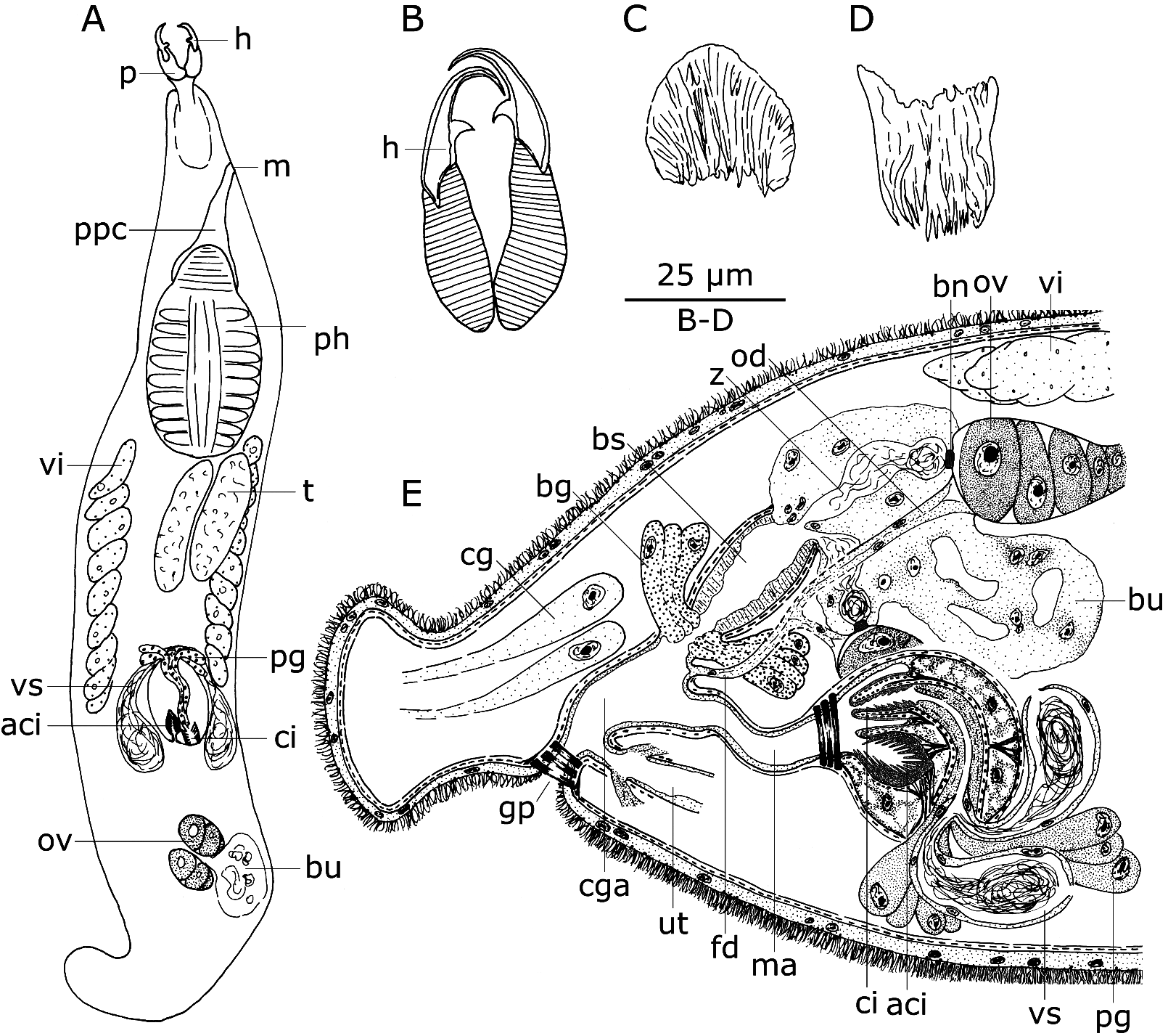
( Fig. 7 View FIGURE 7 )
Localities. iSimangaliso Wetland Park, Eastern Shores , Cape Vidal (28°07’45.0’’S, 32°33’37.0’’E), bay protected by reef, detritus-rich sand from open beach, on the waterline, and in between rocks on cape, December 6, 2009 (type locality) GoogleMaps ; same locality, medium coarse-grained sand from sublittoral (1,5 m deep) between reef and beach, December 6, 2009 GoogleMaps .
Material. Several animals studied alive. Four whole mounts, all designated paratypes (HU, nos 579–582) and eight serially-sectioned individuals, one of them designated holotype ( SMNH, type-8857), six others paratypes (HU, nos. 583–589).
Etymology. Species name refers to the tree genus Cupressus (Cypress) , of which most species have a shape similar to that of the accessory cirrus of this species of Baltoplana .
Diagnosis. Species of Baltoplana with 29-µm-long proboscis hooks, carrying one denticle each. Male copulatory organ with 23-µm-long, straight, armed cirrus and a single, 23-µm-long, spiny accessory cirrus.
Description. The animals lack eyes, are colourless to pale reddish brown and 1–1.4 mm long (measured on the whole mount). At approximately 90%, the body shows a constriction, creating a distinct tail region, in which finegrained, eosinophilic caudal glands are present. The epidermis is syncytial, 2–3 µm thick, with 4–5 µm-long cilia. Rhabdites are rather small and ovoid and sparsely distributed over the entire body.
The proboscis is very similar to that of Baltoplana magna Karling, 1949 , described in detail by Karling (1949). In whole-mounted specimens it is approximately 47 µm long, which is 1/23 to 1/29 of the total body length. The proboscis hooks ( Fig. 7 View FIGURE 7 A–B: h) are slender, 24–33 µm long ( = 29 µm; n = 8) and bent over the most distal 1/3. Close to their base, these hooks carry an inwardly bent, 4–5 µm-long denticle ( = 5 µm; n = 7; could not be measured on one of the hooks).
The pharynx is situated in the first half of the body and is 1/3 to 1/4 of the body length long. Its detailed structure fits the description and figures given for B. magna by Karling (1949).
Testes and ovaries are paired. The testes are situated closely behind the pharynx. The ovaries vary in shape from small and ovoid to rather large and elongated, probably because of different degrees of maturity of the female genital system. They are situated in the caudal third of the body. In most individuals the proximal part of the ovaries points towards the caudal side, whereas in a few it is directed rostrally. The narrow vitellaria extend from the caudal end of the pharynx to the proximal end of the male atrial system.
The common genital pore is situated at approximately 85%, just rostrally from the tail constriction, and can be closed by a strong sphincter. The common genital atrium is lined with a low, anucleated epithelium and surrounded by inner circular and outer longitudinal muscles. It is elongated rostro-dorsally and receives the male system from the rostral side and the female system from the dorsal side. The uterus enters the atrium from the rostral side, ventrally to the male atrium.
The paired seminal vesicles are lined with a low, nucleated epithelium and lack any muscle layer. Both vesicles narrow distally to form seminal ducts, which fuse upon entering the male copulatory organ. This organ is of the duplex-type (terminology of Karling 1956), ovoid in shape and surrounded by outer longitudinal and inner circular muscles. The latter are stronger distally, forming a large sphincter at the transition with the male atrium. At the junction of both seminal ducts, the epithelium shows a large number of nuclei. Together with the seminal ducts, a large bundle of extracapsular, fine-grained, basophilic glands ( Fig. 7 View FIGURE 7 E: pg) enters the copulatory organ. Their gland necks run parallel to the ejaculatory duct, of which the exact trajectory is hard to discern. Gland necks and ejaculatory duct are surrounded by inner circular and outer longitudinal muscles and open into a spiny cirrus ( Fig. 7 View FIGURE 7 D, 7E: ci), which is formed by the inner lining of a penis papilla. The outer lining of the papilla also consists of pseudocuticularised epithelium, but without any spines. In mounted specimens, this cirrus is more or less cupshaped, 20–26 µm long ( = 23 µm; n = 3) and 16–19 µm wide ( = 18 µm; n = 3) at its widest point. A blind, accessory cirrus ( Fig. 7 View FIGURE 7 C, 7E: aci) is situated ventrally or latero-ventrally to the primary cirrus and has the shape of a young cypress tree, which is especially obvious in live specimens. It is 19–26 µm long ( = 23 µm; n = 4) and 19–27 µm wide at its widest point ( = 22 µm; n = 4), and carries spines that are longer than the ones from the primary cirrus. The accessory cirrus is surrounded by weak circular muscles and can be retracted by strong longitudinal muscles, which are attached to the proximal part of the cirrus and extend to the copulatory bulb’s inner wall.
The ovaries have a double connection with the common genital atrium: one through the oviducts and following female duct, the second one through two insemination canals connecting to the bursal stalk. The oviducts are slender, lack any muscle layer and are lined with a high, nucleated epithelium, filling the entire lumen. Their exact trajectory and especially the location of the bifurcation of the female duct into both oviducts are obscured by the large bursa. Close to the connection between ovary and oviduct, a pseudocuticular bursal nozzle ( Fig. 7 View FIGURE 7 E: bn) is present in the most proximal part of the two sperm-filled insemination canals ( Fig. 7 View FIGURE 7 E: z), which run through the large bursa. Distally these canals fuse and open into a wide, muscular bursal stalk ( Fig. 7 View FIGURE 7 E: bs). This stalk is lined with a high, anucleated epithelium, which has a somewhat striated appearance, and is surrounded by inner circular and outer longitudinal muscles. It opens into the common genital atrium, together with a large bundle of coarsegrained, basophilic glands ( Fig. 7 View FIGURE 7 E: bg), just dorsally to the female duct.
Discussion. In contrast with members of the taxon Cheliplana Beauchamp, 1927 , representatives of the taxon Baltoplana Karling, 1949 are characterised by the presence of paired ovaries and insemination canals (see the diagnosis by Karling 1983). The overall structure of the genital system of B. cupressus n. sp. is very similar to that of B. magna Karling, 1949 , the only species of Baltoplana that is described in detail ( Karling 1949). However, the male genital system of both species is clearly different. Whereas the cirrus is rather long and can be coiled proximally in B. magna ( Karling 1949) , it is much shorter in B. cupressus n. sp.. Furthermore, the former species possesses two accessory cirri (diverticles in Karling 1949), while the latter only has one. The two other known species of Baltoplana , B. valkanovi Ax, 1959 and B. bisphaera Jouk & De Vocht, 1989 , can also easily be distinguished by the structure of their copulatory organs. In B. valkanovi no accessory cirri are present ( Ax 1959), whereas two are present in B. bisphaera . However, the latter species lacks an armed cirrus ( Jouk & De Vocht 1989).
Although not known for B. valkanovi , B. cupressus n. sp. is the only species of Baltoplana that has proboscis hooks carrying denticles. Within the taxon Cheliplanidae , denticles on the proboscis hooks are present in all species of Cheliplanilla Meixner, 1938 and in one out of 33 species of Cheliplana , i.e. Cheliplana marcusi ( Karling, 1956) Karling, 1983 .
| SMNH |
Saskatchewan Museum of Natural History |
No known copyright restrictions apply. See Agosti, D., Egloff, W., 2009. Taxonomic information exchange and copyright: the Plazi approach. BMC Research Notes 2009, 2:53 for further explanation.
|
Kingdom |
|
|
Phylum |
|
|
Class |
|
|
Order |
|
|
Family |
|
|
Genus |