
EPIALTINAE MacLeay, 1838
|
publication ID |
https://doi.org/10.1080/00222933.2014.925596 |
|
persistent identifier |
https://treatment.plazi.org/id/03FBE76C-0E2D-2177-FE00-2677A925FB18 |
|
treatment provided by |
Felipe |
|
scientific name |
EPIALTINAE MacLeay, 1838 |
| status |
|
Subfamily EPIALTINAE MacLeay, 1838 View in CoL
Menaethius monoceros Latreille, 1825 View in CoL
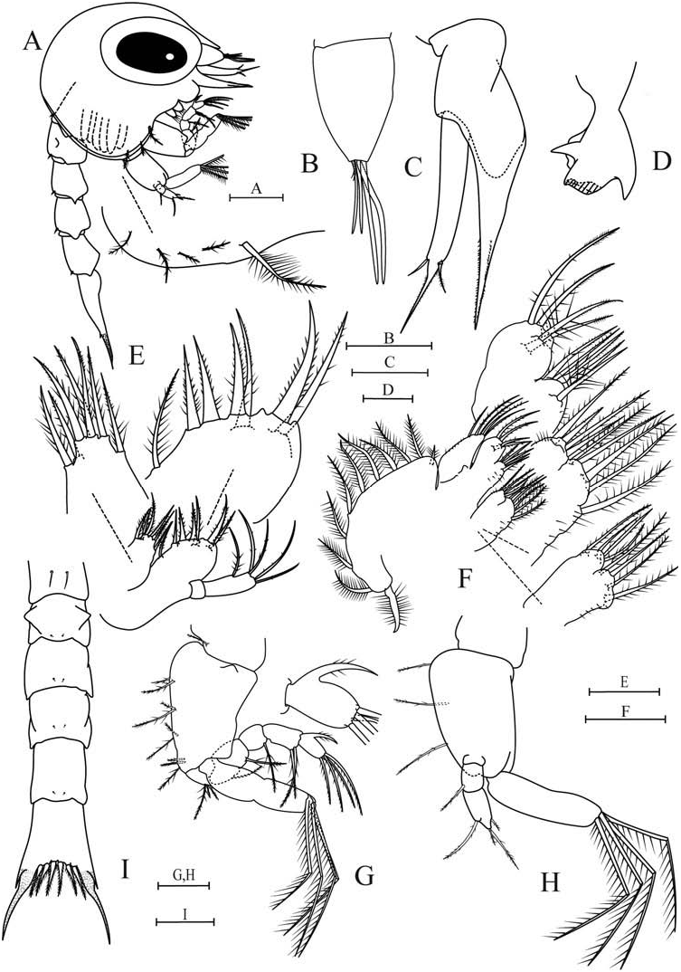
First zoea ( Figure 1 View Figure 1 )
Carapace ( Figure 1A View Figure 1 ). Without dorsal and lateral spines, rostral spine greatly diminished. Ventral margin with densely plumose ‘anterior seta’ ( Clark et al. 1998) posterior to scaphognathite notch, followed by 2–4 smaller plumose setae; carapace otherwise without setae. Eyes sessile. Frontal area bearing a dorsal organ ( sensu Martin and Laverack 1992; Lerosey-Aubrill and Meyer 2013).
Antennule ( Figure 1B View Figure 1 ). Unsegmented, smooth, conical. Terminally bearing 2 long, 2 shorter aesthetascs, and 1–2 short simple setae.
Antenna ( Figure 1C View Figure 1 ). Biramous, protopod long and pointed, bearing 2 rows of sharp spinules; endopod bud present; one-segmented exopod slightly longer than protopod with long spinulated distal process; pair of setae about one third from tip, one serrulate and one simple.
Mandible ( Figure 1D View Figure 1 ). With medial toothed molar process and enlarged lateral incisor processes; marginal teeth between molar and incisor processes. Palp absent.
Maxillule ( Figure 1E View Figure 1 ). Coxal endite bearing 7 setae, 4 graded plumodenticulate terminally, 3 plumodenticulate subterminally. Basial endite with 4 terminal plumodenticulate cuspidate setae and 3 subterminal plumodenticulate setae. Two-segmented endopod, proximal segment without seta, distal segment with 5 plumodenticulate setae, 1 subapical and 4 terminal. Exopod seta absent.
Maxilla ( Figure 1F View Figure 1 ). Coxal endite bilobed, proximal lobe with 4–5 setae, 3–4 plumose and 1 plumodenticulate; distal lobe with 3–4 setae, 1–2 plumose and 2 plumodenticulate. Basial endite bilobed, proximal and distal lobes with 5 and 4 plumodenticulate setae, respectively. Microtrichia present on both endites. Unsegmented endopod unilobed, with 5 plumodenticulate setae, 3 apical and 2 subapical; microtrichia on distal margin. Scaphognathite marginally with 9–11 densely plumose setae, including distal process.
Maxilliped I ( Figure 1A, G View Figure 1 ). Coxa with plumodenticulate seta. Basis with 9 plumodenticulate setae arranged 2 + 2 + 2 + 3. Endopod 5-segmented with 3, 2, 1, 2, 4 plumodenticulate setae and an accessory spine on the distal segment. Incompletely bisegmented exopod with 4 terminal plumose natatory setae.
Maxilliped II ( Figure 1A, H View Figure 1 ). Coxa without seta. Basis with 3 plumodenticulate setae. Endopod three-segmented, with 0, 1, 4 plumodenticulate setae, distal segment with 2 subapical, 2 apical setae. Incompletely bisegmented exopod with 4 terminal plumose natatory setae.
Maxilliped III ( Figure 1A View Figure 1 ). Present as a small undifferentiated bud.
Pereiopods ( Figure 1A View Figure 1 ). Present as small buds, chela indistinct.
Pleon ( Figure 1A, I View Figure 1 ). Five somites. Somite 1 with pair of middorsal long simple setae, somites 2–5 each with pair of shorter posteromedial simple setae. Posterolaterally, somite 2 with blunt process, somites 3–5 with short lateral projections; somite 2 with pair of dorsolateral processes. Pleopod buds rudimentary.
Telson ( Figure 1I View Figure 1 ). Bifurcated, without median notch, three pairs of plumodenticulate setae on inner margin; each furcal shaft proximally bearing lateral spine, furcal shafts and spines covered in rows of spinules to just below tips.
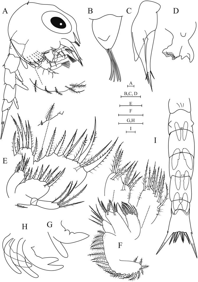
Second zoea ( Figure 2 View Figure 2 )
Carapace ( Figure 2A View Figure 2 ). Eyes mobile. Ventral margin with densely plumose anterior seta followed by 3–4 plumose setae; pair of small plumodenticulate setae dorsally.
Antennule ( Figure 2B View Figure 2 ). With 6–7 long aesthetascs and a short simple seta, endopod bud present.
Antenna ( Figure 2C View Figure 2 ). Endopod bud enlarged to just beyond middle of protopodite.
Mandible ( Figure 2D View Figure 2 ). Palp bud present.
Maxillule ( Figure 2E View Figure 2 ). Basial endite with 9–10 setae, additional graded plumodenticulate seta terminally, additional plumodenticulate setae subterminally; optional very small plumodenticulate seta on proximal margin; exopod pappose seta present.
Maxilla ( Figure 2F View Figure 2 ). Basial endite with 5 plumodenticulate setae on proximal lobe and distal lobe; Scaphognathite with 19–21 marginal plumose setae.
Maxilliped I ( Figure 2A View Figure 2 ). Exopod with 6 plumose natatory setae.
Maxilliped II ( Figure 2A View Figure 2 ). Exopod with 6 plumose natatory setae.
Maxilliped III ( Figure 2A,G View Figure 2 ). Exo-, endo- and epipod buds distinct.
Pereiopods ( Figures 2A,H View Figure 2 ). Longer, chela apparent.
Pleon ( Figures 2A, I View Figure 2 ). Additional sixth somite. Posterolaterally, somites 1 and 6 with blunt processes, somites 2–5 with enlarged lateral projections; somite 1 with three middorsal long simple setae. Pair of unsegmented biramous pleopods on somites 2–5, endopods very small.
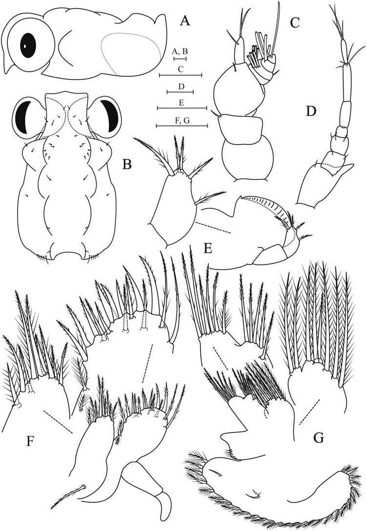
Megalopa ( Figures 3 View Figure 3 and 4 View Figure 4 )
Carapace ( Figures 3A, B View Figure 3 ). Longer than wide, subrectangular; rostral spine very short, ventrally deflected; hepatic region projected, forming 1 knob-like lateral expansion, gastric region well developed, divided in 2 regions, protogastric and metagastric; protogastric region swollen bearing the dorsal organ medially; branchial, cardiac and intestinal regions well defined, inflated. Posterior margin with 10 simple setae, surface covered mostly with simple setae as shown.
Antennule ( Figure 3C View Figure 3 ). Three-segmented peduncle, proximal segment without setae, middle and distal segments with one simple seta each; endopod unsegmented with 1 subterminal and 2 terminal long simple setae. Four-segmented exopod, first segment without seta, second segment with 6 aesthetascs and simple seta, third segment with 4 aesthetascs, distal segment with aesthetasc-like subapical seta.
Antenna ( Figure 3D View Figure 3 ). Segments 1–7, progressing proximally to distally, each with 1, 2, 3, 0, 0, 4, 4 simple setae, respectively. Basal segment with distinct exopod process.
Mandibles ( Figure 3E View Figure 3 ). Asymmetrical, scoop-shaped process with cutting edge and small acute tooth; two-segmented palp bearing 5 plumodenticulate setae on the distal segment.
Maxillule ( Figure 3F View Figure 3 ). Coxal endite with 5 graded plumodenticulate setae apically, 3 plumodenticulate setae, and a plumose seta subterminally. Basial endite with 17 setae, 7 plumodenticulate cuspidate terminally, 8 plumodenticulate subterminally, 2 plumose setae on proximal margin. Epipod plumodenticulate seta present; two-segmented endopod lacking outgrowths.
Maxilla ( Figure 3G View Figure 3 ). Coxal endite bilobed; proximal lobe with 6 setae, 5 plumose setae and 1 plumodenticulate seta; distal lobe bearing 2 plumose setae. Basial endite with 5 and 6 plumodenticulate setae on proximal and distal lobes, respectively. Endopod reduced, with microtrichia on distal margin, without setae. Scaphognathite with 28–31 marginal plumose setae; blade with 3 simple setae.
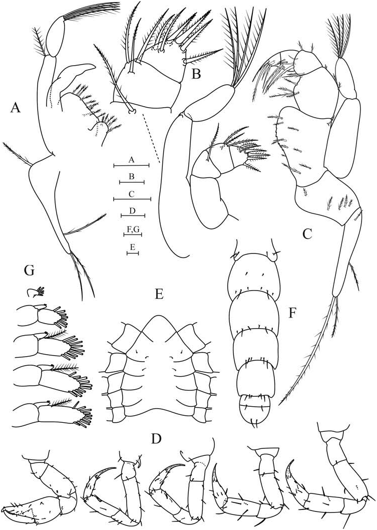
Maxilliped I ( Figure 4A View Figure 4 ). Coxa with 6 plumodenticulate setae, basis bearing 10–11 plumodenticulate setae; endopod without setae; exopod with pappose seta distally on proximal segment, 4 plumose setae on distal segment; epipod with 4 plumodenticulate setae, 1 proximal, 3 distal.
Maxilliped II ( Figure 4B View Figure 4 ). Coxa and basis not clearly differentiated; four endopod segments proximally to distally with 0, 1, 3 and 6 plumodenticulate setae, respectively; exopod with naked proximal segment and 4 plumose setae on distal segment; epipod not present on examined specimens.
Maxilliped III ( Figure 4C View Figure 4 ). Coxa with 1 + 4 plumodenticulate setae, basis fused to ischium, endopod proximally to distally with 10–11 + 1, 5–6 + 3, 4 + 2, 4 and 4 plumodenticulate setae; ischium with protuberances indicative of crista dentata; bisegmented exopod, naked proximal segment, distal segment with 4 plumose setae apically; epipod with short plumodenticulate seta proximally, 3 long plumodenticulate setae distally.
Pereiopods ( Figure 4D View Figure 4 ). Cheliped with mostly simple setae; pereiopods 2–5 mostly with simple setae; basischial segments without spines; coxa of pereiopod 2 with prominent spine; dactyls of pereiopods 2–5 with rows of spinules as shown; 1–2 serrulate setae near tip of dactyls on ventral margin.
Sternum ( Figure 4E View Figure 4 ). Segments 1–3 completely fused into single plate with bellshaped anterior margin, without setae; segment 4 with 2 pairs of simple setae; segment 5 with 1 pair of simple setae; subsequent segments without setae.
Pleon ( Figure 4F View Figure 4 ). Somites 1–5 proximally to distally with 4, 8, 6, 6, 6 simple setae dorsally and laterally, sixth somite with 2 simple setae.
Pleopods ( Figure 4G View Figure 4 ). Four pairs of pleopods, exopods each with 11, 11, 10, 8 plumose natatory setae, respectively; endopod with 2 cincinnuli each. Uropod reduced, segments fused, with 5 plumose setae distally, endopod absent.
Telson ( Figure 4F View Figure 4 ). Rounded posteriorly, bearing a pair of simple dorsal setae.
No known copyright restrictions apply. See Agosti, D., Egloff, W., 2009. Taxonomic information exchange and copyright: the Plazi approach. BMC Research Notes 2009, 2:53 for further explanation.
|
Kingdom |
|
|
Phylum |
|
|
Class |
|
|
Order |
|
|
Family |
EPIALTINAE MacLeay, 1838
| Colavite, Jessica, Santana, William & Pohle, Gerhard 2014 |
Menaethius monoceros
| Latreille 1825 |