
Neostempellina simantoneoa ( Sasa, Suzuki et Sakai, 1998 ) Orel, 2023
|
publication ID |
https://doi.org/ 10.11646/zootaxa.5254.4.8 |
|
publication LSID |
lsid:zoobank.org:pub:77D0E29C-B330-40C4-8927-AA7F1B86410E |
|
DOI |
https://doi.org/10.5281/zenodo.7734323 |
|
persistent identifier |
https://treatment.plazi.org/id/03FC304A-C365-FFA7-FF73-077BFA6DF9E7 |
|
treatment provided by |
Plazi |
|
scientific name |
Neostempellina simantoneoa ( Sasa, Suzuki et Sakai, 1998 ) |
| status |
comb. nov. |
Neostempellina simantoneoa ( Sasa, Suzuki et Sakai, 1998) View in CoL , comb. nov.
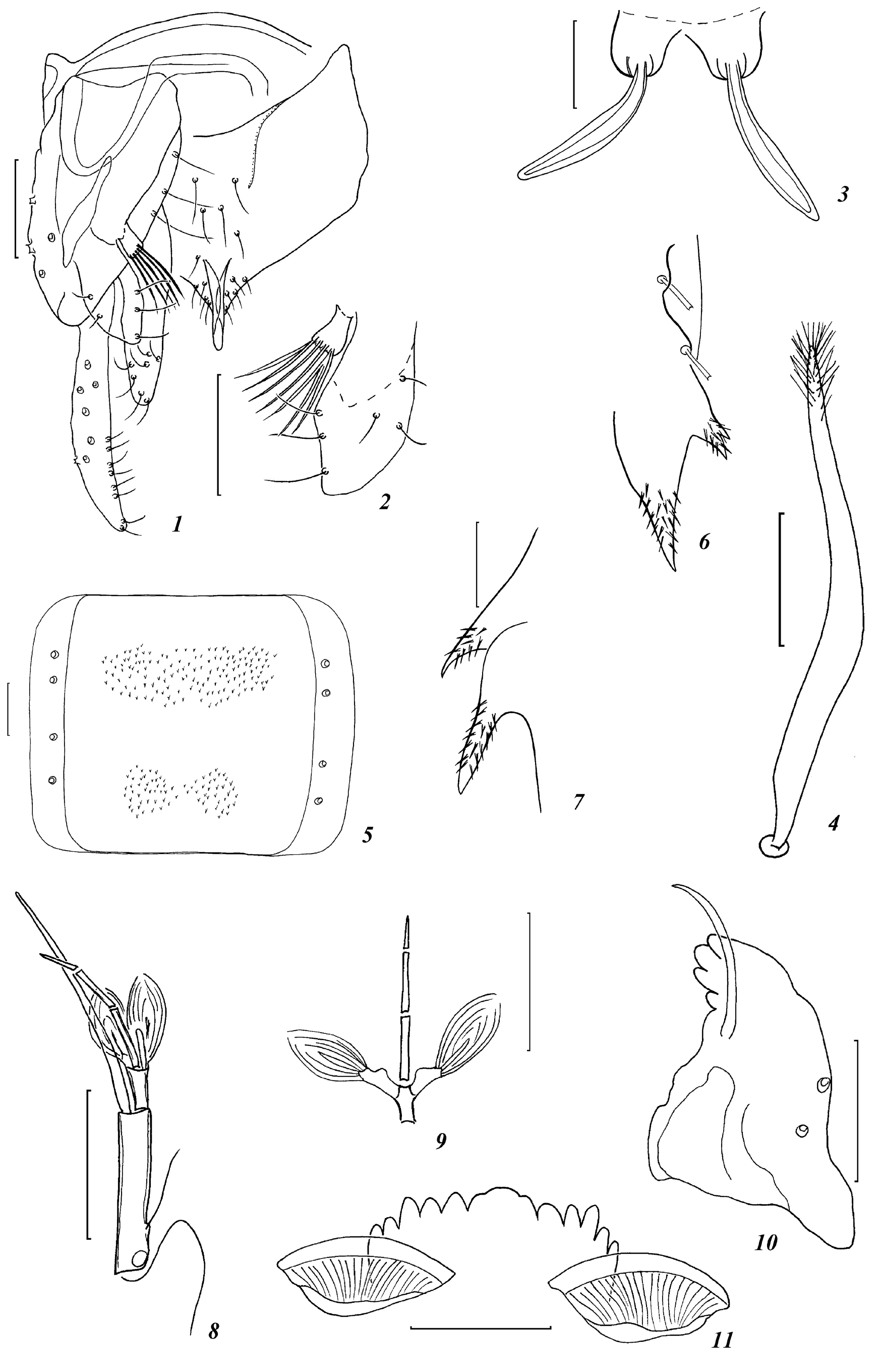
Figs 1–26 View FIGURES 1–11 View FIGURES 12–17 View FIGURES 18–26 .
Micropsectra simantoneoa Sasa, Suzuki et Sakai, 1998: 62 View in CoL , Fig. 15 View FIGURES 12–17 .
Zavrelia simantoneoa ( Sasa, Suzuki et Sakai, 1998) : Kobayashi 2014: 41.
Neostempellina sp. Makarchenko et al. 2005: 414; Orel 2016: 193.
Material examined. Mature pupa (pharate male), RUSSIA, PRIMORYE TERRITORY, Khasansky District, Barabashevka River , 01.vii.2002 ( T.Tiunova); 2 pupae with larval exuviae, 3 larvae, Lazovsky District , Proselochnaya River , 1 km from the cordon, 3–4.vii.2007 (O. Orel) .
Diagnosis. The male has morphometric features consistent with the diagnosis of the genus Neostempellina , namely, pubescent eyes, 10–11-segmented antenna, frontal tubercles absent, tibial spurs absent, anal tergite bands of V-type, median setae present, anal crests present, spinulea absent, superior volsella oval, digitus absent, median volsella short.
The structure of the pupa of this species supplements the diagnosis of the genus Neostempellina : cephalic tubercles well-developed, trapezoidal, frontal setae taeniate, elongated; thoracic horn elongate, gradually tapering towards apex and with short setae apically; Pedes spurii B absent; pedes spurii A well-developed on sternite IV; tergites II–V with a large rectangular to quadrate field of shagreen; segment VIII with large anal spurs.
The larva of this species conforms to the diagnosis of the genus Neostempellina , namely, the head capsule with numerous granulations and tubercles and/or spines; antennal pedestal with a broad spur and a larger multispined process; antennal segment 2 apically with a pair of Lauterborn organs on short pedicels; pecten epipharyngis of 3 apically serrated scales; premandible with 3 large apical teeth and 1 small basal tooth; brush present; mentum with 1 wide median tooth and 6 pairs lateral teeth; procerci strongly sclerotized, with numerous spines and projections; and the curved, tapered, tubular transportable case.
Description. Pharate male (n=1).
General colour yellowish brown.
Head. Eyes pubescent. Frontal tubercles absent. Temporal setae 5. Clypeus with 7 setae. Flagellomeres (1–11) dark brown (flagellomeres clearly separated), 584 μm long; ultimate flagellomere 184 μm long. AR 0.46. Maxillary palp lost.
Thorax. Ground colour of scutum pale yellow, mesonotal stripes and postnotum brown. Aps 0, Ac 5, Dc 6, Pa 1. Scutellum yellow, with 2 setae.
Wings 0.76 mm (not spread), covered with setae apical third.
Legs ( Table 1 View TABLE 1 ) yellowish brown. Spur on ta 1 6 μm long. Combs of ti 2 and ti 3 without spurs. BR 1 2.6, BR 2 4.0, BR 3 4.6. Sensilla chaetica on ta 1 of P 2 absent.
Hypopygium ( Figs 1–2 View FIGURES 1–11 ). Anal tergite with V-type bands and 7 median setae. Anal point 15 μm long and 6 μm wide, widest at its base and tapering towards rounded apex, with anal crests (30 μm long), spinulae absent, 8–10 lateral setae on each side of anal point. Gonocoxite 81 μm long, with 3 setae along inner margin. Transverse sternapodeme 24 μm wide, phallopodeme 39 μm long. Superior volsella oval (30 μm long, 15 μm wide), bearing 3 strong inner and 3–4 fine dorsal setae, microtrichia absent. Digitus and basal seta absent. Stem of median volsella short, 9 μm long and 6 μm wide, with several slender, lanceolate setae. Inferior volsella gradually narrows towards its apex, 51 μm long, with 10 setae. Gonostylus slender, slightly curved inwards, 60 μm long 15 μm wide. HR 1.35.
Pupa (n=4)
Cephalothorax. Length of thorax 0.64–0.69 mm. Frontal tubercles well-developed, 18–24 μm long, 30–36 μm wide. Frontal setae taeniate, 75–81 μm long and 12–15 μm wide, consisting of two parts: an inner core and a transparent shell ( Figs 3 View FIGURES 1–11 , 21 View FIGURES 18–26 ). Thorax granulose along median suture. Thoracic horn elongate, gradually tapering towards apex (159–189 μm long and 15–21 μm wide), apically covered with short setae ( Figs 4 View FIGURES 1–11 , 13 View FIGURES 12–17 ). Antepronotals 2 (median and lateral, taeniate). Precorneals 2–3 (135–171 μm long, taeniate). DC 1–4 simple, 15–39 μm long. Distance between DC 1 and DC 2 3–6 μm, DC 3 and DC 4 3–12 μm, DC 2 and DC 3 93–99 μm. Wing sheath 270–276 μm long, 75 μm wide. Prealar tubercle absent.
Abdomen ( Figs 5 View FIGURES 1–11 , 14–17 View FIGURES 12–17 ) 0.30–0.35 mm long. Hook rows with 53–63 hooks, 90–111 μm wide. Tergite I bare; II–VI with median shagreen; VII in proximal part with two patches of shagreen, with transverse stripe of shagreen in distal part; VIII with anterolateral and posterolateral patches of shagreen. Pedes spurii B absent; pedes spurii A well-developed on sternite IV. Sternite VI with anterolateral and posterolateral patches of shagreen or with three shagreen patches in the proximal part and with posterolateral patches of shagreen; VII with pair anterolateral patches of shagreen and posterior transverse band of spines ( Fig. 5 View FIGURES 1–11 ); VIII with anterolateral and posterolateral patches of shagreen or only with posterolateral patches of shagreen or shagreen absent. Laterosternites II–VI with shagreen. Segment VIII with 2–3 strong dark spurs (12–39 μm long) covered with small spines ( Figs 6–7 View FIGURES 1–11 , 17 View FIGURES 12–17 ). Segment II–IV with 3 L setae, V with 3 LS setae, VI–VII with 4 LS setae, and VIII with 3 LS setae. Anal lobe well developed with complete fringe of 16–20 taeniate setae in single row, dorsal setae absent. Anal segment 99–120 μm long and 171–201 μm wide.
Fourth instar larva (n=5)
Colouration white (in alcohol), with yellowish brown head capsule.
Head. Dorsal surface granulated; S 3 bifid. Frontoclypeal apotome apically with crown of 5 long spines.
Antenna ( Figs 8–9 View FIGURES 1–11 , 21–22 View FIGURES 18–26 ) situated on tall pedestal with pronounced distal spur (15–24 μm long and 12–15 μm wide) and multispined process (39–54 μm long). Antenna 5-segmented, total length 86–90 μm, length of each segment (in μm): 39–41; 11–12; 12–15; 15; 6–9. AR 0.76–0.84. Ring organ located at base of basal segment; seta strong, 18–24 μm long, located 15 µm from base of basal segment and extends beyond the middle of segment. Lauterborn organ 30 μm long, placed apically on short pedicels (7.5 μm long), not reaching apex of fourth segment. Blade 60–63 μm long, situated apically on basal segment, extending to antennal apex. At the apex of the basal segment there is a style 12 µm long.
Labrum ( Figs 18–19 View FIGURES 18–26 ). S I 15–18 μm long, comb-like; S II 24–27 μm long, plumose, situated on tall pedestal 9–15 μm long; S III 24–30 μm, simple; S IV A, B 4.5 and 6 μm long, respectively, finger-shaped. Labral lamella 18 μm long and 24 μm wide. Pecten epipharyngis consisting of 3 separate, distally serrated scales. Premandible 42–48 μm long, with 3 large apical and 1 small basal teeth; brush present.
Mandible ( Fig. 10 View FIGURES 1–11 , 20 View FIGURES 18–26 ) 69–72 μm long, 45 μm wide, with pale dorsal tooth, with brownish apical and two inner teeth. Seta subdentalis 39–45 μm long, curved. Seta interna consisting of 4 plumose branches.
Maxillary palp ( Fig. 20 View FIGURES 18–26 ) 3-segmented; basal segment 15 μm long, ring organ located near base, with several simple setae and sensilla, second segment 9 μm and third 4.5 μm long.
Mentum ( Figs 11 View FIGURES 1–11 , 24 View FIGURES 18–26 ) 60 μm wide; 1 median tooth (15 μm) and 6 pairs lateral teeth (if the teeth of the mentum are worn, then the medial tooth merges with the first lateral teeth and we see one wide pale medial tooth with two notches and 5 lateral teeth); medial and first lateral teeth lighter than other lateral teeth. Ventromental plate 42–45 μm wide, 21–24 μm high; distance between ventromental plates 15 µm.
Body. Posterior parapods with 15 simple hooks ( Fig. 25 View FIGURES 18–26 ). Procerci sclerotized and modified (99 μm long and 66 μm wide), covered with spines of different lengths; anal setae also modified, 5 wide, apically branched setae and 3 thinner setae split at the end ( Figs 25–26 View FIGURES 18–26 ). Anal tubules conical, 45–54 μm long.
Remarks. The male of N. simantoneoa collected in Primorye Territory practically does not differ from Japanese specimens of morphometric parameters ( Sasa et al. 1998, http://www. type.kahaku.go.jp/TypeDB/mediaDetail?cls= diptera&pkey=diptera-000707&lCls=m_diptera&lPkey=275053&detaillnkIdx=0; Kobayshi 2014).
The males of N. simantoneoa can be distinguished from all known species of the genus by the following combination of morphological features: eye hairy, antenna with 11 flagellomeres, frontal tubercles absent, AR 0.41– 0.46; LR P1 1.83–1.90; anal tergite bands of H-type, anal point relatively short tapered to apex, superior volsella oval with 3 inner and 2–3 dorsal setae, digitus absent, median volsella with a short rectangular stem, gonostylus slender, slightly curved inward.
The pupa of N. simantoneoa is most similar to Tanytarsini gen.? sp.? Pe 1 ( Langton & Visser 2003), but differs from the latter by the following features: cephalic tubercles well-developed, trapezoidal, tergite VII with pair anterolateral patches of shagreen and posterior transverse band of shagreen, Pedes spurii B on segment II absent, segment VIII with 1 strong apical spur and 1–2 smaller, more anterior spurs. Whereas pupa the Tanytarsini gen.? sp.? Pe 1 has very small conical cephalic tubercles, tergite VII with anterior and posterior transverse bands of spines, pedes spurii B on segment II hardly perceptible, segment VIII with 1 strong apical spur.
The pupa of N. simantoneoa is also similar to N. reissi Caldwell, 2000 on the thoracic horn form, but differs in the following morphological features: cephalic tubercles well-developed, trapezoidal, frontal setae taeniate, tergite VIII with anterolateral and posterolateral patches of shagreen. Whereas pupa N. reissi Caldwell has small cephalic tubercles, slender frontal setae, shagreen on tergite VIII is located medially and anterolaterally (see Table 2 View TABLE 2 ).
The larva of N. simantoneoa differs from the larvae of N. reissi and N. thienemanni by the following combination of characters: frontoclypeal apotome apically with crown of 5 long spines, S 3 bifid; antenna 5-segmented, width of distal spur equal to or slightly greater than width of basal segment, seta of basal segment strong, extends beyond the middle of the segment, Lauterborn organ placed apically on short pedicels, not reaching the apex of fourth segment, blade long, extending to antennal apex; premandible with 3 large apical and 1 small basal teeth; inner margin of mandible (mola) without spines.
Distribution. Japan (Shimanto River, Prefecture Kohchi, Shikoku Island), Russian Far East (Primorye Territory).


| T |
Tavera, Department of Geology and Geophysics |
No known copyright restrictions apply. See Agosti, D., Egloff, W., 2009. Taxonomic information exchange and copyright: the Plazi approach. BMC Research Notes 2009, 2:53 for further explanation.
|
Kingdom |
|
|
Phylum |
|
|
Class |
|
|
Order |
|
|
Family |
|
|
Genus |
Neostempellina simantoneoa ( Sasa, Suzuki et Sakai, 1998 )
| Orel, Oksana V. 2023 |
Zavrelia simantoneoa ( Sasa, Suzuki et Sakai, 1998 )
| Kobayashi, T. 2014: 41 |
Neostempellina sp.
| Orel, O. V. 2016: 193 |
| Makarchenko, E. A. & Makarchenko, M. A. & Zorina, O. V. & Sergeeva I. V. 2005: 414 |
Micropsectra simantoneoa Sasa, Suzuki et Sakai, 1998: 62
| Sasa, M. & Suzuki, H. & Sakai, T. 1998: 62 |