
Paradoxitettigia longicaudica, Wang & Huang & Shi, 2020
|
publication ID |
https://doi.org/ 10.11646/zootaxa.4750.1.8 |
|
publication LSID |
lsid:zoobank.org:pub:82B45F55-7D69-4CAB-ACE4-F45DF6F5BD5B |
|
DOI |
https://doi.org/10.5281/zenodo.3706187 |
|
persistent identifier |
https://treatment.plazi.org/id/03FC8796-430B-FF92-409F-FE31FD3BFE1C |
|
treatment provided by |
Plazi |
|
scientific name |
Paradoxitettigia longicaudica |
| status |
sp. nov. |
Paradoxitettigia longicaudica View in CoL sp. nov.
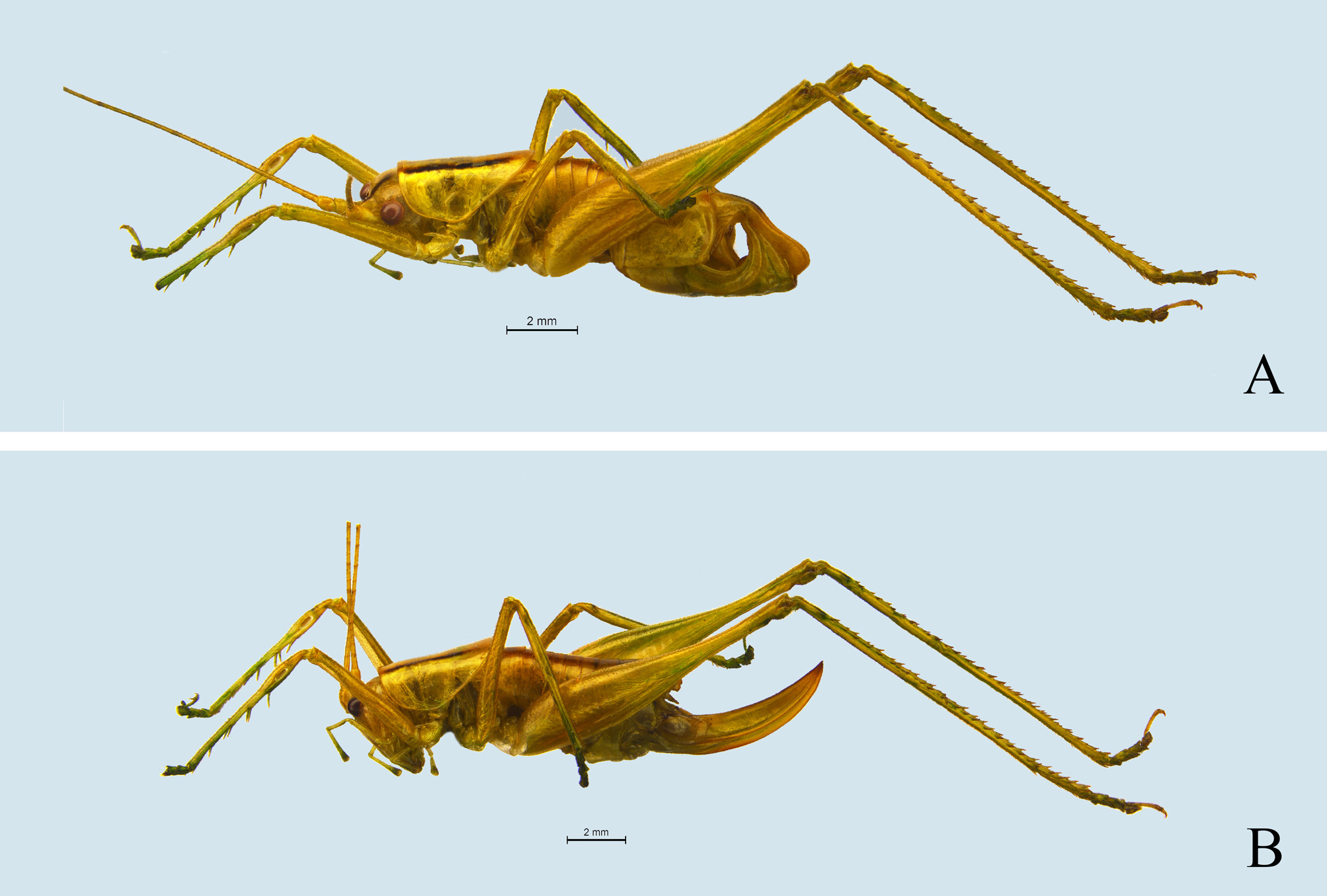
( Fig. 1–2 View FIGURE 1 View FIGURE 2 )
Description. Male. Body small, medium size for the tribe Meconematini , robust. Fastigium verticis conical, stubby, apex obtusely rounded, with a thin median sulcus. Eyes oval, protruding forwards and outwards. Maxillary palpus slender, apical segment longer than subapical one, slightly swollen apically.
Pronotum short, posterior margin reaching middle area of first abdominal tergite, anterior margin faintly straight while posterior margin arc-shaped; disc smooth, only posterior transverse sulcus distinct ( Fig. 1A View FIGURE 1 ); lateral lobe longer than deep, posterior margin tapering, without humeral sinus ( Fig. 1B View FIGURE 1 ). Thoracic auditory spiracle exposed.
Tegmina somewhat short, apices not reaching posterior margin of pronotum, visible in lateral view in some specimens, while some slightly surpassing posterior margin of pronotum, visible in dorsal view; apices of tegmina obtusely rounded ( Fig. 1A View FIGURE 1 ); hind wings absent.
All femora unarmed ventrally, genicular lobes with apices obtuse. Fore coxae with 1 short spine; tibiae with 5–6 spines respectively on both sides of ventral surface, tibial tympana open on both sides, oval. Middle tibiae with 5 inner and 6 outer spines on ventral surface. Hind tibiae with 2 inner and 3 outer spines on ventral surface as well as 26–30 spines on both sides of dorsal surface, bearing 2 pairs of ventral apical spurs and 1 pair of dorsal apical spurs.
The length of first to eighth abdominal segments is shorter than half of abdomen ( Fig. 2A View FIGURE 2 ). Ninth abdominal tergite rather strikingly swollen, of which posterior margin concave ( Fig. 1 View FIGURE 1 C–D). Tenth abdominal tergite obviously prolonged posteriorly, 1/3 basal area slightly broad while 2/3 apical area narrow, lateral margins almost parallel, with a deep longitudinal groove in the midline ( Fig. 1 View FIGURE 1 D–E); ventral surface near middle area with a moundy process ( Fig. 1H View FIGURE 1 ); apex of tenth abdominal tergite blunt, gently expanded ventrally ( Fig. 1C View FIGURE 1 ), centre of posterior margin spilt. Cerci short, conical, apices subacute; inner surface of subapical areas with 1 spiniform process, of which terminal subacute ( Fig. 1I View FIGURE 1 ). Tenth abdominal sternite fused with subgenital plate (with trace of fusion), broad, flat in ventral view, rectangular, basal area concave ( Fig. 1F View FIGURE 1 ); lateral surfaces of apical half folded dorsad and strongly expanded, triangular, apical area narrow, terminal obtusely rounded; styli absent ( Fig. 1C, E View FIGURE 1 ). Genitalia sclerotized, not surpassing posterior margin of subgenital plate, apical area expanded, centre of dorsal margin conspicuously concave ( Fig. 1G View FIGURE 1 ).
Female. Pronotum is similar to that of male; posterior margin of lateral lobe gradually narrowing; humeral sinus absent. Tegmina short, deposited laterally; some specimens exposed, visible in dorsal view; while tegmina of some specimens completely covered by pronotum. Appearance of first to seventh abdominal segments alike. Seventh abdominal sternite triangular, posterior margin bluntly rounded ( Fig. 1J View FIGURE 1 ). Eighth abdominal tergite faintly prolonged posteriorly, posterior margin arc-shaped ( Fig. 1K View FIGURE 1 ); lateral margins strongly prolonged ventrally ( Fig. 1L View FIGURE 1 ). Lateral margins of ninth abdominal tergite prolonged posteriorly ( Fig. 1M View FIGURE 1 ); tenth abdominal tergite elongated posteriorly, basal area somewhat broad then tapering apically, with a U-shaped concavity in the middle on posterior margin ( Fig. 1K View FIGURE 1 ). Cerci conical, apices acute. Subgenital plate short and broad, slightly concave on posterior margin ( Fig. 1L View FIGURE 1 ). Ovipositor stout, mildly bent dorsad, with tip subacute; dorsal and ventral margins smooth ( Fig. 1M View FIGURE 1 ).
Coloration. Body green when alive, while yellowish green when dried and pinned ( Fig. 2 View FIGURE 2 A–B). Eyes brown. Fastigium verticis brown. Dorsal area of head with 1 longitudinal light brown stripe in the midline. Disc of pronotum with 1 broad longitudinal light brown stripe, 3/4 anterior area of which with a black brown edge on each lateral margin, outer margins with 1 longitudinal light yellow stripe respectively ( Fig. 1A View FIGURE 1 ). Dorsal surface of abdomen with 1 longitudinal light brown stripe. Basal area of tenth abdominal tergite with a black brown spot in the middle ( Fig. 1D, K View FIGURE 1 ). Inner spiniform process of male cercus black brown ( Fig. 1I View FIGURE 1 ). Apices of all third segment of tarsi and claws brown, apices of dorsal spines on hind tibiae brown.
Measurements (mm). Body: ♂ 12.7–13.3, ♀ 10.6–13.2; pronotum: ♂ 3.8–4.1, ♀ 4.4–4.5; hind femora: ♂ 9.4– 9.6, ♀ 10.3–10.9; ovipositor: 5.6–6.3. Material examined. Holotype: ♂, Hengshan, Hengyang, Hunan, 21 August, 2018, coll. Tao Wang. Paratypes : 3♂ 4♀, same data as holotype. Other specimens : 9♂ 3♀, Hengshan, Hengyang, Hunan, 21 August, 2018, coll. Tao Wang; 2♂ 1♀, Hengshan, Hengyang, Hunan, 23 August, 2018, coll. Tao Wang. Distribution. China (Hunan). Etymology. The name of new species is derived from male tenth abdominal tergite prolonged posteriorly. Latin “ long -” means long, and Latin “ caud -” means caudal.

No known copyright restrictions apply. See Agosti, D., Egloff, W., 2009. Taxonomic information exchange and copyright: the Plazi approach. BMC Research Notes 2009, 2:53 for further explanation.