
Mesoleptobasis Sjöstedt 1918
|
publication ID |
https://doi.org/ 10.5281/zenodo.188653 |
|
DOI |
https://doi.org/10.5281/zenodo.6214942 |
|
persistent identifier |
https://treatment.plazi.org/id/03FC87D3-EB00-FF84-01B2-FB4B836540E0 |
|
treatment provided by |
Plazi |
|
scientific name |
Mesoleptobasis Sjöstedt 1918 |
| status |
|
Mesoleptobasis Sjöstedt 1918 View in CoL
Mesoleptobasis Sjöstedt 1918: 21 View in CoL (description); — St. Quentin 1960: 45, 51 (generic key and characterization); — Davies 1981: 2; — Davies & Tobin 1984: 77 (catalog); — Bridges 1994: III.30 (catalog); — Steinmann 1997: 288 (catalog); — Tsuda 2000: 39 (catalog); — Lencioni 2006: 18 (key), 41 (notes); — Heckman 2008: 305 (key to neotropical genera).
Type species. Mesoleptobasis incus Sjöstedt 1918 by monotypy.
[ NOTE: Davies & Tobin (1984) followed by Tsuda (2000), Steinmann (2007), and Heckman (2008) listed this species as Mesoleptobasis inca apparently to conform to Art. 31.2.1 of the Code. The species epithet incus is a noun in apposition (f., Latin: anvil), probably referring to the shape of the pronotum, and therefore should not be changed [Arts. 32. 2.2 and 31.2.3].
Other species included. M. acuminata Santos 1961 , M. cantralli Santos 1961 , M. cyanolineata ( Wasscher 1998) , M. elongata sp. nov.
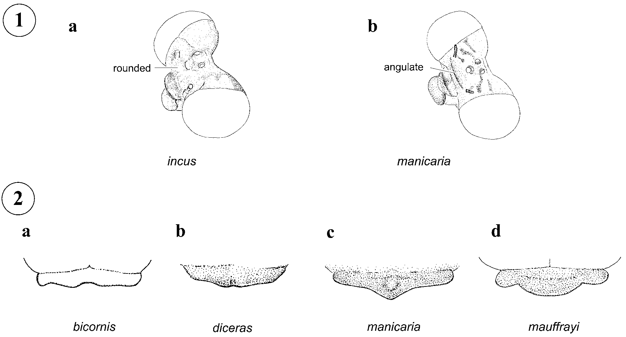
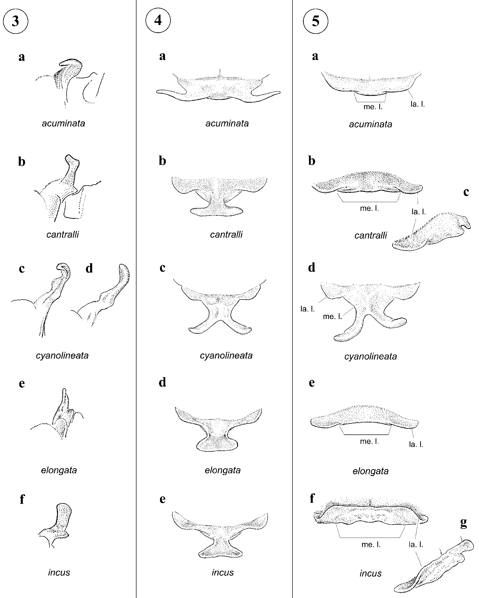
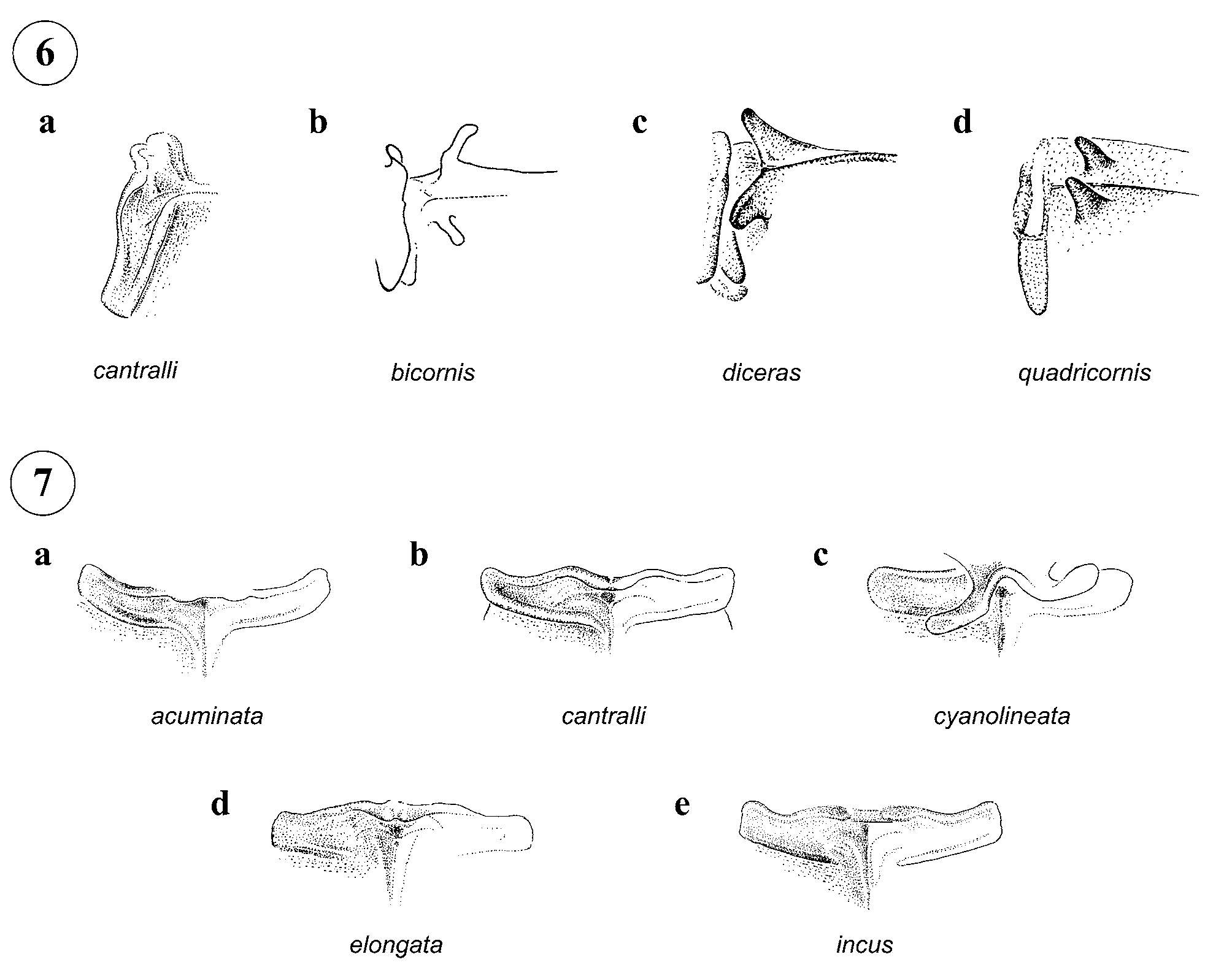
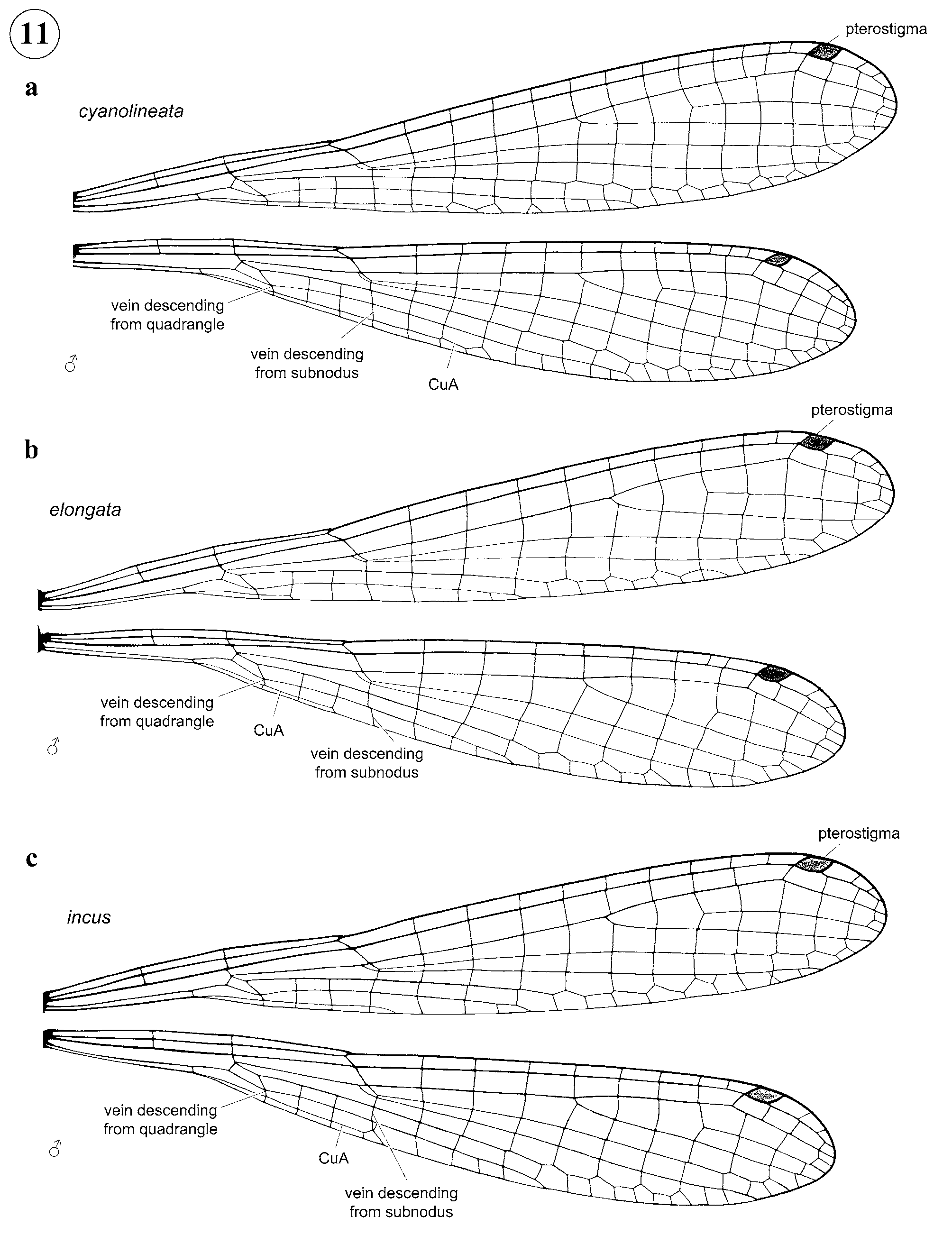
Characterization. Medium sized (36–45 mm), largely pale coenagrionids with a long abdomen (ratio of 5.1–6.36 to length of head plus thorax); head and dorsum of abdomen orange to brown or black with some metallic iridescence, pale areas yellow or light blue. Blue postocular spots present or absent and pale occipital bar absent. Pterothorax lacking dark mid-dorsal and metapleural stripes; in mature specimens brown mesepisternal and metepisternal stripes and pale antehumeral, mesepimeral, and metepisternal stripes present ( Fig. 8 a); in immature specimens pterothorax entirely pale brown or orange. Frons rounded; location of most posterior point of head at level of eyes ( Fig. 1 a). Posterior lobe of prothorax in male with long projections, which may be represented by lateral processes ( Fig. 4 a) or by a medial bifurcate process ( Figs. 4 b–e); pronotum in female projected as in male (in M. cyanolineata ; Fig. 5 d) or slightly trilobate (in M. acuminata , M. cantralli , M. elongata , and M. incus ; Figs. 5 a–c, e–g). Hind femur short, not reaching anterior margin of S1; metatibial spurs shorter than twice intervening spaces ( Fig. 8 a); pretarsus with vestigial supplementary tooth represented by an obtuse low prominence ( Fig. 9 View FIGURE 9 a). CuA extending from two to four ( Figs. 10 View FIGURE 10 a) or six or seven cells (in M. cyanolineata and females of M. acuminata ; Figs. 11 View FIGURE 11 a, 12a, 13a) distal to vein descending from subnodus to one or two cells proximal to vein descending from subnodus ( Figs. 10 View FIGURE 10 b, 11b, c); CuP reaching posterior margin of wing; vein descending from quadrangle forming an unbroken line to wing margin ( Figs. 10–13 View FIGURE 10 View FIGURE 11 View FIGURE 12 View FIGURE 13 ). Genital ligula distal segment with inner fold; with sclerotized areas present as spine-like projections on lateral margins ( Fig. 15); with two pairs of lateral lobes, a smaller latero-basal one and a larger latero-medial one; apex transverse ( M. acuminata ; Fig. 14 View FIGURE 14 a) or deeply bifid ( Figs. 14 View FIGURE 14 b–e). Postero-dorsal margin of male S10 projected caudally and with a pair of medial lobe-like projections ( M. cantralli , M. elongata , and M. incus ; Figs. 17 View FIGURE 17 b, d, e, 18b, d, e, 19a–c) or emarginate and lacking lobe-like projections ( M. acuminata and M. cyanolineata ; Figs. 17 View FIGURE 17 a, c, 18a, c). Male cercus entire, approximately horizontal with tip bent ventrally, shorter than to subequal to S10 ( Figs. 17–19 View FIGURE 17 ), with an elongate membranous depression on dorsal surface only in M. acuminata ( Fig. 17 View FIGURE 17 a); male paraproct forcipate, much longer than S10, with tip recurved medio-ventrally ( Figs. 17–19 View FIGURE 17 ). Female lacking vulvar spine on S8 ( Figs. 16 View FIGURE 16 a, b) or with a very small spine in some females of M. cyanolineata (reported by Wasscher 1998 for allotype) and M. incus ( Fig. 16 View FIGURE 16 e); postero-dorsal margin of S9 with denticles; ovipositor extending beyond tips of cerci for a distance shorter ( Figs. 16 View FIGURE 16 a, c, e) to subequal ( Fig. 16 View FIGURE 16 d) or longer ( Fig. 16 View FIGURE 16 b) than cerci length.
Diagnosis. Within New World Coenagrionidae , Mesoleptobasis shares only with Aceratobasis and Metaleptobasis the combination of a pretarsus with vestigial supplementary tooth represented by an obtuse low prominence or else absent ( Fig. 9 View FIGURE 9 ) and a vein descending from quadrangle forming an unbroken line to wing margin ( Figs. 10–13 View FIGURE 10 View FIGURE 11 View FIGURE 12 View FIGURE 13 ). However, it differs from both Aceratobasis and Metaleptobasis by its rounded frons ( Fig. 1 a), highly modified pronotum with long processes at least in males ( Figs. 3 View FIGURE 3 , 4, 5 d), pterothoracic color pattern lacking dark mid-dorsal stripe ( Fig. 8 a), and genital ligula with a small inner fold and with spinelike projections on lateral margins distal to flexure ( Fig. 15), this last character being unique for the genus. In Aceratobasis and Metaleptobasis the frons is angled ( Fig. 1 b), the pronotum is devoid of long projections ( Fig. 2 View FIGURE 2 ), and the distal segment of genital ligula lacks an inner fold, lateral lobes, and any sclerotized projections distal to flexure ( Cumming 1954; Rácenis 1955; von Ellenrieder & Garrison 2008). CuA in Metaleptobasis extends more than seven cells posterior to the vein descending from the subnodus and males of all species have long mesepisternal horns ( Fig. 8 b), which in females are similar in size and length ( Figs. 6 View FIGURE 6 b, c) or may be reduced to tubercles in some species ( Fig. 6 View FIGURE 6 d). In Mesoleptobasis CuA never extends more than 7 cells distal to the vein descending from the subnodus ( Figs. 10–13 View FIGURE 10 View FIGURE 11 View FIGURE 12 View FIGURE 13 ) and the mesepisterna are always smooth ( Figs. 6 View FIGURE 6 a, 7). Mesoleptobasis further differs from Aceratobasis by having male cerci entire and shorter than long, forcipate paraprocts ( Figs. 17–19 View FIGURE 17 ); cerci in Aceratobasis are branched with a ventro-basal spur or spine and are longer than the paraprocts.
Distribution. Amazon forest in Venezuela, Guyana, Surinam, Brazil, and Peru ( Fig. 21 View FIGURE 21 ). Specimens are infrequently collected, and it is therefore likely that distribution ranges are larger than those indicated by existing specimens.
Habitat. Adults found within forest, in dark or shaded areas flying near the ground; some specimens of M. cyanolineata were captured at night attracted to lights ( Wasscher 1998); breeding habitat and larvae unknown.


No known copyright restrictions apply. See Agosti, D., Egloff, W., 2009. Taxonomic information exchange and copyright: the Plazi approach. BMC Research Notes 2009, 2:53 for further explanation.
|
Kingdom |
|
|
Phylum |
|
|
Class |
|
|
Order |
|
|
Family |
Mesoleptobasis Sjöstedt 1918
| Garrison, Rosser W. & Ellenrieder, Natalia Von 2009 |
Mesoleptobasis Sjöstedt 1918 : 21
| Heckman 2008: 305 |
| Lencioni 2006: 18 |
| Tsuda 2000: 39 |
| Steinmann 1997: 288 |
| Davies 1984: 77 |
| Davies 1981: 2 |
| St 1960: 45 |
| Sjostedt 1918: 21 |