
Mesoleptobasis elongata, Garrison, Rosser W. & Ellenrieder, Natalia Von, 2009
|
publication ID |
https://doi.org/ 10.5281/zenodo.188653 |
|
DOI |
https://doi.org/10.5281/zenodo.6214954 |
|
persistent identifier |
https://treatment.plazi.org/id/03FC87D3-EB0D-FF91-01B2-FA19832743BB |
|
treatment provided by |
Plazi |
|
scientific name |
Mesoleptobasis elongata |
| status |
sp. nov. |
Mesoleptobasis elongata View in CoL sp. nov.
Figs. 3 View FIGURE 3 e, 4d, 5e, 7d, 11b, 13b, 14d, 15d, 16d, 17d, 18d, 19b, d, 20a, 21
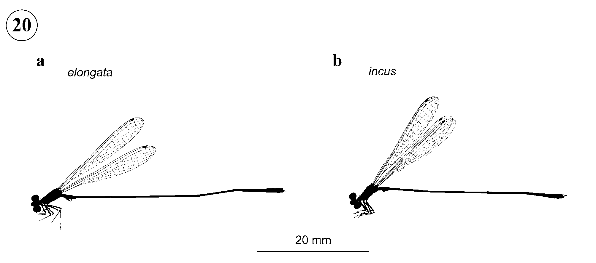
Etymology. From elongatus (Lat.), referring to its long abdomen which distinguishes it from its congeners ( Fig. 20 View FIGURE 20 a).
Types. Holotype 3 (from Boven Coesewijne, Para, Surinam, 5°22'N, 55°32'W) in RMNH (examined).
Specimens examined. Total: 2 3, 1 Ƥ. Surinam, Para District: 1 3 holotype, 1 Ƥ allotype, Boven Coesewijne (5°22'N, 55°32'W), 0 6 viii 1960, leg. J. Belle (RMNH). Brazil, Amazonas State: 1 3 paratype, Paraná Costa da Ilha de Curari (Rio Solimões), canopy fogging (with pyrethrum), project TRS #04, tray #322, white water inundation forest, varzea (3°25'S, 60°15'W), 0 3 viii 1914, leg. Adis, T. Erwin, Montgomery, et al. (RWG).
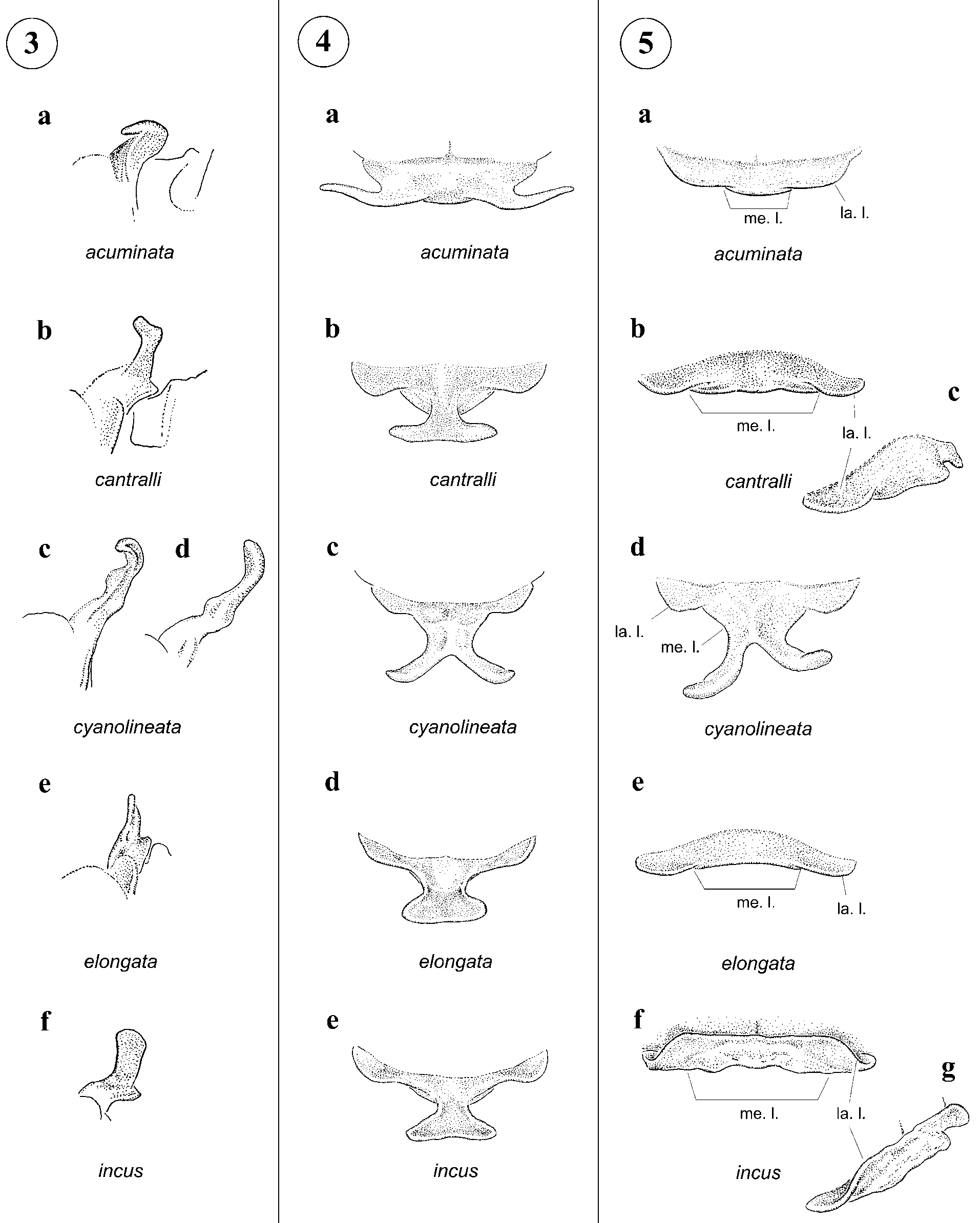
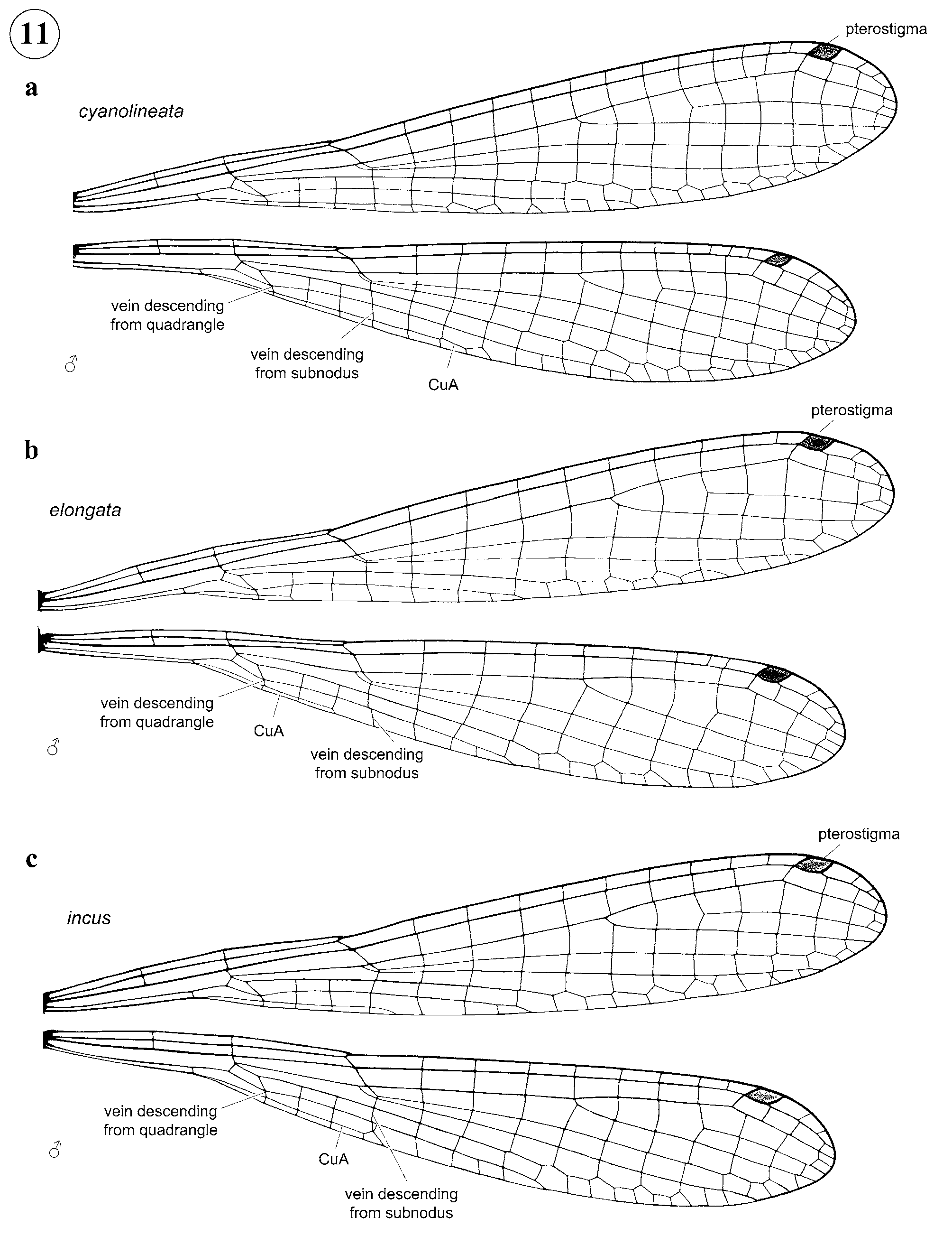
Description. Male holotype ( Fig. 20 View FIGURE 20 a). — Head: Labium ivory, remainder of head including epicranium dull olive green, paler on rear of head, dark brown along base of postfrons, base of antennae, postfrontal suture, and irregular diffuse spot between ocelli and antenna; postocular spots absent. — Thorax: Prothorax dull olive blue, middle lobe light brown; pterothorax entirely olive blue with diffuse brown thoracic stripes along medial portion of mesepisternum and mesopleural suture, remainder of thorax becoming paler laterally; hind lobe of prothorax with a medial bifurcate process ( Fig. 4 d), with apices directed laterally ( Fig. 3 View FIGURE 3 e) forming a transverse line between them. Costal side of Fw pt longer than basal side, its posterior margin slightly convex. CuA relatively short, ending one cell proximal to vein descending from subnodus. Wings hyaline, Px Fw 10; Px Hw 8 (left)/ 9 (right); RP 2 originating at Px 5 in Fw, at Px 4 in Hw; pterostigma 0.4 mm long. Legs ivory with wash of brown at flexor surfaces of femora, spines black — Abdomen: S1-7 brown dorsally, pale olive blue laterally, S3-7 with pale basal ring, S8-10 dull orange brown. Genital ligula in ectal view ( Fig. 14 View FIGURE 14 d) with distal margin deeply bifid and lacking lateral emarginations; in lateral view ( Fig. 15 d) with basal lobe sclerotized, long, pointed, and directed posteriorly, and dorsal margin of lateral lobe lacking a single small sclerotized spine. Posterior margin of S10 ( Figs. 17 View FIGURE 17 d, 18d) projected medio-dorsally, with a pair of postero-lateral small lobe-like processes. Male cercus lacking a membranous area dorsally, approximately semicircular, as long as wide; in lateral view ( Fig. 18 d) smoothly curved, with tip directed postero-ventrally; paraproct slender and narrower than half of S10 height at base in lateral view; base of paraproct with a thumblike tubercle ( Fig. 18 d).
Dimensions. Hw 18.4; abdomen 40; total length 45.
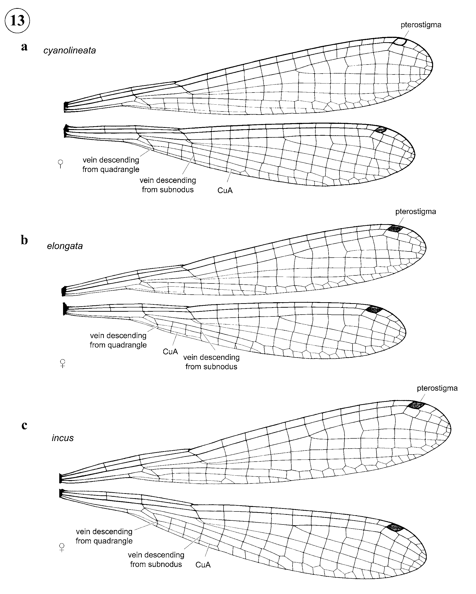
Female allotype. Head: similar to male but head entirely dark olive becoming paler along genae and lacking dark suture and epicranial spots. — Thorax: as in male except hind lobe of pronotum ( Fig. 5 e) weakly trilobate, transverse, gently concave medially in dorsal view and with middle lobe erect; mesostigmal plates as in Fig. 7 d. Costal side of FW pt longer than basal side, its posterior margin slightly convex. CuA relatively short, ending one cell proximal to vein descending from subnodus in Fw, at level of subnodus in Hw ( Fig. 13 View FIGURE 13 b). Wings hyaline, Px Fw 11 (left)/ 10 (right); Px Hw 9 (left)/ 10 (right); RP 2 originating at Px 5 in Fw, at Px 4 in Hw; pterostigma 0.5 mm long. — Abdomen: as in male but shorter, more robust, and S8-10 dark brown above and pale green laterally; ovipositor surpassing tip of cerci for a distance subequal to length of cerci ( Fig. 16 View FIGURE 16 d).
Dimensions. — Hw 19.2; abdomen 32; total length 38.
Variation in male paratype. As in holotype but overall body coloration paler with more of an orange tinge on olivaceous areas, CuA relatively short, ending two cells proximal to vein descending from subnodus in Fw, one cell proximal in Hw ( Fig. 11 View FIGURE 11 b), Px Fw 9; Px Hw 8 (left)/ 7 (right); RP 2 originating at Px 5 in Fw, at Px 4 in Hw; pterostigma 0.4 mm long.
Dimensions. Hw 18.0; abdomen 37; total length 42.
Diagnosis. Morphology of genital ligula, male cerci and paraprocts, and female ovipositor ally this species to M. cantralli and M. incus , but the longer abdomen of male ( Fig. 20 View FIGURE 20 a) relative to the other two species (as in Fig. 20 View FIGURE 20 b) readily separate them. It can be further diagnosed from its congeners by: male prothorax with a medial bifurcate process with arms directed laterally forming a transverse line between them ( Fig. 3 View FIGURE 3 e; shared with M. cantralli and M. incus ); female prothorax lacking processes, with posterior margin slightly trilobate, with medial lobe not surpassing lateral lobes posteriorly; lateral lobes not bent anteriorly ( Fig. 5 e; shared with M. cantralli ). Costal side of Fw pt longer than basal side, its posterior margin slightly convex in both sexes ( Figs. 11 View FIGURE 11 b, 13b; shared with M. cantralli , M. cyanolineata , and M. incus ). CuA relatively short ( Figs. 11 View FIGURE 11 b, 13b; shared with M. cantralli and M. incus ), ending one or two cells proximal to vein descending from subnodus in male and one cell proximal to vein descending from subnodus in female. Genital ligula in ectal view with distal margin deeply bifid and lacking lateral emarginations ( Fig. 14 View FIGURE 14 d; shared with M. cantralli , M. cyanolineata , and M. incus ); in lateral view with basal lobe sclerotized, long, pointed, and directed posteriorly ( Fig. 15 d; shared with M. cantralli and M. incus ). Posterior margin of S10 projected medio-dorsally, with a pair of postero-lateral small lobe-like processes ( Figs. 17 View FIGURE 17 d, 19b; shared with M. cantralli and M. incus ). Male cercus lacking a membranous area dorsally (shared with M. cantralli , M. cyanolineata , and M. incus ), approximately semicircular, as long as wide ( Figs. 17 View FIGURE 17 d, 19b, d; shared with M.
incus ); in lateral view smoothly curved, with tip directed postero-ventrally ( Fig. 18 d); paraproct slender and narrower than half of S10 height at base in lateral view ( Fig. 18 d; shared with M. cantralli and M. incus ); base of paraproct with a thumb-like tubercle ( Fig. 18 d; shared with M. cantralli ). Ovipositor surpassing tip of cerci for a distance as long as length of cerci ( Fig. 16 View FIGURE 16 d; shared with M. cantralli ).
Remarks. Despite careful examination we did not observe any spines such as those present in M. cantralli ( Fig. 15 b) and M. incus ( Fig. 15 e) on the dorsal margin of lateral lobe of genital ligula; this area appears to be slightly darker and thickened in the holotype.
Distribution. Para State in Surinam and Amazonas State in Brazil ( Fig. 21 View FIGURE 21 ). The paratype male was canopy-fogged.


No known copyright restrictions apply. See Agosti, D., Egloff, W., 2009. Taxonomic information exchange and copyright: the Plazi approach. BMC Research Notes 2009, 2:53 for further explanation.
|
Kingdom |
|
|
Phylum |
|
|
Class |
|
|
Order |
|
|
Family |
|
|
Genus |