
Mycetophylax morschi ( Emery, 1888 )
|
publication ID |
https://doi.org/10.11646/zootaxa.2052.1.1 |
|
DOI |
https://doi.org/10.5281/zenodo.4675137 |
|
persistent identifier |
https://treatment.plazi.org/id/03FC87DA-F510-C57E-42B2-FDFAFD78FD32 |
|
treatment provided by |
Plazi |
|
scientific name |
Mycetophylax morschi ( Emery, 1888 ) |
| status |
comb. nov. |
Mycetophylax morschi ( Emery, 1888) new combination
( Figs. 2 View FIGURE 2 , 6 View FIGURE 6 c, d, g, 7 View FIGURE 7 a)
Cyphomyrmex morschi Emery, 1888 View in CoL ("1887"): 9, (worker) Syntypes, Brazil, Rio Grande do Sul, São Lourenço (von Ihering), (MZSP, examined); Kempf, 1972: 93 (catalogue); Bolton, 1995: 168 (catalogue), Klingenberg & Brandão, 2005: 44 ( syntype worker in MZSP); new combination in Mycetophylax View in CoL .
Cyphomyrmex View in CoL sp. (in part): Mayr 1887: 556 (key)
Cyphomyrmex ( Mycetoritis) (sic) personatus Santschi, 1923: 268 : (gyne) Argentina: Prov. de Buenos Aires, Monte Hermose, no coll. data (C. Bruch); ( type specimens not localized); Kempf, 1964: 25: synonym of Cyphomyrmex morschi. Kempf, 1972: 93 View in CoL (catalogue); Bolton, 1995: 168 (catalogue)
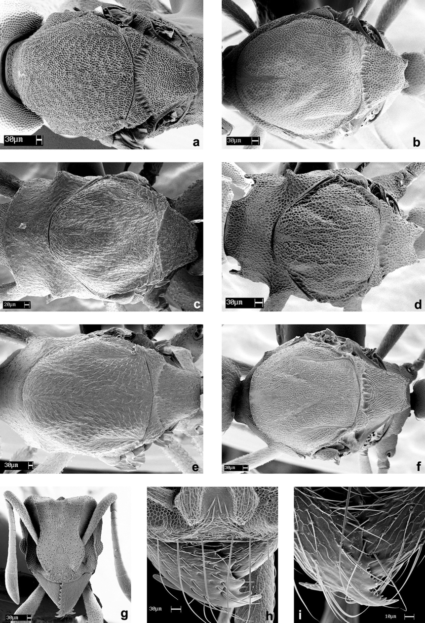
Worker ( Figs. 2 View FIGURE 2 a, b, g, 6 g and 7 a)
Range of measurements (mm) and indices of examined specimens (N = 10): IOD 0.53–0.67; HL 0.6–0.71; CI 82–97; SL 0.49–0.60; SI 79–98; ML 0.20–0.28; MI 32–42; WL 0.80–1.00; PrW 0.40–0.50; PL 0.10–0.20; PPL 0.20–0.30; GL 0.54–0.70; FL 0.66–0.93; TL 2.54–3.03.
Color brown to light brown. Mandibles, antennae and legs opaque brown to yellowish. Surface of frons with small irregular pores; a single seta coming out of each of them (only visible with higher magnifications). Mandibular disc almost entirely covered by very fine longitudinal rugae. Whole body, legs and antennae covered by bright, short and sparse appressed white hairs; only the area between frontal carinae and preocular carinae hairless. Hairs of masticatory border of mandible somewhat longer than in the other areas. Sculpture areolate. The individual submitted to SEM is covered by a thin layer of "dirt”, detritus adhering at the integument, leaving the sculpture with a punctate appearance ( Fig. 6 View FIGURE 6 g).
Head longer than wide, subrectangular; posterior portion slightly wider than anterior one. Compound eyes with eight ommatidia at maximum length and six ommatidia at maximum width. Mandibles with nine triangular teeth, the four most apical larger and the remaining slightly smaller. Anterior margin of clypeus gently rounded and with a short and hardly visible median seta. Frontal lobes maximum expansion less than half the distance between the median line and external border of the head, external margins gently rounded. Frontal and lateral carinae almost reaching the posterolateral lobes, forming a scrobe-like impressed area, where the ants accommodate the antennal scapes. Lateral carinae distinct for half of head length, posterior part of carinae obsolete, faintly developed. Antennal scapes straight, surpassing the posterolateral corners by a distance similar to their diameter, when laid back. Antennae ending in a three-segmented club, last antennal segment as long as the three preceding together. Vertexal margin concave, impressed, posterolateral corners rounded.
Pronotum in lateral view with the dorsum marked by a very low median tubercle, a pair of low posterior tubercles, and short, blunt pronotal inferior spines. Mesonotum with a median rounded tubercle. Metapropodeal impression distinct. Propodeum in lateral view with basal face slightly convex; declivous face almost straight, armed with a pair of small blunt spines, occasionally these spines appearing as protuberances. Peduncle of petiole reduced, node of petiole as long as high, ending in two lateral lobes posteriorly. In dorsal view, posterior portion of postpetiole with a deep impression; posterior margin slightly convex, almost straight.
Gyne ( Figs. 2 View FIGURE 2 c, d, h, 6 View FIGURE 6 c and 7 View FIGURE 7 a)
Range of measurements (in mm) and indices of examined specimens (N = 13): IOD 0.66–0.72; HL 0.74–0.78; CI 87–92; SL 0.54–0.64; SI 79–94; ML 0.35–0.40; MI 47–52; WL 1.04–1.14; PL 0.20–0.24; PPL 0.26–0.30; GL 0.84–0.98; TL 3.45–3.90.
Color brownish to dark brown. Mandibles, antennae and legs yellowish, according to the age of individuals. Most characters as in conspecific workers. Compound eyes at maximum length with 15 ommatidia and at maximum width eleven ommatidia. In lateral view, scutum flattened, covering more than half of the pronotum. Pronotum with anterior lateral pronotal protuberances, a low median elevation, and distinct blunt inferior pronotal spines. Parapsidial lines indistinct, glabrous and parallel in relation to the main body axis. Notaulices shallowly impressed, scutum-scutellar sulcus distinctly impressed. Scutellum concave at anterior margin in dorsal view and twice the width of the area anteriorly than posteriorly. Posterior margin angled, concave in the middle. Katepisternum subtriangular, anepisternum subrectangular, both divided by a distinct suture. Propodeum declivous and basal face almost straight, with small blunt spines, directed back- and upwards.
Male (undescribed) ( Figs. 2 View FIGURE 2 e, f, i, 6 View FIGURE 6 d and 7 View FIGURE 7 a)
Range of measurements (in mm) and indices of examined specimens (N = 10): IOD 0.44–0.52; HL 0.52–0.62; CI 79–87; SL 0.54–0.68; SI 117–113; ML 0.22–0.28; MI 38–48; WL 0.84–1.04; PL 0.18–0.22; PPL 0.18–0.24; GL 0.70–0.84; TL 2.69–3.15.
Color yellowish to brownish. Mandibles, antennae (base and funiculus) and legs yellowish, lighter in young individuals. Sculpture like in the conspecific workers, but for the gaster, which sculpture is fainter in males.
Head longer than wide, compound eyes occuping a fourth of its lateral margin, in full face view, with 18 ommatidia at maximum length and 16 ommatidia at maximum width. Anterior margin of clypeus almost straight, with one visible median seta twice as long as the mandible hairs. Middle portion of clypeus ending in a straight suture at the level of the antennal insertions, followed by the relatively large, impressed, frontal area. Mandibles with three apical teeth followed by a diastema and two to three smaller teeth; last tooth as a denticle. Frontal lobes covering half of the antennal insertions in full face view. Antennae 13-segmented. Antennal scapes slightly curved, flattened at base, rounded and thicker apically. Apical segment of funiculus as long as the two anterior segments together. Frontal carinae extending back up to the level of the median ocellus, disappearing near the vertexal margin. Lateral carinae following the internal margin of the compound eyes anteriorly, ending little after the level of the posterior margin of the compound eyes; both carinae converging but not touching posteriorly. Area between frontal and lateral carinae impressed and free of hairs. Vertex flattened, slightly and evenly concave, laterally marked by defined angles produced as blunt denticles.
In lateral view scutum covering half of the pronotum. Pronotum with small, acute, triangular, inferior spines and a pair of well developed, acute, lateral spines. Notaulices, in dorsal view, as wide impressed sutures with slightly transverse rugae at its posterior portion. Parapsidial lines very close to the lateral margins, gently rounded, diverging anteriorly. Prescutellum relatively wide. Scutum-scutellar sulcus deeply impressed with transversal rugae. Anterior margin of scutellum rounded; in dorsal view. Katepisternum and anepisternum divided by a suture with transverse rugae. Katepisternum subquadrate, antero-inferior margin rounded; anepisternum subtriangular, with anterior vertex rounded. Mesocoxa occupying only the posterior third of katepisternum inferior margin. Propodeum dorsal and declivous faces almost vertical, with short, backwards directed, spines.
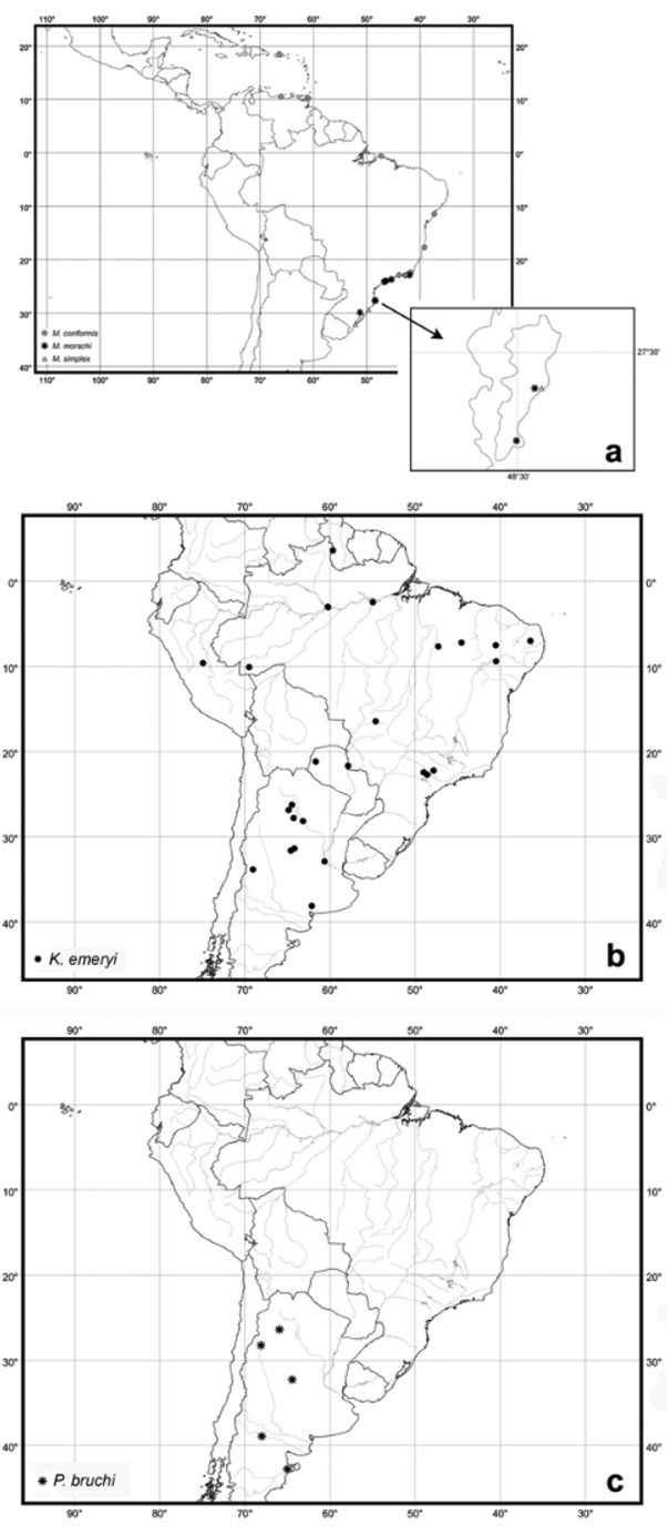
Examined material: BRAZIL: Rio Grande do Sul: Morrete, Fazenda Oliveira , xii.1975 ( V. P. Daniel) [# 12212 ], 1 w ( MZSP) ; without data, ( H. v. Ihering) [ 11419 ], 1 w ( Syntype), ( MZSP) ; Rio de Janeiro: Cabo Frio , viii.1926 ( T. Borgmeier), 1 w ( MZSP) ; Santa Catarina: Florianópolis, Praia da Joaquina , 3.v.1991 ( A. Bonnet & B. C. Lopes) 2 w ( MZSP ), 8.iv.2003 ( C. Klingenberg, R. R. Silva & B. C. Lopes) 29 w, 16 q, 9 m ( MZSP) ; Pântano do Sul , 9.iv.2003 ( C. Klingenberg, R. R. Silva & B. C. Lopes) 9 w, 2 q, 4 m ( MZSP, SMNK) ; São Paulo: Itanhaém , vi.1914 ( Luederwaldt), [ 18862 , 18867 ], 3 w ( MZSP) ; Itanhaém , 15–19.vii.1961 ( A. Guedes & F. Grossmann), 3 w ( MZSP) ; Mongaguá, Praia Grande , 18.vi.1960 ( W. W. Kempf), 1 w ( MZSP) ; São Sebastião , 30.i.1955 ( B. Fleddermann), 1 w [# 4] ; São Vincente, Praia Grande , 18.xii.1955 ( W. W. Kempf) [# 1496 ], 1 w ( MZSP) ; Caraguatatuba , 22.v–1.vi.1962 ( K. Lenko) 1 q ( MZSP) .
Comments. Mycetophylax morschi is the only species in the genus which bears a lateral depression on the head, similar to an antennal scrobe, a very common character of Cyphomyrmex . However, in M. morschi , the scrobe-like depression shelters only the anterior portion of the antennal scapes, by virtue of its laterally expanded frontal carina. In Cyphomyrmex the scrobe lodges all or most of the scape as the frontal carina is expanded over the whole scrobe. We consider that M. morschi and Cyphomyrmex acquired scrobe-like structures independently.
A further strong argument for transferring C. morschi to Mycetophylax is the nesting biology and habitat choice of these ants. As in M. conformis and M. simplex , the nests of M. morschi are exclusively found in the sandy soil of the South Atlantic beaches. The nest architecture and colony size are also similar in the three species (average of 120 workers per colony, Klingenberg et al., 2007), while most Cyphomyrmex species have smaller colonies (see Mueller & Wcislo, 1998; Murakami et al. 2000, Schultz et al. 2002) and seldom occupy sandy dunes. In the results of Schultz & Brady´s (2008) molecular phylogenetic study of the Attini, C. morschi and M. conformis grouped together in all topologies and methods of analysis, corroborating our findings based on our current morphological study.

No known copyright restrictions apply. See Agosti, D., Egloff, W., 2009. Taxonomic information exchange and copyright: the Plazi approach. BMC Research Notes 2009, 2:53 for further explanation.
|
Kingdom |
|
|
Phylum |
|
|
Class |
|
|
Order |
|
|
Family |
|
|
SubFamily |
Myrmicinae |
|
Tribe |
Attini |
|
Genus |
Mycetophylax morschi ( Emery, 1888 )
| Klingenberg, Christiana & Brandão, Roberto F. 2009 |
Cyphomyrmex ( Mycetoritis ) (sic) personatus
| Bolton 1995: 168 |
| Kempf 1972: 93 |
| Kempf 1964: 25 |
| Santschi 1923: 268 |
Cyphomyrmex
| Mayr 1887: 556 |
Cyphomyrmex morschi
| Cyphomyrmex morschi Emery, 1888 |