
Paramycetophylax bruchi ( Santschi, 1916 ) Santschi, 1916
|
publication ID |
https://doi.org/ 10.11646/zootaxa.2052.1.1 |
|
DOI |
https://doi.org/10.5281/zenodo.4675147 |
|
persistent identifier |
https://treatment.plazi.org/id/03FC87DA-F51A-C570-42B2-FF77FECBFA63 |
|
treatment provided by |
Plazi |
|
scientific name |
Paramycetophylax bruchi ( Santschi, 1916 ) |
| status |
comb. nov. |
Paramycetophylax bruchi ( Santschi, 1916) new combination
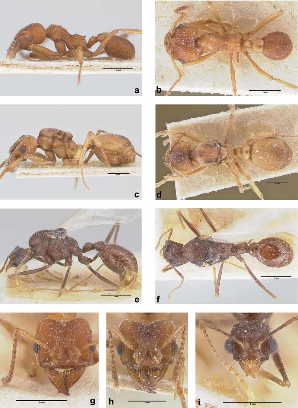
( Figs. 5 View FIGURE 5 , 7 View FIGURE 7 c)
Sericomyrmex bruchi Santschi, 1916: 383 , (worker) Holotype, Argentina: Puerto Madryn (Biraben) (NHMB, examined); Santschi 1922: 355 combination in Myrmicocrypta (Mycetophylax) View in CoL ; Santschi 1923: 268 combination in Mycetophylax View in CoL ; Kusnezov 1956: 24 combination in Paramycetophylax ; Weber 1958: 262 combination in Mycetophylax View in CoL ; Kempf, 1972: 145 (catalogue); Bolton 1995: 268 (catalogue) new combination.
Mycetophylax bruchi var. pauper Santschi, 1923: 268 View in CoL replacement name, junior secondary homonym of Mycetophylax bruchi var. simplex Santschi, 1922: 355 View in CoL (worker) Type, Argentina: Neuquén, (Dr. Carette col.) (MZSP, NHMB examined); Kempf 1972: 145 (catalogue); Bolton 1995: 269 (catalogue); new synonym.
Myrmicocrypta (Mycetophylax) cristulata Santschi 1922: 356 (worker, queen, male) Syntypes, Argentina: Tucumán, El Bañado, Valle Santa Maria, Ing. Weiser col. (NHMB, examined); Santschi 1929: 304 combination in Mycetophylax View in CoL ; Bucher, 1974: 63, Kempf 1972: 145 (catalogue), Bolton 1995: 268 (catalogue); new synonym.
Mycetophylax cristulatus var. emmae Santschi 1929: 304 View in CoL (worker) Syntypes, Argentina, Catamarca, Nacimientos, (Weiser col.) (NHMB, MZSP examined); Kempf 1972: 145 (catalogue), Bolton 1995: 268 (catalogue), Klingenberg & Brandão, 2005: 45 (syntype worker in MZSP); new synonym.
Worker ( Figs. 5 View FIGURE 5 a, b, g, 7 View FIGURE 7 c)
Range of measurements (in mm) and indices of examined specimens (N = 13): IOD 0.88–1.18; HL 0.82–1.10; CI 101–113; SL 0.67–0.87; SI 57–80; ML 0.47–0.58; MI 47–58; WL 1.25–1.73; PrW 0.53–0.70; PL 0.25–0.37; PPL 0.23–0.37; GL 0.90–1.18; FL 0.93–1.39; TL 3.93–5.82.
Measurements (in mm) and indices of Holotype (worker without gaster): IOD 1.00, HL 0.91, CI 110, SL 0.76, SI 76, ML 0,53, M 58, WL 1.48, PrW 0.62, PL 0.30, FL 0.93.
Color yellow to reddish-brown. Masticatory and external borders of mandible, margins of clypeus and carinae brownish. Under optical microscope, body sculpture densely reticulate with exception of dorsal discs of mandibles, where sculpture is finely striate. Whole body sparsely covered by golden shiny appressed hairs. Anterior margin of clypeus with five to nine fine, stiff, and long setae, reaching half the length of the mandibles; three median setae longer than lateral ones.
Head wider than long (see CI). Compound eyes set close to the middle of the head, with eleven ommatidia at maximum width and eight ommatidia at maximum length. Mandibles with eight to ten teeth, the two most apical teeth bigger than the others, followed by five to seven smaller triangular teeth and a last denticle. Anterior margin of clypeus slightly concave, almost straight, which bears a 6–8 long setae psamophore. In frontal view, clypeus attaining posteriorly the level of half the frontal lobes, in a rounded suture, followed by a weakly impressed triangular frontal area. Triangular shaped frontal lobes fully covering the antennal insertions. Glabrous area between antennal insertions and lateral carinae ending posteriorly at the level of posterior margin of the compound eyes. Sharp lateral carinae, almost vertical, marginate the anterior border of compound eyes. Vertexal margin concave, with a median impression and forming two lobes. Antennal scapes flattened, slightly curved; depending on degree of curving, reaching or slightly surpassing the posterolateral corners of the head. Apical end of funiculus with a three segmented club, wider than preceeding segments. The apical segment of funiculus as long as previous two segments together. Ventral face of head conspicuously flat.
Mesosoma. Pronotum with a pair of anterior blunt and low spines, and a pair of inferior spines, square in lateral view. Dorsal face of mesonotum with a small blunt low median protuberance anteriorly. Inferior margin of mesosoma bordered by a sharp translucent carina. In side view, dorsal face of mesonotum slightly concave in the middle, metapropodeal suture straight; propodeum with a pair of triangular anterior protuberances at the basal face, a pair of divergent, short, blunt narrow triangular spines at the meeting of basal and declivous faces, and declivous face almost vertical. Petiole compact; in lateral view peduncle very short, and dorsal margin of node gently sloping until two posterior low triangular corner-like protuberances, with a weakly developed ventral process. Postpetiole in dorsal view subquadrate, with rounded margins. In lateral view, sternite of postpetiole well defined, covering 2/3 of tergite surface.
Gyne ( Figs. 5 View FIGURE 5 c, d, h, 7 c)
Measurements (in mm) and indices of examined specimen (N = 1): IOD 1.50; HL 1.36; CI 110; SL 1.01; SI 67; ML 0.57; MI 42; WL 2.05; PL 0.52; PPL 0.49; GL 2.10; TL 7.09.
Color, pilosity and main morphological character traits of head, propodeal spiracle, petiole, postpetiole and gaster conspecific with the workers. Mandibles with nine teeth; apical tooth bigger than all others, followed by a smaller second apical tooth, six equally developed triangular teeth and a small basal denticule. Compound eyes with 16 ommatidia at maximum width and 21 ommatidia at maximum length. Posterior fourth of head with three equally developed ocelli. Most apical funicular segment slightly shorter than the two anterior together.
Mesosoma. In lateral view anterior margin of pronotum and anterior face of scutum almost vertical, dorsal face of scutum flat almost concealing the pronotum in dorsal view, anterior margin of scutum rounded. In dorsal view, posterior margin almost straight, slightly rounded. Parapsidial lines visible due to the darker color of the parapsidial region and median portion of scutum. Notaulices obsolete. Prescutum narrow, at middle portion anterior and posterior margin not touching, axillae subtriangular. Scutum-scutellar sulcus impressed, convex and rounded. Scutellum trapezoid, anterior margin double the width of the slightly convex posterior margin. Metanotum reduced, appearing only as small, flattened disc in dorsal view. Katepisternum subquadrate to subtriangular; anepisternum two thirds of size of katepisternum, subquadrate, both divided by a distinct groove and ending posteriorly in a carina. Propodeum basal face straight in lateral view, oblique, with a sharp and produced triangular spine. Petiole, postpetiole and gaster as in the workers. Spiracle of first gastral segment indistinct.
Male ( Figs. 5 View FIGURE 5 e, f, i, 7 c)
Range of measurements (in mm) and indices of examined specimens (N = 3, in two speciemens it was not possible to measure the mandible length, therefore range for mandible length, mandibular index and total length are not given): IOD 0.7–0.71; HL 0.68–0.74; CI 101; SL 0.77–0.86; SI 108–123; ML 0.31; MI 42; WL 1.6–1.77; PL 0.37–0.43; PPL 0.26–0.29; GL 1.59–1.73; TL 5.05.
Color dark brown. Funiculus, mandibles, pretarsi and tarsi brownish to yellowish. Integument and pilosity like in the conspecific workers. Integument of gaster shiny, with an almost vestigial reticulation. Head slightly wider than long (see CI), with the postero-lateral angles almost straight. Compound eyes with 21 ommatidia at maximum length and 20 ommatidia at maximum width. Mandibles slender and elongated with only two apical teeth, followed by a straight margin. Anterior margin of clypeus straight with three long setae. Clypeus posterior area attaining the level of half the antennal insertions, ending in a distinct triangular suture, followed by a narrow, impressed triangular area. Frontal lobes reduced, covering only half the antennal insertions. Lateral carinae barely surpassing the level of the posterior margin of compound eyes. Vertexal margin almost straight, posterolateral corners of the head almost rectangular. Antennal scapes straight, with half the length of all other antennal segments together. First funicular segment as long as the two next segments together. Ventral portion of head convex behind the buccal cavity, ending postero-ventrally in a sharp angle.
Mesosoma. In lateral view scutum fully covering the pronotum. Scutum dorsal margin rounded in lateral view; in dorsal view with a middle shallow impression. Anterior margin of scutum rounded in dorsal view. Posterior margin convex. Parapsidial lines parallel to the median body axis. Median portion of prescutum narrow but anterior and posterior margin not touching, axillae subtriangular. Scutum-scutellar sulcus distinct. Scutellum bulging, strongly rounded in lateral view; scutellum anterior margin slightly convex, posterior margin rounded. Anepisternum and katepisternum divided by a sinous groove, development varies among the specimens. Anepisternum subtriangular with four to five transversal rugae dorsally. Katepisternum subquadrate and antero-ventral margin sinuous. Propodeum basal face slightly convex in lateral view, with a pair of small blunt spines; declivous face slightly concave. Petiolar dorsal margin rounded in side view, without spines, only two lobes in dorsal view. Ventral process vestigial. Postpetiole almost twice as wide as petiole in dorsal view, wider posteriorly; posterior margin of postpetiole with a median depression.
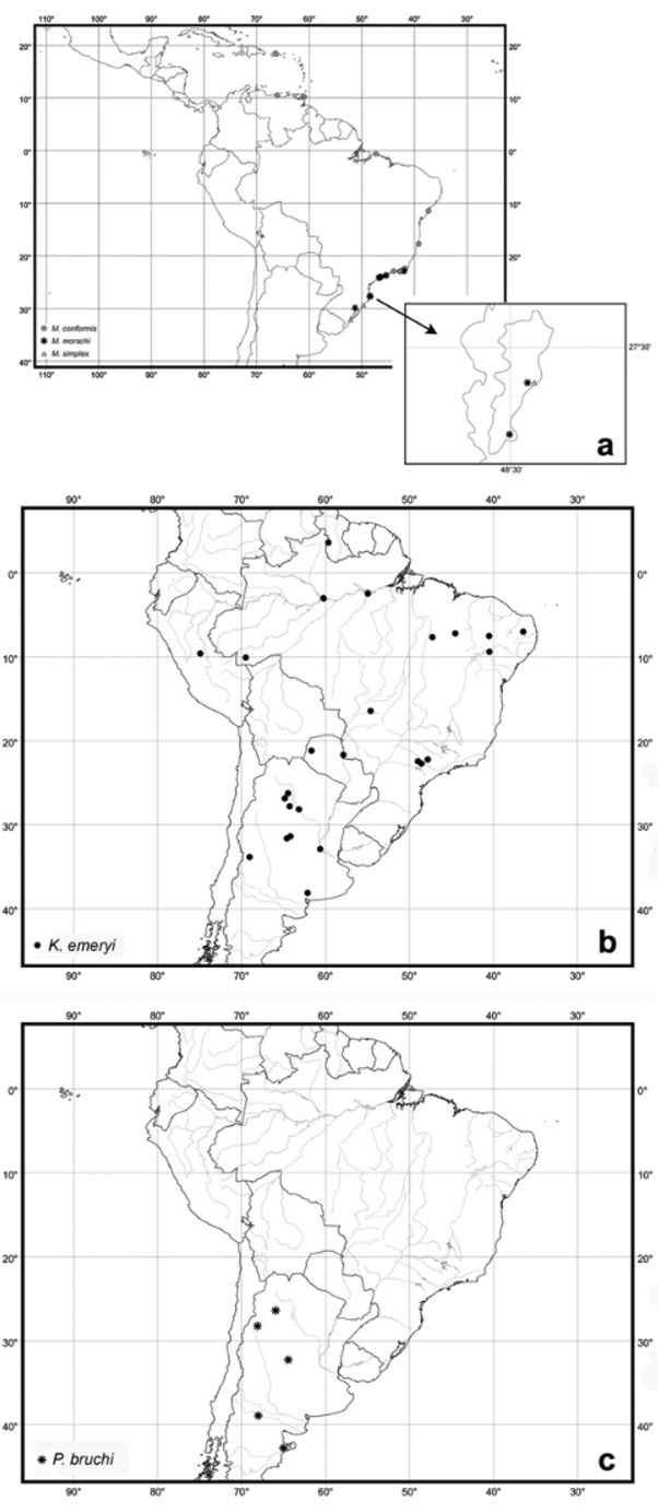
Examined material: ARGENTINA: Neuquén, no coll. data 2 w ( NHMB), v.1925 (Dr. Carette), [ #1425 ], 1 w ( MZSP) ; Nacimientos , 2.xii.1922 (Weiser), 3 w ( NHMB) 1 w ( MZSP) (Cotype) ; Puerto Madryn (Biraben), 1 w ( NHMB) ; El Bañado, Valle Sta. Maria (Weiser), 3 w, 3 m, 1 g ( NHMB) ; Tucumán, Siete de Abril , Depto. Burruyacú, 2.vi.1965 (E. Bucher), 2 w ( MZSP) .
Comments. The shape of the workers antennal scapes varies among individuals; it was not possible to determine a typical shape for the species: some specimens have straight scapes, surpassing the posterolateral corners of the head while others have curved scapes, only reaching the posterolateral corners. Santschi (1922) commented on the variation in P. simplex (= Mycetophylax bruchi var. pauper Santschi, 1923 ) with some individuals with slightly longer antennal scapes and without greyish powder covering (“pruinosité”). We suspect that he actually observed the symbiont bacteria Streptomyces covering the Attini ants in some individuals, which can cause such an appearance. Currie et al. (1999) described this phenomenon in Acromyrmex ants.
In his description of Myrmicocrypta (Mycetophylax) cristulatus, Santschi (1922) mentioned larger dimensions in comparison with M. bruchi . But he was not able to define any clear morphological difference between the species, except for the shape of the postpetiole. In 1929 Santschi published the description of Mycetophylax cristulatus var. emmae . Again he justified the description of the variety because of its different dimensions and color.
In his key, published in 1922, Santschi recognized similarities between M. bruchi and M. cristulatus , as well as differences and based his arguments for distinguishing them on the dimensions of the postpetiole. In M. cristulatus Santschi argued that the postpetiole seems to be wider than long, whereas in M. bruchi the postpetiole seems to be as long as wide. From our measurements of all available specimens we find that this character is not efficient for species separation. The same is true for the separation of M. bruchi from its variety M. bruchi var. simplex (latter renamed as pauper ).
However, Santschi was never able to show distinct morphological differences between the species bruchi and cristulatus and among their variations. Our observations show that the examined individuals present variations in body dimensions and color, especially in the length and shape of the antennal scape. These variations are gradual, so that the recognition of distinct forms is not possible. We propose then the synonymy of M. cristulatus and all varieties under P. bruchi .
In two of the three examined males, the inferior vein of the radial cell does not touch the costa of the forewing. So the apicalmost part of the radial cell appears as not fully closed.
There is no recent information published about the natural history of the species. Only Bucher (1974) briefly commented on the nest architecture of P. bruchi and K. emeryi ; both species nesting in sandy soil, in places clear of vegetation.
The species P. bruchi is known only from continental Argentina. A detailed map of distribution records is given in Fig. 7 View FIGURE 7 c.
No known copyright restrictions apply. See Agosti, D., Egloff, W., 2009. Taxonomic information exchange and copyright: the Plazi approach. BMC Research Notes 2009, 2:53 for further explanation.
|
Kingdom |
|
|
Phylum |
|
|
Class |
|
|
Order |
|
|
Family |
|
|
SubFamily |
Myrmicinae |
|
Tribe |
Attini |
|
Genus |
Paramycetophylax bruchi ( Santschi, 1916 )
| Klingenberg, Christiana & Brandão, Roberto F. 2009 |
Mycetophylax cristulatus var. emmae
| Klingenberg 2005: 45 |
| Bolton 1995: 268 |
| Kempf 1972: 145 |
| Santschi 1929: 304 |
Mycetophylax bruchi var. pauper
| Bolton 1995: 269 |
| Kempf 1972: 145 |
| Santschi 1923: 268 |
| Santschi 1922: 355 |
Myrmicocrypta (Mycetophylax) cristulata
| Bolton 1995: 268 |
| Bucher 1974: 63 |
| Kempf 1972: 145 |
| Santschi 1929: 304 |
| Santschi 1922: 356 |
Sericomyrmex bruchi
| Bolton 1995: 268 |
| Kempf 1972: 145 |
| Weber 1958: 262 |
| Kusnezov 1956: 24 |
| Santschi 1923: 268 |
| Santschi 1922: 355 |
| Santschi 1916: 383 |