
Tyrannosaurus, Osborn, 1905
|
publication ID |
https://doi.org/10.5281/zenodo.3724478 |
|
DOI |
https://doi.org/10.5281/zenodo.3728575 |
|
persistent identifier |
https://treatment.plazi.org/id/03FC87E7-FFF7-C646-FF44-F890FB1BFCD3 |
|
treatment provided by |
Jeremy |
|
scientific name |
Tyrannosaurus |
| status |
|
In a number of cases areas of origin from the pubis and ischium are atbsolutely determinable in saurischian dinosaurs; and in every case they agree closely with the conditions found in the Crocodilia . The tendinous origin of flexor tibialis internus 3 is seen on almost every theropod pelvis; it was apparently a large muscle. That of flexor tibialis externus 1 is shown on seyeral specimens. These two origins are also to be observed on a specimen of Diplodocus in this museum.
In Tyrannosaurus an outward curving of the antero-ventral corner of the ischium is comparable to the region from which adductor 1 arises in the alligator.
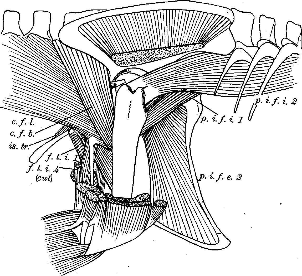
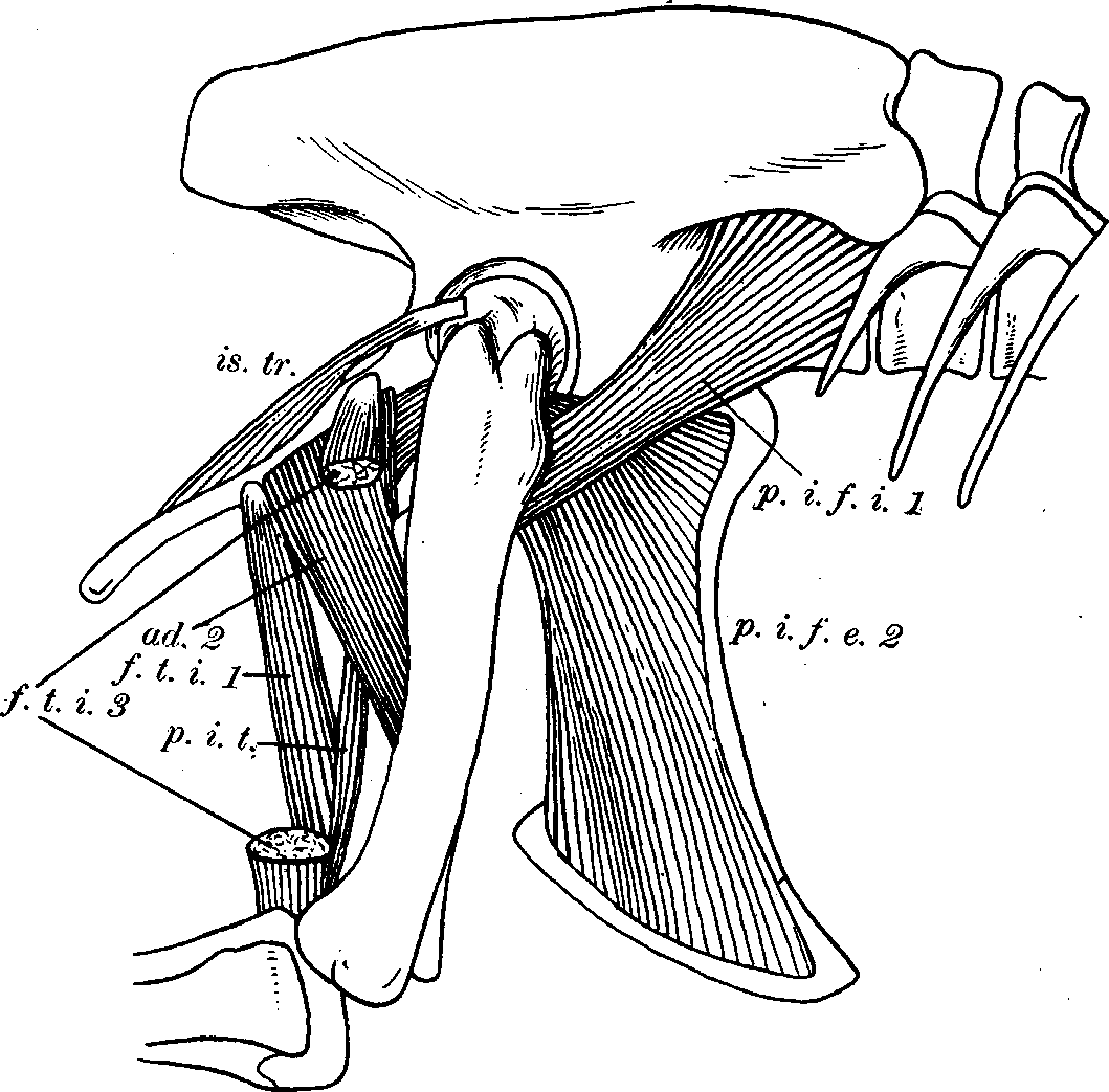
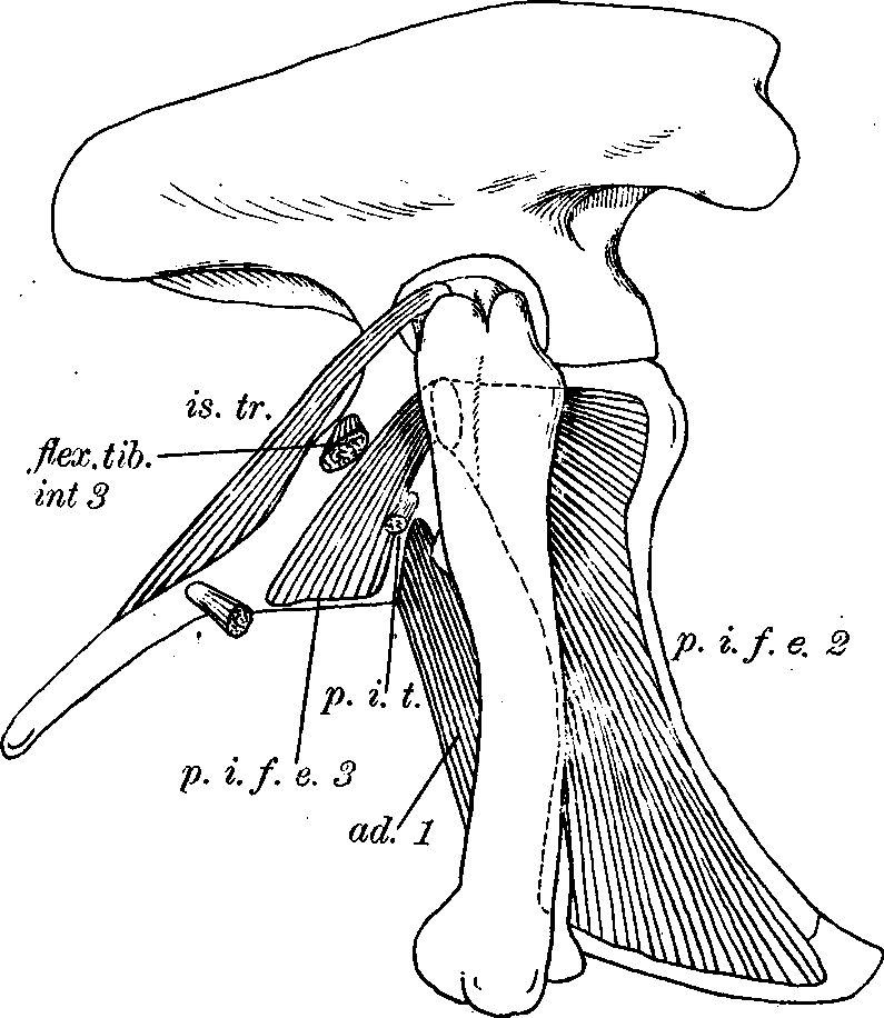
And in a mounted specimen of Tyrannosaurus the origin of pubo-ischio-tibialis may be seen. With these landmarks the areas of origin of pubo-ischiofemoralis externus 3 and adductor 2 may be determined with reasonable accuracy. A tuberosity on the pubis (or pubis and ilium) is usually present and indicates the site of the ambiens.
In figures 4-8 a View Fig. 4 View Fig. 5 View Fig. 6 View Fig. 7 View Fig. 8 restoration of the pelvic musculature of Tyrannosaurus has been attempted. It is, of course, impossible to be sure of a number of points, such as the subdivisions of the ilio-tibialis or ambiens or the arrangement of the tendons at the knee. Where there has been doubt the crocodilian arrangement has been followed as being the most probable in view of the close similarity in all known points. The muscles were restored in clay on a reduced model of the pelvic region. In the figures the axial musculature has been omitted to avoid too great a complexity. Its relations to the girdle are simple.
The dorsal musculature was primitively continuous, universally attaching itself to the inner surface of the ilium above the ribs and to the anterior (ilio-costalis) and posterior margins of this bone. This was true of the sauropods and some theropods, as Ornitholestes . In Tyrannosaurus the two ilia are applied to the spines, separating caudal and trunk portions. Huge rugose surfaces on the posterior end of the ilium of Brontosaurus and Tyrannosaurus and surfaces on a line with these on the transverse processes of the caudal vertebrae indicate strong ligaments running longitudinally in this region and separating the dorsal tail musculature (dorsalis caudae) from the ventral (ilio-ischio-caudalis). The ilio-caudalis undoubtedly attached to the lower edge of this ligament and perhaps along its lower margin reached the ilium.
Antero-ventrally, the rectus attached to the extremity of the pubis, or rather its cartilaginous extension, and probably, as mentioned above, to the anterior edge of the body of the pubis. The obliquus externus probably attached tendinously in the neighborhood of the ambiens, as is' usually the case in reptiles, and (through its connection with the rectus) to the pubis. Postero-ventrally, the ischio-caudalis arose from the distal portion of the ischium, as did undoubtedly part of the cloacal musculature.
A number of criticisms may be made of the restorations of the musculature of Triassic dinosaurs by Von Huene (1908); as I have mentioned most of these are due to our inadequate knowledge of the Crocodilia . On the ilium (p. 291) no origin is afforded for ilio-tibialis (except as we consider ilio-fibularis 1 as part of this muscle). Ilio-fibularis was probably farther posterior and ventral. Ilio-femoralis (externus) was probably larger. On the pubis (p. 292) the "pubo-tibialis" insertion may have been either for part of ambiens or a tendon of the obliquus: there is no pubo-tibialis in the Crocodilia nor in birds. In regard to the ischium (p. 293) there is never; of course, any origin of the extensor iliotibialis from this bone. Pubo-ischio-femoralis externus (3) and ischiofemoralis (adductor 1) are correctly placed. Flexor tibialis internus 2 is incorrectly located, and lo.cations for pubo-ischio-tibialis and flexor tibialis internus 1 and 3 of the writer are not indicated. Pubo-ischiofemoralis posterior, if equivalent to the writer's ischio-trochantericus, should be on the upper internal instead of the ventral external portion of the bone. The ischio-caudalis should be restricted to the distal portion of the bone.
The femur (p. 295). The ilio-femoralis (externus), "quadratus liimborum," and pubo-ischio-femoralis posterior are shown attaching to the greater trochanter. The first is undoubtedly correct. "Quadratus lumborum" (pubo-ischio-femoralis internus 2) runs beneath the iliofemoralis in A direction opposite to that of the red arrow and probably inserted about, but not on, the trochanter. Caudo-ilio-femoralis (coccygeo-femoralis brevis) inserted more ventrally, about in the position of Von Huene's pubo-ischio-femoralis internus 3. The area occupied by femora-tibialis, to judge by either Crocodilia or birds, was much greater. I know of no part of pubo-ischio-femoralis internus likely to insert so far proximally as that shown in figure b. Of the muscles shown inserting on the fourth trochanter, probably only caudo-femoralis (coccygeo-femoralis internus longus) should be placed there. The pubo-ischiofemoralis externus and that portion of pubo-ischio-femoralis which equals ischio-trochantericus inserted proximally to this trochanter. The adductors (ischio-femoralis and pubo-ischio-femoralis posterior in part) inserted about in the area marked " pubo-ischio-femoralis posterior zum Teil."
Most of the modifications of the tentative restoration of Ornitholestes by Gregory and Camp (1918), which might be suggested, are merely ones of nomenclature.
The question as to whether pubo-ischio-trochantericus internus (= pubo ischio-femoralis externus 1 of the Crocodilia ) extended down the inner side of the pubis has been discussed above. "Adductor longus" = pubo-ischio-tibialis. The ridge below it was probably occupied by adductor 1.,The space labeled ischio-femoralis was occupied by pubo-. ischio-femoralis 3 and this probably extended anteriorly to include the excavation in the ischium, which is thus (as explained above) a close parallel to the "obturator fenestra." The posterior area marked puboischio-femoralis externus is that for adductor femoris 2, except that the latter muscle probably did not extend so far distally. No area is marked off for flexor tibialis internus 1. It seems probable that ilio-femoralis extended farther posteriorly.
Few criticisms may be made of the later restorations by Dr. Gregory shown in figure 1. Ilio-tibialis, ilio-femoralis, ilio-fibularis, flexor tibialis externus, caudi-ilio-femoralis (= coccygeo-femoralis brevis), "quadratus lumborum" (=pubo-ischio-femoralis internus), ambiens, the pubic head of pubo-ischio-femoralis externus, pubo-ischio-femoralis posterior (= ischio-trochantericus), and the iliac and one ischiadic head of flexor tibialis internus, I believe to be all correctly located. The question as to an internal pubic muscle has been mentioned previously. Adductor 2 probably occupied the proximal part of the area given the ischiadic head of pubo-ischio-femoralis externus. That muscle occupied most of the flat surface marked ischio-femoralis, the ischio-femoralis (adductor 1) lying slightly more anteriorly. Pubo-ischio-tibialis and flexor tibialis internus 1 are not located; the head of the latter muscle shown arising from beneath the posterior part of the ilium is probably incorrect.


No known copyright restrictions apply. See Agosti, D., Egloff, W., 2009. Taxonomic information exchange and copyright: the Plazi approach. BMC Research Notes 2009, 2:53 for further explanation.
|
Kingdom |
|
|
Phylum |
|
|
Class |
|
|
Order |
|
|
Family |