
Levensteiniella manusensis, Wu & Xu, 2018
|
publication ID |
https://doi.org/ 10.11646/zootaxa.4388.1.7 |
|
publication LSID |
lsid:zoobank.org:pub:33075379-7E70-4B6F-B4B6-94900A44B5D1 |
|
DOI |
https://doi.org/10.5281/zenodo.5971640 |
|
persistent identifier |
https://treatment.plazi.org/id/03FCB24D-FFFA-C178-FF48-FF3B94B6FED1 |
|
treatment provided by |
Plazi |
|
scientific name |
Levensteiniella manusensis |
| status |
sp. nov. |
Levensteiniella manusensis View in CoL sp. nov.
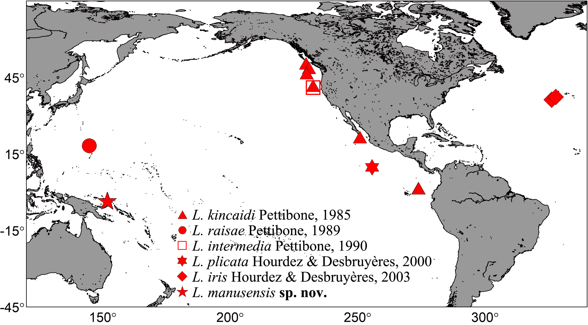
Figures 1–4 View FIGURE 1 View FIGURE 2 View FIGURE 3 View FIGURE 4 ; Table 1
Type material. All type specimens were collected by the ROV FaXian, except the paratype MBM286002 View Materials , which was obtained by a TV grab, from hydrothermal vents in the Manus Back-Arc Basin of the Western Pacific . Holotype: MBM285996 View Materials , Dive 40, 151°51'52.328''E, 3°40'47.684''S, 1940 m, 18 June 2015 GoogleMaps .
Paratypes: MBM285997 (n=3), same collection information as above. MBM285998 (n=1), Dive 39, 151°51'47.613''E, 3°40'54.605''S, 1853 m, 17 June 2015; MBM285999 (n=2), Dive 33, 151°40'20.851''E, 3°43'42.065''S, 1707 m, 12 June 2015; MBM286000 (n=4), same collection information as above; MBM2860001 (n=1), same collection information as above; MBM286002 (n=1), 151°40'19.56''E, 3°43'40.8''S, 1708 m, coll. TV grab, 21 June 2015.
Type locality. Hydrothermal vent in the Manus Back-Arc Basin, Western Pacific (151°51'52.328''E, 3°40'47.684''S, 1940 m). Most specimens were collected in the surrounding areas of the mussel ( Bathymodiolus manusensis Hashimoto & Furuta, 2007 ) bed and the community of tube-worms ( Arcovestia ivanovi Southward & Galkin, 1997 ).
TABLE]. Comparisons of main features of the six species of Levensteiniella . Abbreviation: Segments, number of segments; Lt, total length of bođy; Wm, maximum wiđth of bođy with chaetae
incluđeđ; VP, ventral papillae; PCL, postchaetal lobe of notopođium.
Elytra Filiform micropapillae on Micro- anđ macropapillae Oval micropapillae on Smooth on surface, with Smooth on surface, with Micro- anđ macropapillae on surface anđ borđer; some on surface anđ posterior surface anđ thickeneđ large međian longituđinal thickeneđ bulbous surface anđ thickeneđ with enlargeđ bases on borđer bulbous projections on folđ on surface projections on posterior bulbous projections on posterior borđer posterior borđer borđer posterior borđer Diagnosis. Body with 24–28 segments, 6.5–57 mm long, maximum width 4.5–19.6 mm (including chaetae). Elytra with thickened bulbous projections along posterior border, and micro- and macropapillae on surface. Notochaetae either smooth or serrated with widely spaced spines along curved side, tips bare and blunt. Supraacicular neurochaetae slender, distal spinous region with two rows of long, widely spaced spines, and proximal spinous region with two rows of transverse teeth. Subacicular neurochaetae stouter, distal spinous region knifelike, with minute, close-set spines along curved side and slightly hooked smooth tips, and proximal spinous region with a row of transverse teeth. Two pairs of elongate ventral papillae on segments 11 and 12 present only in males, first pair distally dichotomous. Pharynx with seven pairs of papillae, and two pairs of jaws without distinct teeth.
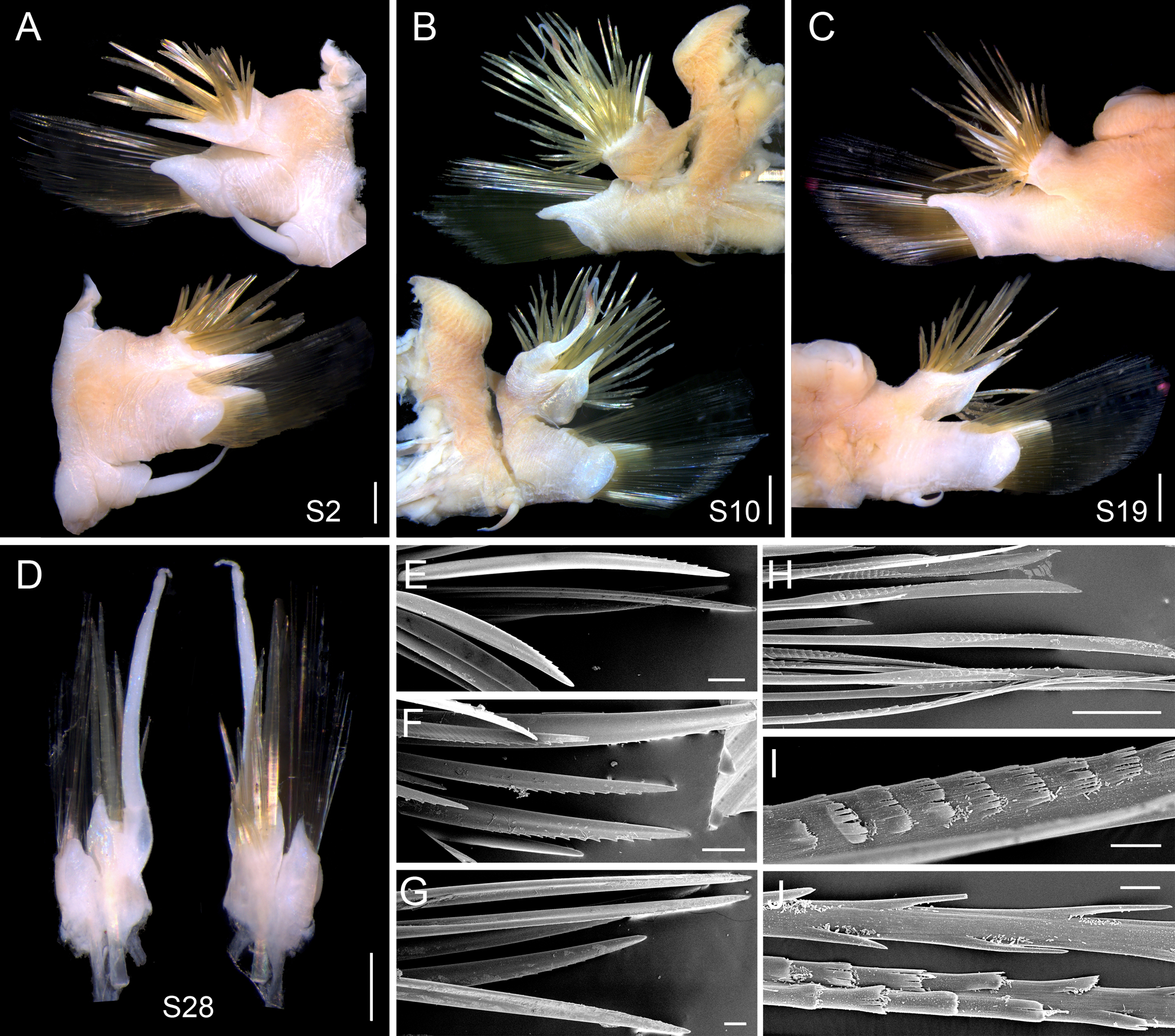
Description. Holotype well-preserved with 28 segments, length 43 mm, maximum width 19.6 mm including chaetae and 13.6 mm excluding chaetae. Paratypes with 24–27 segments, length 6.5 to 34 mm, maximum width 4.5 to 19 mm including chaetae and 3.4–14 mm excluding chaetae.
Living specimens purple red, parapodia with golden chaetae ( Fig. 2A, B View FIGURE 2 ). Body oval and flattened, tapering slightly anteriorly and more so posteriorly; parapodia almost as long as trunk width ( Fig. 2A–J View FIGURE 2 ). Eleven pairs of elytra, attached to prominent elytrophores located on segments 2, 4, 5, 7, and alternate segments to 21, completely covering dorsum ( Fig. 2A View FIGURE 2 ); thick and stiff, round to subreniform, with thickened bulbous projections along posterior border ( Figs 2A View FIGURE 2 ; 3G–L); micro- and macropapillae on surface, filiform to oval, mainly scattered in posterior part ( Fig. 3J–L View FIGURE 3 ), extending anteriorly in small elytra ( Fig. 3L View FIGURE 3 ), detached in larger elytra ( Fig. 3G, H, I View FIGURE 3 ). Elytrophores large and prominent ( Figs 2C View FIGURE 2 ; 3A). Dorsal cirri present on segments lacking elytra, with cirrophores cylindrical, bulbous basally, attached to posterodorsal sides of notopodia; styles with long slender tips, extending beyond tips of chaetae ( Fig. 4B, D View FIGURE 4 ). Dorsal tubercles nodular ( Figs 2C View FIGURE 2 ; 3A; 4B).
Prostomium wider than long, deeply bilobed, cephalic peaks (anterior lobes) triangular with delicate frontal filaments ( Fig. 2E–J View FIGURE 2 ; frontal filaments detached in holotype). Ceratophore of median antenna cylindrical, inserted in anterior notch; style subulate with a long terminal filament ( Fig. 3A View FIGURE 3 ). Palps stout, tapered with slender tips ( Fig. 3A, B View FIGURE 3 ; right palp detached in holotype). Lateral antennae and eyes absent. Tentacular segment (peristomium) achaetous, dorsally invisible, ventrally forming bilobed facial tubercle and lips surrounding mouth ( Fig. 3B View FIGURE 3 ). Tentaculophores cylindrical, inserted laterally to prostomium; two pairs of tentacular cirri with long slender tips, slightly shorter than palps, dorsal pair much longer than ventral pair ( Fig. 3B View FIGURE 3 ).
Second (buccal segment) bearing first pair of elytrophores, and biramous parapodia with notopodia and neuropodia equal in length ( Fig. 4A View FIGURE 4 ); ventral cirri (buccal cirri) morphologically similar to ventral tentacular cirri, attached basally on bulbous cirrophores, much longer than subsequent ventral cirri ( Figs 3B View FIGURE 3 ; 4A). Pharynx with seven pairs of similar-size, conical papillae and two pairs of slender jaws without distinct teeth ( Fig. 3E View FIGURE 3 ).
Biramous parapodia well developed, notopodia slightly shorter than neuropodia except for segment 2 ( Fig. 4A–D View FIGURE 4 ). Notopodia cylindrical with a projecting acicular lobe on lower side and numerous notochaetae forming a radiating bundle. Notochaetae golden, much stouter than neurochaetae, short to longer ( Fig. 4A–D View FIGURE 4 ), straight or slightly curved with bare blunt tips, smooth or serrated with widely spaced spines along curved side ( Fig. 4E–G View FIGURE 4 ). Neuropodia including a conical prechaetal lobe with a projecting acicular process, and a short rounded postchaetal lobe ( Fig. 4A–D View FIGURE 4 ). Neurochaetae numerous, long and slender, forming a fan-shaped bundle; supra-acicular neurochaetae slender, distal spinous region with two rows of long, widely spaced spines along borders ( Fig. 4H, J View FIGURE 4 ), and proximal spinous region with two rows of transverse teeth ( Fig. 4J View FIGURE 4 ); subacicular neurochaetae stouter than supra-acicular ones, distal spinous region knife-like with minute, close-set spines along curved side, and slightly hooked bare tips ( Fig. 4H View FIGURE 4 ), proximal spinous region with a row of transverse teeth ( Fig. 4I View FIGURE 4 ). Ventral cirri short, tapered, attached on middle of neuropodia ( Fig. 4B–D View FIGURE 4 ).
Pygidium small, rectangular, with a pair of slender anal cirri ( Fig. 3F View FIGURE 3 ). Seven out of 13 specimens with two pairs of long ventral papillae (containing a whitish secretion) on segments 11 and 12 ( Fig. 2 View FIGURE 2 ); the first pair distally dichotomous ( Fig. 3C, D View FIGURE 3 ).
Etymology. The species is named after the location ‘Manus Basin’ where the species was discovered.
Distribution. Hydrothermal vents in the Manus Back-Arc Basin, Western Pacific (water depth: 1707–1940 m).
Remarks. Levensteiniella manusensis sp. nov. has a prostomium with a single median antenna, parapodia without branchiae, and long projecting acicular processes in both rami of parapodia. With these features, the new species is obviously a member of the polynoid subfamily Macellicephalinae , where it belongs to the genus Levensteiniella by having 11 pairs of elytra and up to 28 segments.
Levensteiniella manusensis sp. nov. can be readily distinguished from its congeners by having two pairs of ventral papillae on segments 11 and 12, with the first pair distally dichotomous. Among the five known species of Levensteiniella , L. kincaidi , L. raisae and L. plicata also possess two pairs of ventral papillae, in which, however, no dichotomous branches occur in the first pair (Tab. 1). Levensteiniella manusensis sp. nov. resembles the other two species, L. intermedia and L. iris , in the presence of thickened bulbous projections on the posterior border of elytra (Tab. 1). However, the latter two species have a single pair of ventral papillae on segment 11 (vs. two pairs on segments 11 and 12).
Other diagnostic features of L. manusensis include: the elytra possessing both micro- and macropapillae on the surface and thickened bulbous projections on the posterior border; and the notochaetae either smooth or serrated on the curved side. Among the species of Levensteiniella , both L. raisae and L. intermedia have similar notochaetae as in L. manusensis . Nonetheless, L. raisae possesses no thickened bulbous projections on the posterior border of elytra and L. intermedia has no macropapillae on the surface of elytra (Tab. 1). Based on these distinctions, we provide a key to all species of Levensteiniella .


| ROV |
Museo Civico di Rovereto |
No known copyright restrictions apply. See Agosti, D., Egloff, W., 2009. Taxonomic information exchange and copyright: the Plazi approach. BMC Research Notes 2009, 2:53 for further explanation.
|
Kingdom |
|
|
Phylum |
|
|
Class |
|
|
Order |
|
|
Family |
|
|
Genus |