
Spelaeoblatta myugei, Vidlička & Vršanský & Shcherbakov, 2003
|
publication ID |
https://doi.org/10.1080/713834390 |
|
DOI |
https://doi.org/10.5281/zenodo.4653152 |
|
persistent identifier |
https://treatment.plazi.org/id/03FD878A-A26E-FFDA-5B58-FBD28DA82A34 |
|
treatment provided by |
Carolina |
|
scientific name |
Spelaeoblatta myugei |
| status |
sp. nov. |
Spelaeoblatta myugei View in CoL n. sp.
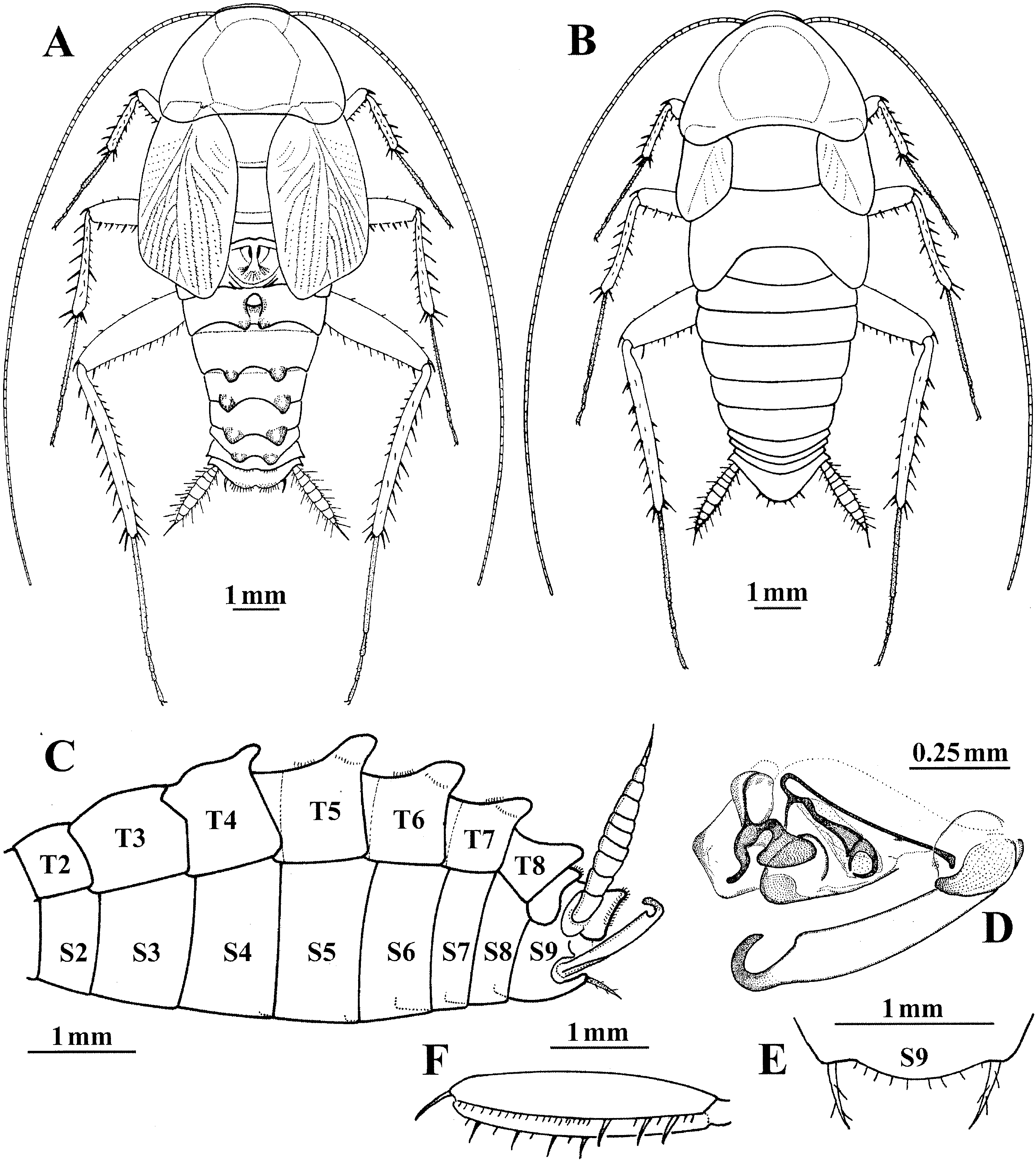
( figures 2–4 View FIG View FIG View FIG )
Type material. HOLOTYPE: ³, north-west Thailand: cave Tham Pha Mon ( Mae Hong Son Province, Nam Lang region , 19°28∞N, 98°14∞E), 20 March 1997, N. Myuge ( H. Miore) leg., coll. Slovak National Museum–Natural History Museum Bratislava ( SNM-NHMB) . PARATYPES: 1 ³, 3 ♀, same data as holotype (Vidlička collection) .
Etymology. The species is named in honour of N. Myuge, who collected this species.
Description
Size (mm). Body length: ³ 10.0–10.75, ♀ 9.9–11.8; pronotum length×width, ³ 2.5×3.9, ♀ 2.4–2.75×3.5–4.1; forewing length, ³ 4.5–5.0, ♀ 1.75–2.0.
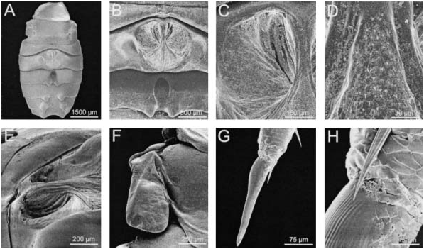
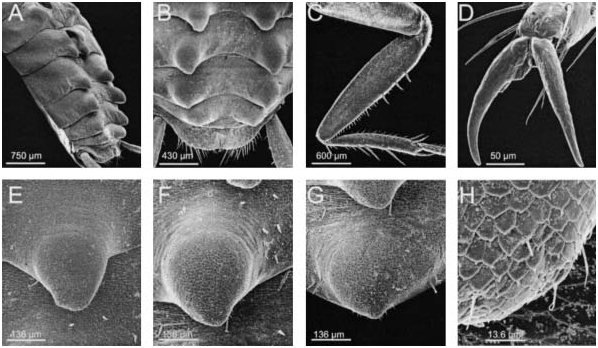
Male ( figures 2A, C, D, E, F View FIG , 3 View FIG A–H, 4A–H). Head longitudinal, oval, partly visible from above; faceted eyes small, strongly reduced, facetes indistinct; ocelli absent; fifth maxillary palpomere shorter than the fourth, apex rounded. Antennae long, slender, about 55 segments, scapus long, pedicel short, third segment as long as scapus, other segments shorter. Pronotum parabolic, lateral hind corner slightly oblong. Tegmina reaching to the base of fourth tergite, obliquely truncate, surface covered by rather sparse trichia. Trichia are present mostly apically on the reduced intercalary veins. Sc is highly expanded and supported by series of branches, clavus is strongly reduced, radial field covers almost the half of the wing’s width, media is four branched. Hind wings reduced, lobate ( figure 3F View FIG ), slightly shorter than metanotum. Anteroventral margin of front femur with three large proximal spines succeeded by a row of piliform spinules terminating in a large distal spine ( type B) ( figures 2F, 1 View FIG View FIG View FIG View FIG
4C); anterior margin of front tibia with several small spines, followed by three large spines near the centre and terminating in two large spines, posterior margin with many piliform spinules, two large spines at the distal end; tarsal claws simple, symmetrical, pulvilli and arolia absent ( figure 4D View FIG ). Ten terga (T1–T10) are visible on the abdomen ( figures 2A View FIG , 3A View FIG , 4B View FIG ). Abdominal terga (T2–T8) are strongly specialized: the posterior margin of T2 is medially strongly concave, T3 contains medially large round glandular pit with longitudinal elevated part in the middle, T4 has a large deep fossa, a pair of large tubercles at each posterior border of T5–T8 (unique in Blattaria; figure 4A, B View FIG , E–H). Hind margin of supraanal plate rounded with a shallow medial depression. Cerci 9–10 segmented, surface dorsally smooth, ventrally with long setae, long sensilla on the apex ( figure 3G, H View FIG ). Subgenital plate is symmetrical with two similar setose styles, interstylar margin convexly rounded ( figure 2E View FIG ). Genitalia are shown in figure 2D View FIG : genital hook projects on the left side, the apex strongly narrow, curved; right phallomere strongly sclerotized, curved to S shape.
Colour: the specimens are pale brownish yellow, nearly translucent. Only maxillae and tubercles on abdomen are darker.
Female ( figure 2B View FIG ). Head exposed, faceted eyes reduced, but present. Pronotum parabolic, lateral hind corner slightly oblong. Tegmina strongly reduced to lateral pads slightly overlapping mesomotum, veins indistinct. Metanotum laterally distinct elongated. Hind margin of supraanal plate convexly rounded with long setae. Subgenital plate with distinct valves. Pattern of front femur same as in the male.
Colour: the specimens are pale brownish yellow, nearly translucent.
Remarks
This species differs from S. thamfaranga described by Roth principally by the presence of large tubercles on the hind margins of abdominal terga 5–8. Roth claimed that the tergal glands were present on the second and third segments, but our observations suggest that he may have missed the narrow first segment. His drawing ( figure 2H View FIG in Roth and McGavin, 1994: 1322) is very similar to our figure 2A View FIG , except for the apparent absence of segment 1.
No known copyright restrictions apply. See Agosti, D., Egloff, W., 2009. Taxonomic information exchange and copyright: the Plazi approach. BMC Research Notes 2009, 2:53 for further explanation.