
Edwardsia aff. tuberculata Duben and Koren, 1847
|
publication ID |
https://doi.org/ 10.11646/zootaxa.4661.3.7 |
|
publication LSID |
lsid:zoobank.org:pub:7A7E6A85-B904-4BC2-90A1-8CA19CA86F24 |
|
persistent identifier |
https://treatment.plazi.org/id/03FD87A5-FFCB-EF6C-99D1-F8DCFC3066C0 |
|
treatment provided by |
Plazi |
|
scientific name |
Edwardsia aff. tuberculata Duben and Koren, 1847 |
| status |
|
Edwardsia aff. tuberculata Duben and Koren, 1847 View in CoL
(New Japanese name: oozutsu-mushimodoki-ginchaku)
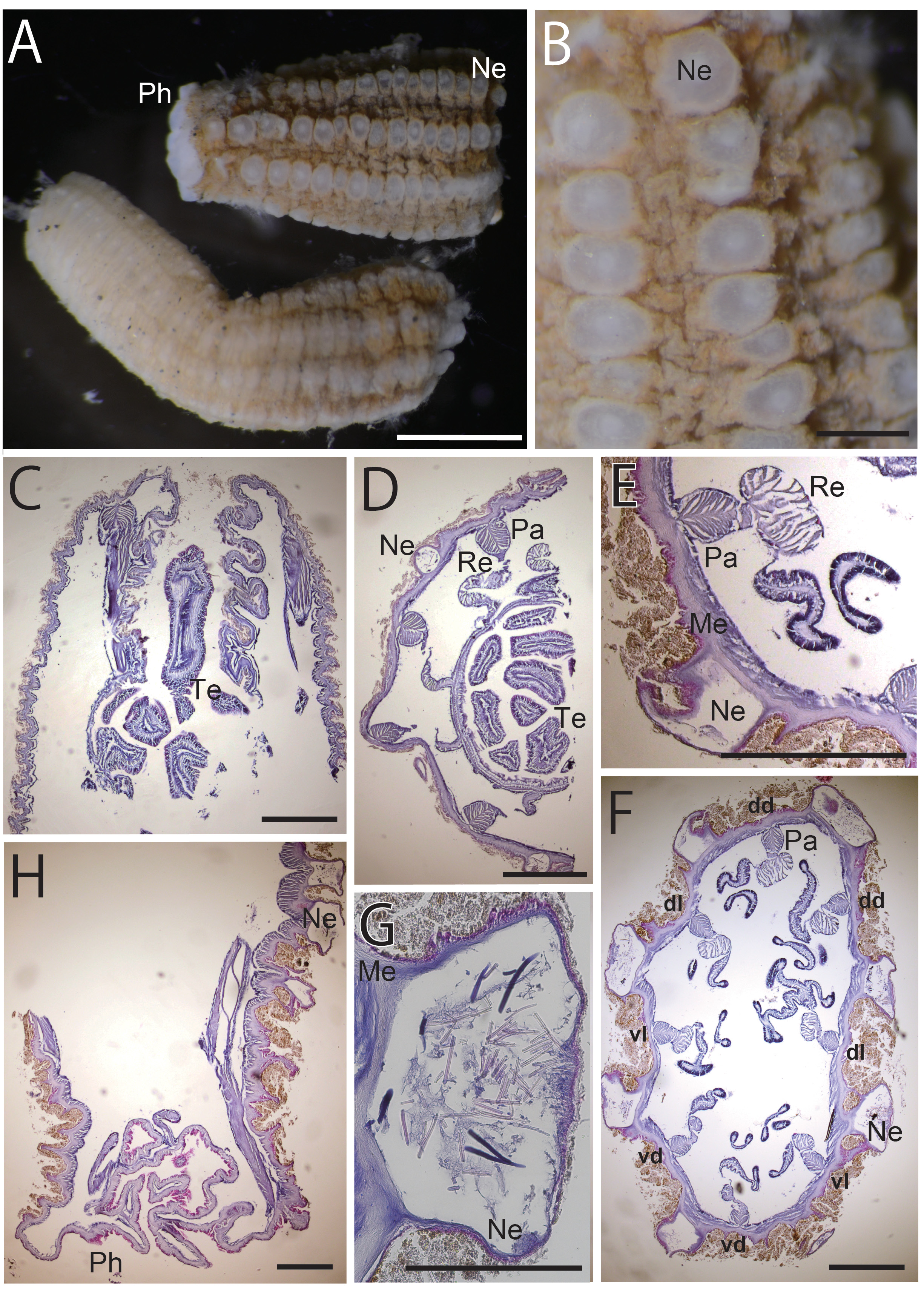
Figs. 2 View FIGURE 2 , 4 View FIGURE 4 ; Table 1 View TABLE 1
Synonymy: see Fautin (2016)
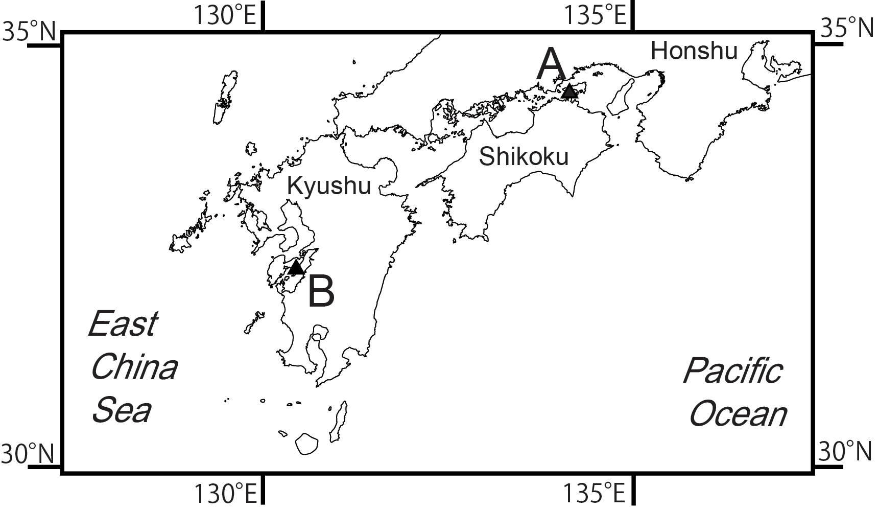
Material examined. NSMT-Co 1654, dissected specimen, histological sections (7 slides), and prepared cnidae (5 slides), November 6, 2014, east of Ogishima Island, Kagawa Pref., Seto Inland Sea (34°24′54′′N, 134°05′30′′E; Fig. 1A View FIGURE 1 ), 16 m depth, collected by Naoto Jimi. NSMT-Co 1655, histological sections of damaged individuals (3 slides), same date, locality, and collector as NSMT-Co 1654.
External anatomy. Column of contracted specimen 20–25 mm in whole length (25.0 mm in NSMT-Co 1654), and 6–7 mm in width (7 mm in NSMT-Co 1654), swelled by huge nemathybomes, cylinder-like in shape but slightly swollen proximally ( Fig. 2A View FIGURE 2 ). Body divided into capitulum, scapus and physa; in our specimen, capitulum shrunken in column, invisible. About half of scapus occupied by nemathybomes, remaining half covered with brownishgrey periderm. Nemathybomes extremely large, like papillae, up to 1.5 mm in diameter ( Fig. 2B View FIGURE 2 ), in eight rows in proximal part, gradually becoming smaller distally ( Fig. 2A View FIGURE 2 ). Aboral physa differentiated from scapus, naked, not rounded but tapered, without nemathybomes ( Fig. 2A View FIGURE 2 ). Tentacles 16 in number, in two cycles of eight, relative size of each cycle unclear because tentacles contracted into scapus.
Internal anatomy. Eight perfect mesenteries, macrocnemes, distributed along whole body from distal to proximal end. These eight are the paired dorsal and ventral directives plus four unpaired lateral mesenteries. Eight tiny microcnemes, without muscles, limited to distal-most part: four between dorsal directives and dorso-lateral mesenteries, two between dorso- and ventro-lateral mesenteries, and two between ventro-lateral mesenteries and ventral directives. All macrocnemic mesenteries bear retractor and parietal muscles. The retractor muscle of each lateral mesentery faces ventrally ( Fig. 2E, F View FIGURE 2 ). Each retractor muscle distinctly developed; diffuse, with approximately 20 simple or slightly branched muscular processes next to actinopharynx ( Fig. 2D View FIGURE 2 ); diffuse or restricted to parietal muscle side, developed like a pennon, with 10–25 simple or slightly branched muscular processes in lower part ( Fig. 2E, F View FIGURE 2 ). Parietal muscles of macrocnemes distinct, semi-elliptic, with 10 slightly branched muscular processes in each side ( Fig. 2 View FIGURE 2 D–F). Actinopharynx very short, grooved but without distinct siphonoglyph. One tentacle in each endo- or exo-coel. Tentacular longitudal muscle ectodermal and distinct. Marginal sphincter muscle and basilar muscle absent ( Fig. 2C, H View FIGURE 2 ). Mesoglea generally thin in mesenteries, and comparatively thick in body wall (Fig, 2D, F). Nemathybomes far thicker than mesoglea, greatly protruding from body wall and containing very large basitrichs ( Fig. 2F, G View FIGURE 2 ). Gonads on mesenteries between retractor muscle and filament; gametocytes immature.
Cnidome. Spirocysts, basitrichs, microbasic b -mastigophores, microbasic p -mastigophores, and microbasic amastigophores ( Table 1 View TABLE 1 , Fig. 4 View FIGURE 4 A–D; NSMT-Co 1654). Two sizes of basitrichs in actinopharynx; three sizes of basitrichs in nemathybomes.
Deriviation of Japanese name. Japanese name “oozutu” means a cannon, because the huge nemathybomes of this species are suggestive of spherical projectiles of cannon, and the cylindrical shape of the body resembles to a cannon barrel. Nemathybome is called as “shihoudan” in Japanese, which is constructed by shihou (= cnidae) + dan (=projectile or bullet), so this Japanese name matches E. aff. tuberculata . “Mushimodoki-ginchaku” means Edwardsia .
Remarks. Edwardsia tuberculata was originally described in Duben and Koren (1847), and more detailed morphological features were described in Carlgren (1921). Morphology of the examined specimens agrees well with the description by Carlgren (1921): shrunken body size is 2 cm in length and 7 mm in width ( Fig. 2A View FIGURE 2 ); a few outstandingly large nemathybomes look like papillae arranged in 8 rows ( Fig. 2B, F, G View FIGURE 2 ); physa is developed but flattened or tapered ( Fig. 2A, H View FIGURE 2 ); retractor muscle is diffused with slightly branched processes and parietal muscle is distinct with 20 processes ( Fig. 2 View FIGURE 2 D–F). The cnidom also almost perfectly corresponds to the description of Carlgren (1921): this species is characterized by large nemathybomes that contain huge basitrichs. The larger type of basitrichs in nemathybomes of our specimen are 116–187 µm in length and 4.8–9.0 µm in width ( Table 1 View TABLE 1 ; Fig. 4C View FIGURE 4 2 View FIGURE 2 ). This matches the nematocysts of E. tuberculata reported by Carlgren (1921), which he measured at 110–190 µm in length and 5–7 µm in width. In filaments, Carlgren (1921) mentioned that some of nematocysts are a little broader than the others. This observation would be convincing if one assumes that the wide nematocyst of Carlgren (1921) is what we identify as microbasic b -mastigophores in our specimen. There were some small differences between the description and our specimens. For example, the retractor muscle of our specimen is composed of 10–25 muscular processes while Carlgren (1921) mentioned more 30 processes, and we found some spirocyst in actinopharynx, although Carlgren (1921) did not mention these.
According to England (1987), in addition to E. tuberculata three other species of Edwardsia have basitrichs over 150 µm in length; E. californica McMurrich, 1913 , E. maroccana Carlgren, 1931 , and E. claparedi (Panceri. 1869) . However, they never exceed 170 µm even in maximum size ( Carlgren, 1931, 1936; England, 1987; Manuel, 1977). The huge basitrichs are, however, now also reported in Edwardsia alternobomen sp. nov., described below, which has basitrichs over 200 µm in length ( Table 1 View TABLE 1 , Fig. 2G2 View FIGURE 2 ).
Edwardsia tuberculata View in CoL is distinguished from the other species with large basitrichs in their nemathybomes by having larger basitrichs than those other species and by some anatomical features. Between mesenteries, E. tuberculata View in CoL consistently has a single row of nemathybomes, whereas E. californica View in CoL has two or three rows on the proximal part ( Carlgren, 1936). Edwardsia tuberculata View in CoL has flattened physa but E. californica View in CoL has very apparent, large, rounded physa. Edwardsia californica View in CoL has a very characteristic restricted retractor muscle with approximately 40 muscular processes, whereas one of our specimens has diffuse retractor muscle composed of about 20 processes. According to Carlgren (1931), Edwardsia maroccana View in CoL has more developed muscles with 30 processes, and it is apparently pinnate near the column, whereas those of E. tuberculata View in CoL do not branch so much. In addition, Carlgren (1931) showed basitrichs of 30–40 µm in length and 6.5 µm in width bearing a shimmering thread in the capsule in the actinopharynx, these are likely a kind of mastigophore, a type of nematocyst not found in actinopharynx of the examined specimen of E. tuberculata View in CoL . The body size of E. tuberculata View in CoL (20–25 mm in length) is completely different from Edwardsia claparedi View in CoL , which reach to 120 mm in length ( Manuel, 1981).
Edwardsia tuberculata View in CoL can also be distinguished from the other species of Edwardsia View in CoL that have two types of basitrichs in their nemathybomes (i.e., E. elegans Verrill, 1869 View in CoL , Edwardsia handi Daly and Ljubenkov, 2008 View in CoL , E. hantueusis England, 1987 , E. sulcata Verrill, 1864 View in CoL ). However, the larger type of basitrichs in nemathybomes of those species barely exceed 100 μm and at most 117 µm for E. handi ( Daly and Ljubenkov, 2008) View in CoL .
Although our specimens clearly align with the description of E. tuberculata View in CoL , this species is distributed only in north western Europe and there is no record from the Pacific or from the eastern Arctic Ocean. Recognizing that expanding the range so much without intermediate populations or an explanation for the long-distance dispersal is problematic, and pending comprehensive analysis through genetics, we mark our specimens as having an affinity for E. tuberculata View in CoL , rather than assigning that name to them definitively.

No known copyright restrictions apply. See Agosti, D., Egloff, W., 2009. Taxonomic information exchange and copyright: the Plazi approach. BMC Research Notes 2009, 2:53 for further explanation.
|
Kingdom |
|
|
Phylum |
|
|
Class |
|
|
Order |
|
|
Family |
|
|
Genus |
Edwardsia aff. tuberculata Duben and Koren, 1847
| Izumi, Takato & Fujita, Toshihiko 2019 |
Edwardsia handi
| Daly and Ljubenkov 2008 |
E. handi (
| Daly and Ljubenkov 2008 |
E. hantueusis
| England 1987 |
Edwardsia maroccana
| Carlgren 1931 |
E. californica
| McMurrich 1913 |
E. californica
| McMurrich 1913 |
Edwardsia californica
| McMurrich 1913 |
Edwardsia claparedi
| Panceri 1869 |
E. elegans
| Verrill 1869 |
E. sulcata
| Verrill 1864 |
Edwardsia tuberculata
| Duben and Koren 1847 |
E. tuberculata
| Duben and Koren 1847 |
Edwardsia tuberculata
| Duben and Koren 1847 |
E. tuberculata
| Duben and Koren 1847 |
E. tuberculata
| Duben and Koren 1847 |
E. tuberculata
| Duben and Koren 1847 |
Edwardsia tuberculata
| Duben and Koren 1847 |
E. tuberculata
| Duben and Koren 1847 |
E. tuberculata
| Duben and Koren 1847 |
Edwardsia
| de Quatrefages 1842 |