
Myrmarachne alticephalon, Yamasaki, Takeshi & Ahmad, Abdul Hamid, 2013
|
publication ID |
https://doi.org/ 10.11646/zootaxa.3710.6.1 |
|
publication LSID |
lsid:zoobank.org:pub:C5F537B3-8112-4CC7-A0AC-B5CA071AD9BA |
|
DOI |
https://doi.org/10.5281/zenodo.6150819 |
|
persistent identifier |
https://treatment.plazi.org/id/03FD87D4-FFB1-3D75-FF6E-C121FF1AF82A |
|
treatment provided by |
Plazi |
|
scientific name |
Myrmarachne alticephalon |
| status |
sp. nov. |
Myrmarachne alticephalon View in CoL sp. nov.
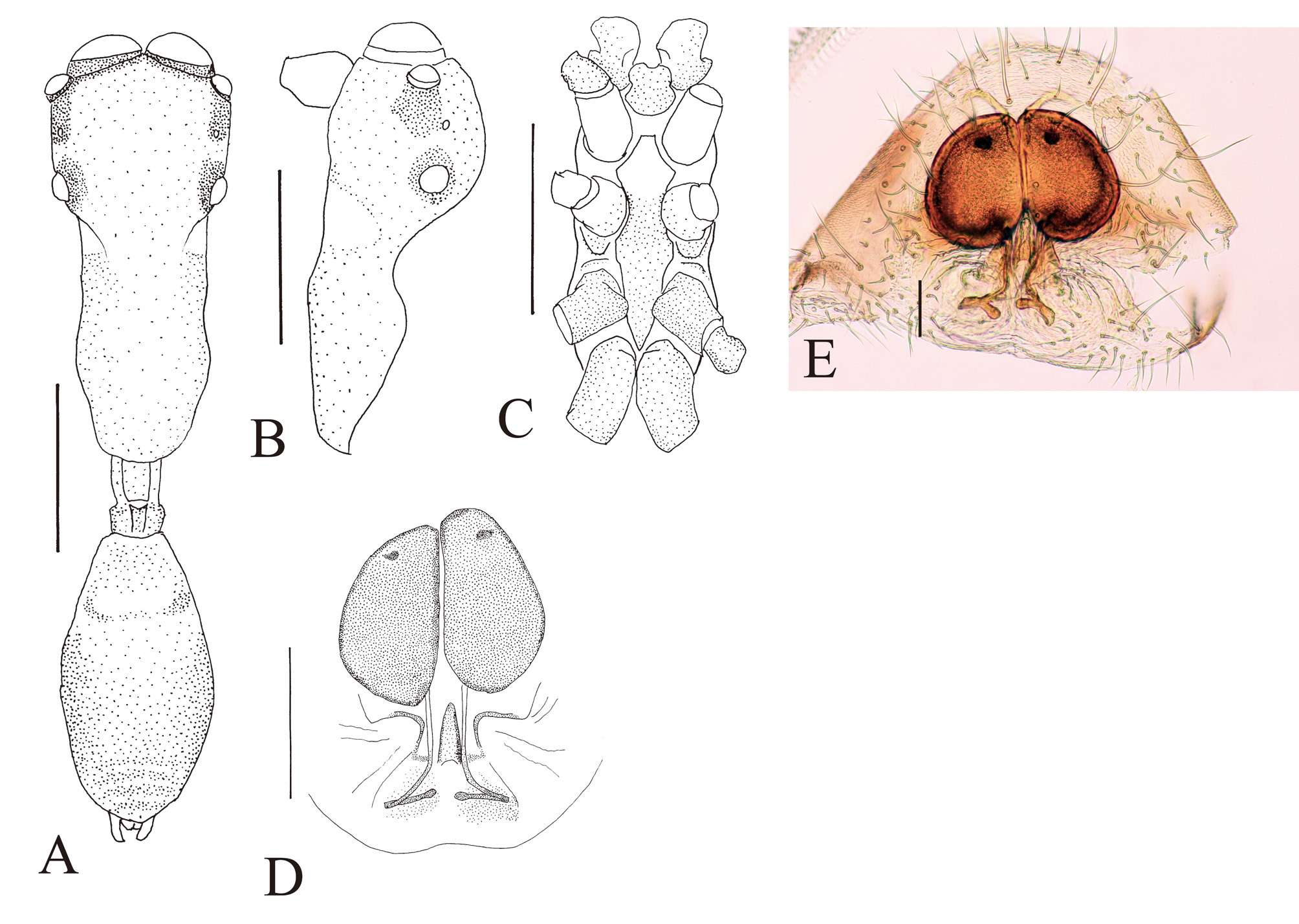
( Figs 4 View FIGURE 4. M A–G, 5A–E)
Type material. Holotype male (UMS), Poring Hot Spring, Kinabalu Park, Sabah, BORNEO, 12 XI 2010, T. Yamasaki leg. Paratypes: BORNEO: 1 male and 2 females (UMS), Danum Valley, Sabah, 15–16 IX 2004, T. Endo leg.; 1 female (UMS), same loc., 15 XII 2006, T. Endo leg.; 1 male and 1 female (BMNH), same loc. as the holotype, 13 XI 2010, T. Yamasaki leg.
Non-type material examined. SUMATRA: 1 female, Forest of Andalas University, Padang, 12 X 2008, Kei. Nakamura leg.; 1 male, Payakumbuh, Padang, 15 X 2008, Sk. Yamane leg.; 1 male, Forest of Andalas University, Padang, 28 XI 2008, Kei. Nakamura leg.; 1 male and 1 female, same loc., 20 VII 2010, Ky. Nakamura leg.; 1 male, same loc., 2 VIII 2010, Ky. Nakamura leg.
Diagnosis. Reddish species, Cephalic part at least twice as high as thoracic part in both sexes ( Figs 4 View FIGURE 4. M B, 5B). In males, chelicera apically swollen and basally narrow, without retrolateral teeth, and inner distal surface black ( Fig. 4 View FIGURE 4. M C). In females, spermathecae very large with very narrow sclerotised copulatory ducts ( Figs 5 View FIGURE 5. M D–E).
Measurements (male/female). Carapace length (2.15)/2.30, width (1.22)–1.24/1.11. Abdomen length (2.25)– 2.30/1.70. Chelicera length (2.75)–2.90. Sternum length (1.20)–1.23/1.18. Width of eye row I (1.24)/1.16; II (1.08)–1.09/0.99; III (1.22)–1.24/1.11. ALE–PLE (0.86)–0.88/0.78; ALE–PME (0.40)–0.43/0.38. Eye size: AME (0.41)–0.43/0.41, ALE 0.20–(0.21)/0.19, PME (0.06)/0.05, PLE (0.21)/0.19.
Male ( Figs 4 View FIGURE 4. M A–G). Cephalic part almost flat dorsally, more than twice higher than thoracic part. Thoracic part convex dorsally. Chelicera apically swollen and basally narrow, with four or five prolateral teeth and without any retrolateral tooth; prolateral teeth confined to anterior part of chelicera; first apical prolateral tooth bifid. Fang almost straight with hooked tip, without tooth-like apophysis. Sternum slender, slightly overlapped by coxae II and III. Abdomen oval, with broad dorsal scutum that is weakly incised on each lateral outline anteriorly.
Palp ( Figs 4 View FIGURE 4. M E–G). Cymbium without apical spine. Tegulum large, oval with strongly curved seminal reservoir in anterior part of tegulum. Embolus forming two large oval coils; embolus coils occupying more than half of venter of cymbium. RTA well developed and strongly curved. Palp in dorsal and ventral views with flange of RTA forming protuberance.
Leg spination. Patella I rv 1; tibia I pv 6, rv 6; metatarsus I pv 2, rv 2; tibia II pv 3, rv 3; metatarsus II pv 2, rv 2. Coloration and pilosity. Carapace brown; cephalic part dorsally covered with fine hairs; thoracic part sparsely covered with fine hairs. Chelicera brown, dorsally covered with sparse greyish hairs; its inner face black. Maxilla and labium brown. Sternum brown; anterior part lighter than posterior part. Coxae pale brown and slightly tinged with black; coxa III darker than other coxae. Dorsum of abdomen covered with fine hairs; dorsal scutum brown; integument except for scutum grey.
Female ( Figs 5 View FIGURE 5. M A–E). Cephalic part convex dorsally, more than twice higher than thoracic part. Thoracic part convex dorsally. Chelicera with seven prolateral and nine retrolateral teeth. Sternum almost as in males. Abdomen oval, weakly constricted anteriorly (more easily observed in profile), without dorsal scutum.
Epigyne ( Figs 5 View FIGURE 5. M D–E). Copulatory atria indistinct. Spermathecae remarkably large, occupying most of epigastric area; sclerotised copulatory ducts very narrow, extending downward from spermathecae; the ducts curved inward proximally. Median pocket elongated, and located between sclerotised copulatory ducts.
Leg spination. Patella I rv 1; tibia I pv 5, rv 6; metatarsus I pv 2, rv 2; tibia II pv 3, rv 3; metatarsus II pv 2, rv 2. Coloration and pilosity. Carapace light brown, covered with fine hairs dorsally; lateral area between cephalic and thoracic parts white; thoracic part almost without hairs. Maxilla and labium pale brown. Sternum pale brown; its anterior part white. Coxae I and II white; coxa III tinged with black; coxa IV pale brown. Abdomen grey, covered with fine hairs.
Etymology. The specific name is derived from the cephalic part much higher than thoracic part (Latin prefix “alti-” (high) and noun “cephalon” (head)).
Distribution. Sumatra, Borneo.
Remarks. Myrmarachne alticephalon has a high cephalic part and low thoracic part. The males in external appearance are very similar to M. turriformis Badcock, 1918 distributed on Asian continent. However, the former is distinguished from the latter by the dentition of the chelicera and the structure of the palp in the males, and by the structure of epigyne in the females. The spermathecae of M. alticephalon are remarkably large, a condition unique among the Asian species of Myrmarachne .
Biology. The species is arboreal, and often occurs in forest canopies. The species often rapidly moves the abdomen dorsoventrally, and sometime keeps the abdomen elevated. The morphology and behaviour of M. alticephalon are similar to those of Camponotus saundersi Emery , and they often occur at the same sites.

No known copyright restrictions apply. See Agosti, D., Egloff, W., 2009. Taxonomic information exchange and copyright: the Plazi approach. BMC Research Notes 2009, 2:53 for further explanation.