
Phanocloidea venezuelica, Hennemann & Conle, 2024
|
publication ID |
https://doi.org/10.11646/zootaxa.5444.1.1 |
|
publication LSID |
lsid:zoobank.org:pub:5DE4A9DD-99F7-4E23-AD50-58DC491BB75E |
|
persistent identifier |
https://treatment.plazi.org/id/4D6E7339-8A95-4A26-B70C-423081E9A595 |
|
taxon LSID |
lsid:zoobank.org:act:4D6E7339-8A95-4A26-B70C-423081E9A595 |
|
treatment provided by |
Plazi |
|
scientific name |
Phanocloidea venezuelica |
| status |
sp. nov. |
Phanocloidea venezuelica sp. n.
urn:lsid:zoobank.org:act:
( Figs. 5 View FIGURE 5 , 70A–C View FIGURE 70 , 72L View FIGURE 72 , 73G, 73J View FIGURE 73 , 74L–N View FIGURE 74 , 75D–F View FIGURE 75 , 87D–E View FIGURE 87 , 91G View FIGURE 91 , 96B View FIGURE 96 , 100M View FIGURE 100 , 126C–D View FIGURE 126 )
Bacteria nodulosa, Zompro, 2000: 171 View in CoL , figs. 1–9. [Description of ♀ and egg—Misidentification]
Phanocloidea nodulosa, Zompro, 2001: 196 View in CoL , figs. 1–2, 70–71 (in part). [Misidentification]
Harman, 2012: 12.
HT, ♂: Venezuela, Amazonas Territory, Dept. Atures, River Parucito , VIII.1992, A.J.E. Harman; A. Harman M. Salton Coll ; ♂ Phanocloidea nodulosa ( Redtenbacher, 1908) ; BMNH(E) 2014–186; NHMUK 014423116 [ NHMUK] .
PT, ♂: Venezuela, Aragua State, River Parucito , 19.VIII.1992, A.J.E. Harman [ NHMUK] .
PT, ♂: Venezuela, Aragua State, River Parucito , 17.VIII.1992, A.J.E. Harman [ NHMUK] .
PT, ♂: Venezuela, Aragua State, River Parucito , 22.VIII.1992, A.J.E. Harman [ NHMUK] .
PT, 3 ♂♂, 3 ♀♀, 5 eggs: Bred England, Essex, Hockley, 1994, A.J.E. Harman ; ♂ Phanocloidea nodulosa ( Redtenbacher, 1908) [ NHMUK] .
PT, 2 ♂♂: ex Zucht: F. Hennemann, urspr.: Venezuela, PSG No. 152, I.1999; Herkunft: SW-Venezuela, Prov. Amazonas, Dept. Atures, Rio Parucito (PSG No. 152), leg. A.J.E. Harman VIII.1992 [ FH, No’s 0400–2 & 3] .
PT, 1 ♀, 2 ♂♂, 200 eggs: ex Zucht: F. Hennemann, urspr.: Venezuela, PSG No. 152, III. 2001; Herkunft: SW-Venezuela, Prov. Amazonas, Dept. Atures, Rio Parucito (PSG No. 152), leg. A.J.E. Harman VIII.1992 [ FH, No’s 0400–1,4, 5, E, ED & MP] .
PT, ♀: ex Zucht: K. Rabaey (Belgien), urspr. Venezuela, PSG 152, 2004; Herkunft: SW-Venezuela, Prov. Amazonas, Dept. Atures, Rio Parucito (PSG No. 152), leg. A.J.E. Harman VIII.1992 [ FH, No. 0400–6] .
PT, 1 ♀, 4 ♂♂: ex Zucht: F. Hennemann 2009, Herkunft: SW-Venezuela, Prov. Amazonas, Dept. Atures, Rio Parucito , leg. A.J.E. Harman VIII.1992, PSG No. 152 [ FH, No’s 0400–7 to 11] .
PT, 5 ♀♀, 2 ♂♂: ex Zucht: Rob Krijns 2009, Herkunft: SW-Venezuela, Prov. Amazonas, Dept. Atures, Rio Parucito , leg. A.J.E. Harman VIII.1992, PSG No. 152 [ FH, No’s 0400–12 to 18] .
Diagnosis. Both sexes are similar to the Venezuelan P. turgida ( Westwood, 1859) and P. muricata ( Burmeister, 1838) from French Guiana, Suriname and NW-Brazil. It however differs from both species by the longer median segment, which is 1.4x longer than the metanotum in ♀♀ and more than twice the length of the metanotum in ♂♂ ( Fig. 73J View FIGURE 73 ; just scarcely longer in muricata and shorter than the metanotum in turgida ) and more elongate, much less globose and entirely unarmed head of both sexes ( Figs. 72L View FIGURE 72 , 73G View FIGURE 73 ). From turgida both sexes of this new species differ by the unarmed medioventral carina of the meso- and metafemora (with several apical spines in turgida ), while ♀♀ may also be distinguished by the unarmed meso- and metasternum ( Fig. 87D View FIGURE 87 ; tubercular to spinose in turgida ) and on average shorter subgenital plate which has the apex triangular (obtusely rounded in turgida ) and ♂♂ by the very different colouration, which includes a broad black dorsal marking on the mesonotum, large triangular lateral lobes of abdominal tergum IX ( Fig. 75D View FIGURE 75 ), elongate vomer with just a single terminal hook ( Fig. 96B View FIGURE 96 ; broad an bidentate in turgida ) and triangular posterior margin of the poculum ( Fig. 75F View FIGURE 75 ). From the generally much larger and slenderer muricata , ♀♀ readily differ by the unarmed mesonotum (prominently spinose in muricata ) and somewhat smaller lobes of the praeopercular organ ( Fig. 91G View FIGURE 91 ) and ♂♂ can be separated by the somewhat smaller lateral dilation of abdominal tergum IX ( Fig. 75D View FIGURE 75 ), triangular posterior margin of the poculum ( Fig. 75F View FIGURE 75 ; broad and obtusely bidentate in muricata ) and different colouration. The eggs ( Fig. 100M View FIGURE 100 ) strongly resemble those of muricata but differ in the somewhat smaller dimensions, proportionally shorter and broader micropylar plate, more scaly capsule sculpturing and smaller opercular crest. Moreover, ♀♀ of this new species are also very similar to the Colombian P. laevigata Conle et al., 2011 in general appearance and in having the head flattened and unarmed, but they can be separated by the somewhat stockier shaped and proportionally shorter body segments (mesothorax 6x longer than prothorax vs. 7.5x in laevigata ), less elongate head, presence of tubercles on the meso- and metapleurae ( Fig. 87D View FIGURE 87 ), having the anal segment broadened posteriorly ( Fig. 74M View FIGURE 74 ; narrowed in laevigata ) and the subgenital much shorter (projecting over tip of abdomen by considerably more than the length of the anal segment in laevigata ).
Etymology. The name refers to the distribution of this new species in South-east Venezuela.
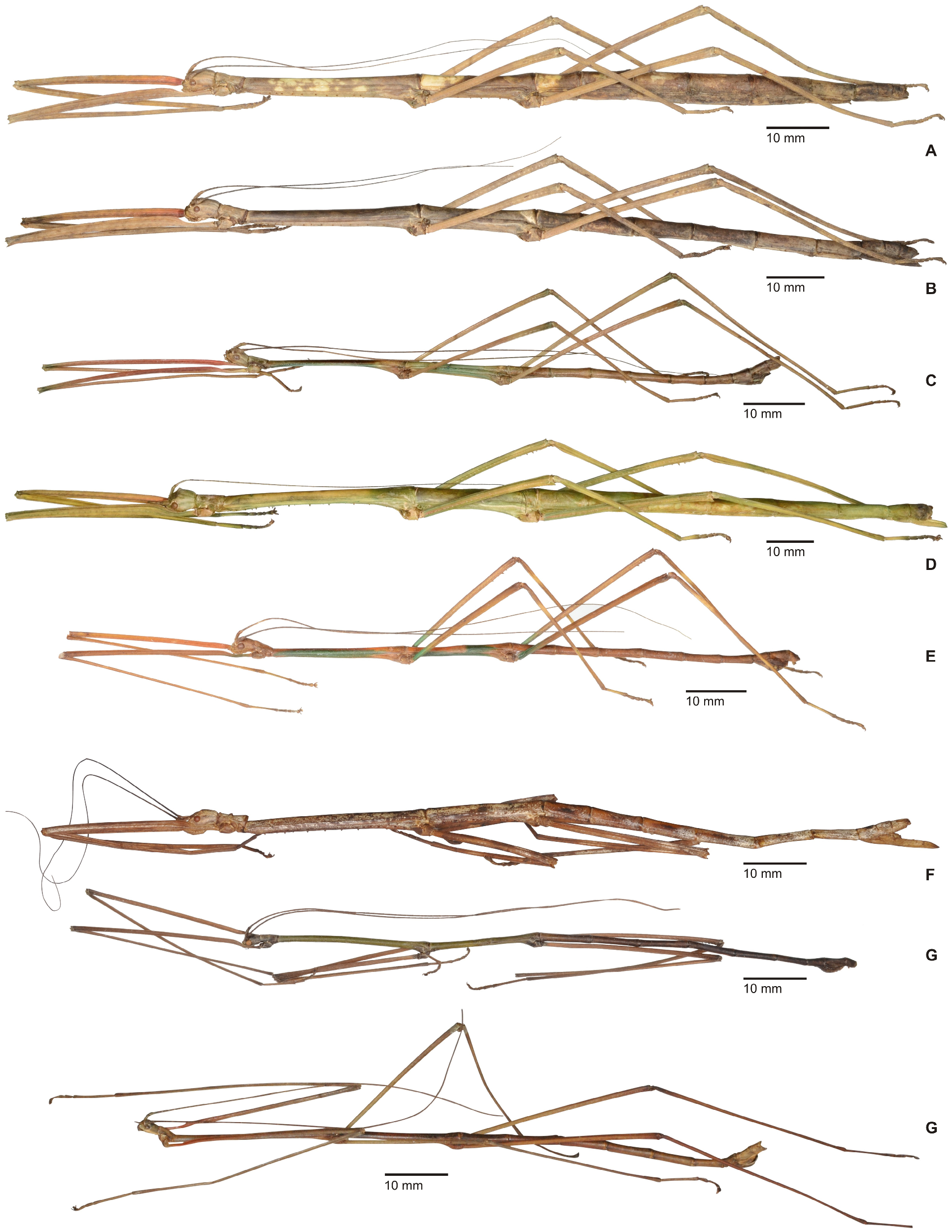
Description. ♀♀ ( Figs. 70A–B View FIGURE 70 , 126D View FIGURE 126 ): Medium-sized (body length including subgenital plate 123.0–166.0 mm) and fairly stocky for the genus, with a median segment that is notably longer than the metanotum, a rather elongate and smooth head, unarmed mesonotum and a subgenital plate that just very slightly projects over the anal segment. Colouration variable and ranging from green over straw and buff to mid brown, often with a conspicuous, triangular white marking near the posterior margin of the median segment ( Fig. 70B View FIGURE 70 ). More rarely conspicuous white dorsal markings may occur on the mesonotum, in the anterior portion of the metanotum and on the five basal abdominal terga ( Figs. 70A View FIGURE 70 , 126D View FIGURE 126 ). Spines of the meso- and metapleurae ochre with black points. Antennae drab dorsally with ventral surface black except for scapus and pedicellus. Bases of profemora red interiorly ( Fig. 72L View FIGURE 72 ).
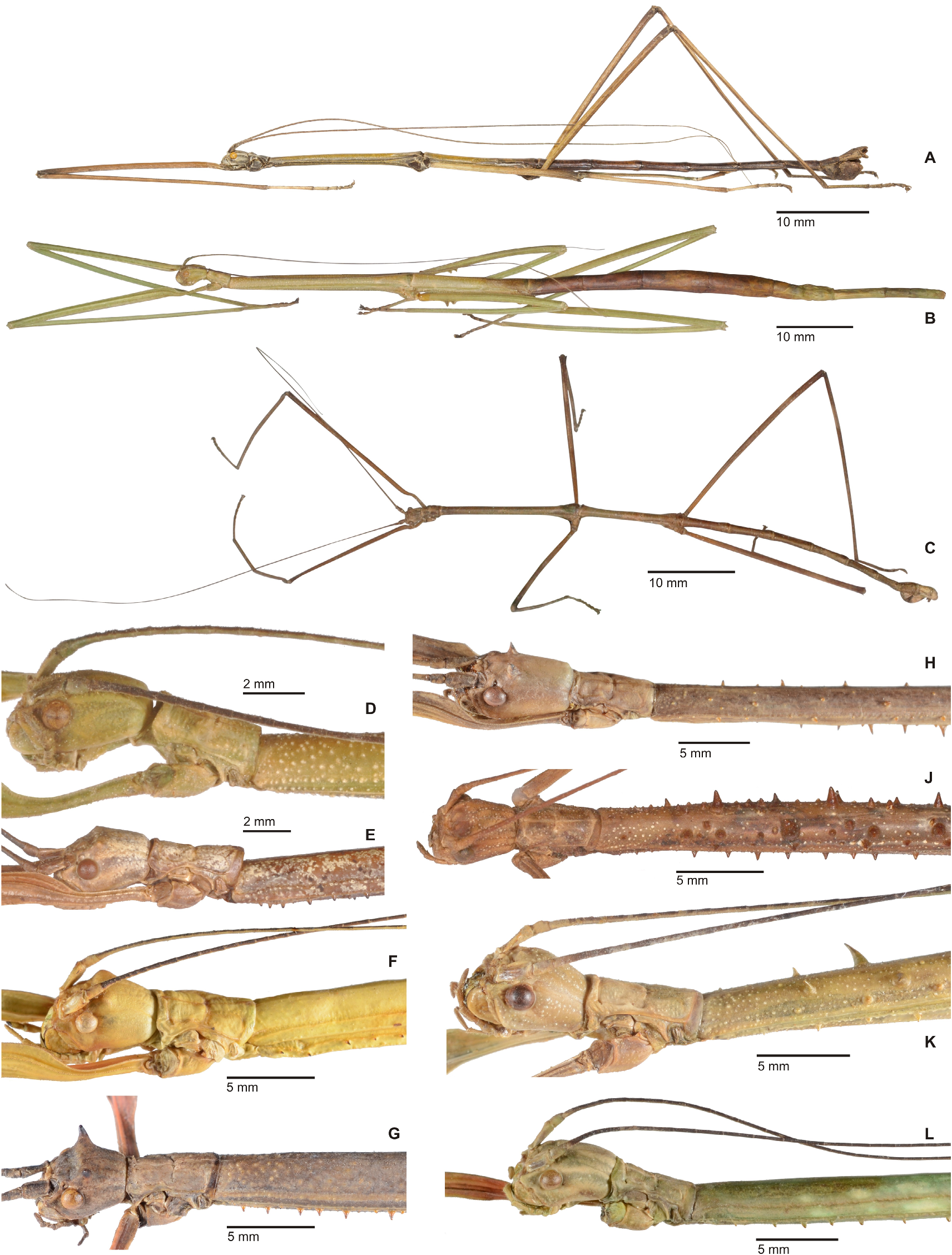
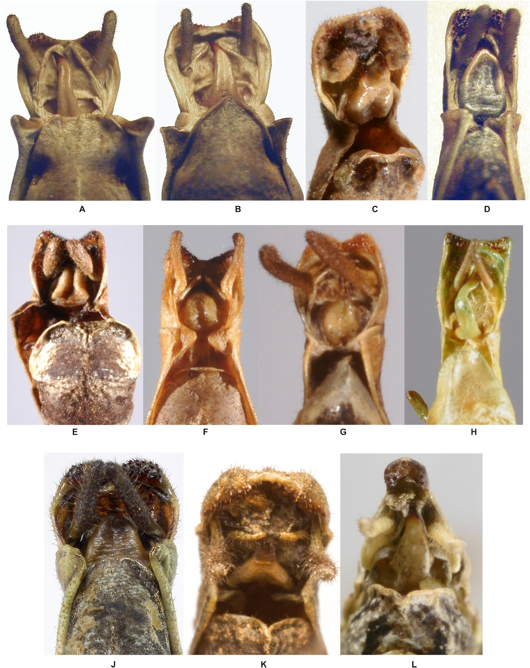
Head ( Fig. 72L View FIGURE 72 ): Roundly rectangular in dorsal aspect, slightly compressed dorsoventrally and suB-oval in cross-section, almost 1.4x longer than wide; vertex very weakly rounded and smooth except for two small, oval median impressions just behind the eyes. Frons with two deep median pits between the bases of antennae. Eyes rather small, circular in outline, moderately projecting and their diameter contained 2.4x in length of genae. Antennae projecting over posterior margin of abdominal tergum III. Scapus almost 2x longer than wide, rectangular in dorsal aspect and distinctly compressed dorsoventrally. Pedicellus about half as long as scapus, somewhat inflated and with apical portion constricted. III slightly longer and much narrower than pedicellus.
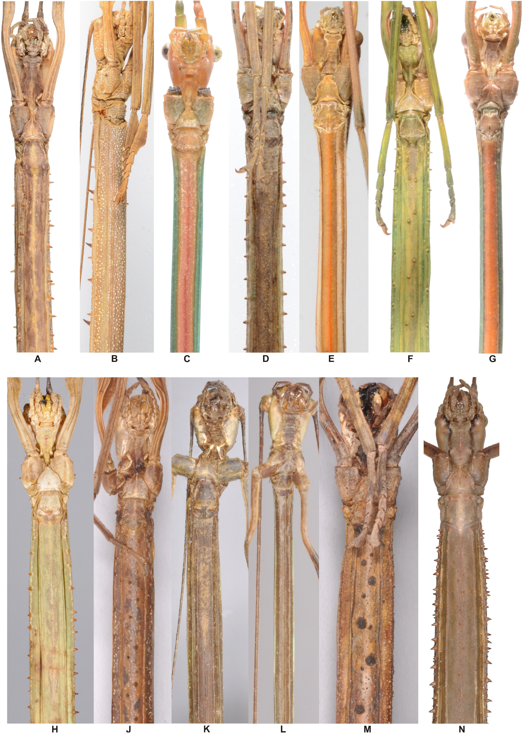
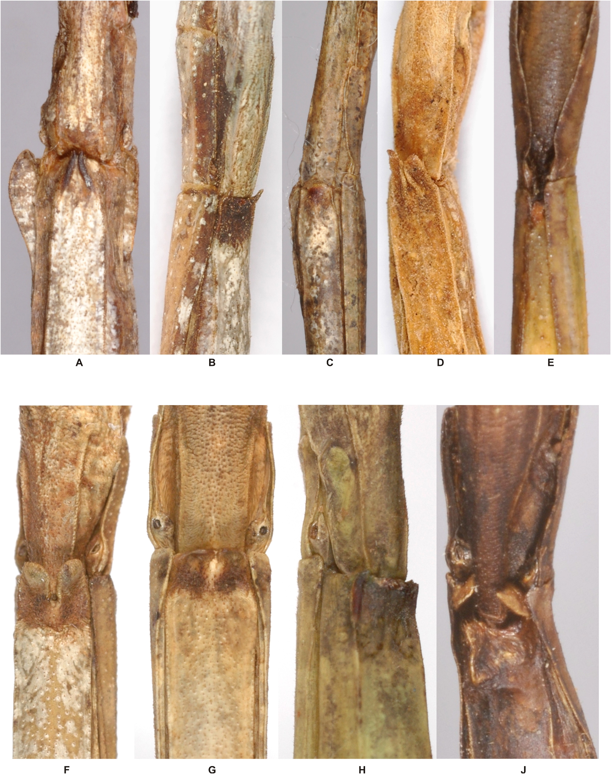
Thorax: Pronotum about as long but slightly narrower than head, basically rectangular and about 1.5x longer than wide, the posterior margin rounded and with a slight median narrowing. Anterior margin somewhat raised and with a median pair of small, obtuse granules; near anterolateral angles with a small but distinct pit. Transverse median sulcus distinct, strongly curved and almost reaching lateral margins of segment, the longitudinal median line slightly impressed. Mesothorax 6x longer than pronotum and uniform in diameter except for a slight widening at posterior margin. Mesonotum with the medio-longitudinal carina weakly indicated in the anterior one-third and with a granulose longitudinal lateral carina parallel to lateral margins; otherwise smooth. Metanotum short and scarcely more than one-quarter then length of mesonotum, 2.5x longer than wide, indistinctly narrowing towards the posterior and with a weakly indicated medio-longitudinal carina. Mesopleurae with a longitudinal row of 12–15 irregularly placed pointed tubercles to small spines ( Figs. 70L View FIGURE 70 , 87D View FIGURE 87 ) and metapleurae with about ten such tubercles or spines; just above with an obtuse, granulose longitudinal bulge. Meso- and metasternum granulose with the granules more numerous and aggregated along longitudinal median line, not tectate or keeled; a fine longitudinal carina however, along lateral margins ( Fig. 87D View FIGURE 87 ).
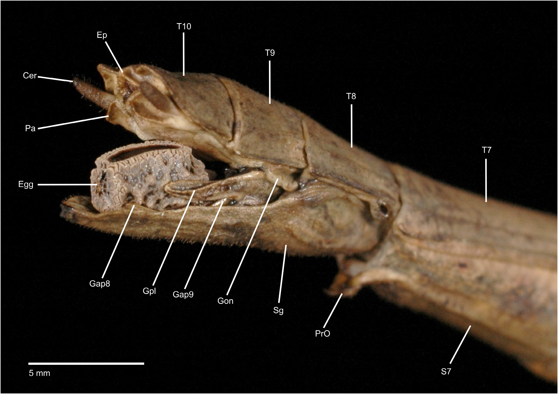
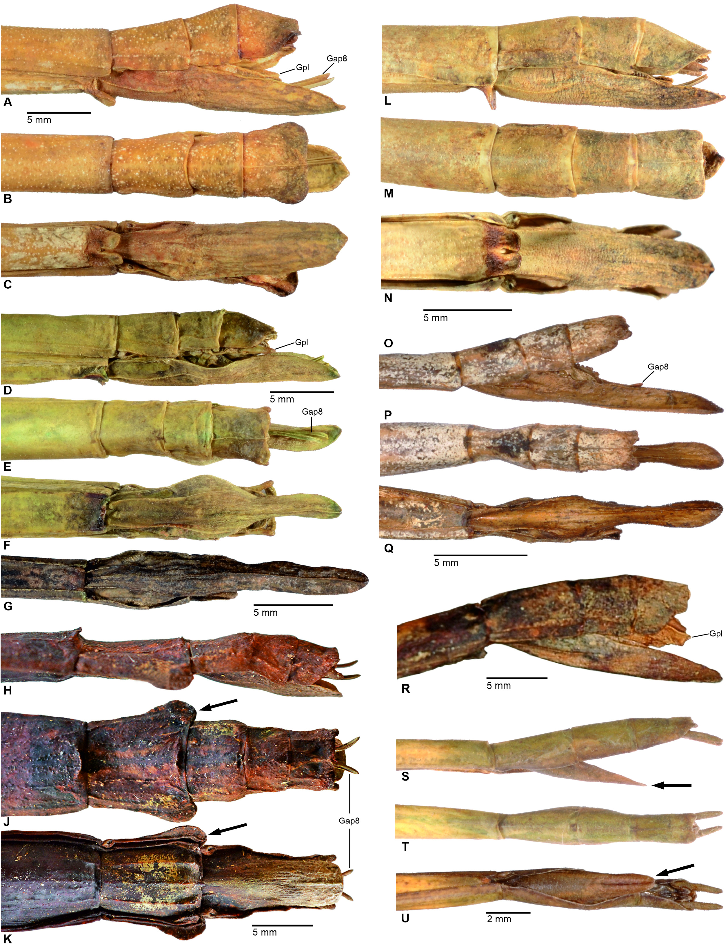
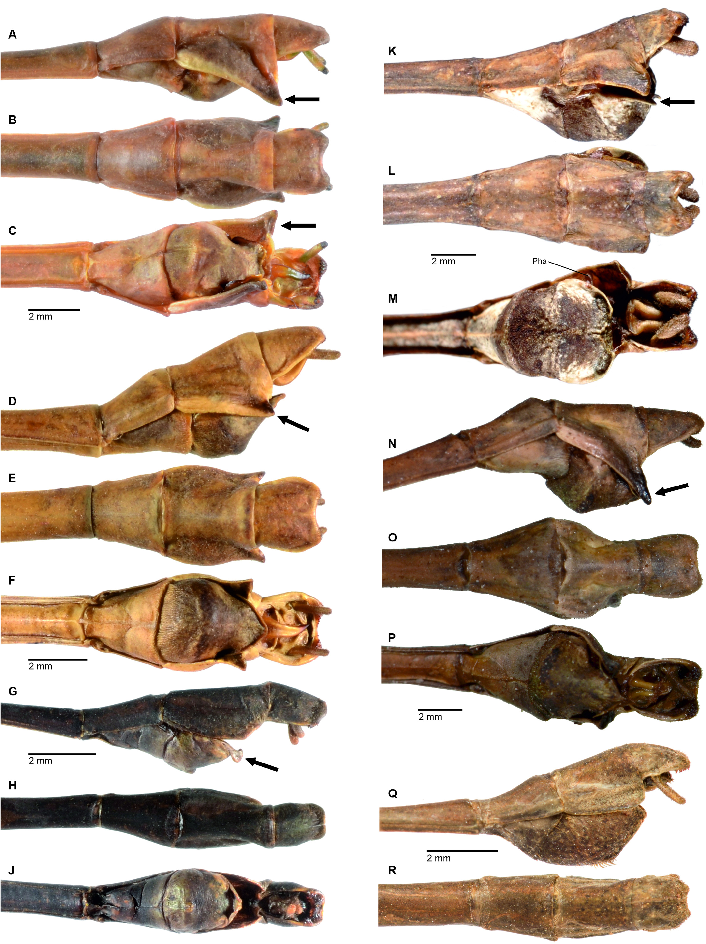
Abdomen: Median segment 1.4x longer than metanotum, about 3.5–4x longer than wide with the lateral margins notably concave; surface smooth. Segment II about three-fifth the length of median segment. Segments II–V slightly increasing and VI–VII decreasing in length with VII slightly shorter than II; II about 2x and V 2.6x longer than wide. II somewhat constricted medially, III–Iv slightly widening and VI–VII narrowing; tergum VII with lateral margins straight. Sterna II–VII minutely granulose. Sternum VII with a prominent praeopercular organ, that is formed by a pair of distinct, transverse, rounded lobes ( Fig. 91G View FIGURE 91 ). Terga VIII–X together as long as V, VIII about half the length of VII but wider and rectangular, IX narrower and shorter than VIII and gently narrowed towards the posterior; both scarcely longer than wide. Anal segment about as long as VIII and with a fine medio-longitudinal carina, slightly gradually widening towards the posterior and the posterior margin gently concave with the outer angles obtusely rounded ( Fig. 74M View FIGURE 74 ); lateral margins angular. Epiproct very small, triangular and weakly projecting beyond posterior margin of anal segment. Gonapophyses VIII elongate, upcurved and roughly reaching to apex of anal segment ( Figs. 5 View FIGURE 5 , 74L View FIGURE 74 ). Gonoplacs with outer lateral surface granulose ( Fig. 5 View FIGURE 5 ). Cerci small, conical, slightly compressed laterally and projecting scarcely over anal segment. Subgenital plate with an obtuse but distinct medio-longitudinal keel in apical one-third and projecting over tip of abdomen by no more than length of anal segment; apex broadly triangular ( Fig. 74N View FIGURE 74 ).
Legs:All long, moderately slender and entirely unarmed. Profemora a little longer than mesothorax, mesofemora longer than metathorax, metafemora projecting over posterior margin of abdominal segment IV and metatarsi slightly projecting beyond apex of abdomen. Medioventral carina of meso- and metafemora distinct, unarmed. Probasitarsus about as long as remaining tarsomeres except claw combined. Dorsal carina gently rounded. Meso- and metabasitarsus somewhat longer than following three tarsomeres combined, dorsal carina lamellate and weakly rounded.
♂♂ ( Figs. 70C View FIGURE 70 ). Medium-sized (body length 78.5–99.0 mm) and moderately slender for the genus with a very long median segment that is more than twice the length of the metanotum, vestigial alae ( Fig. 73J View FIGURE 73 ), a large triangular deflexion of abdominal tergum IX and fairly complex colouration. Head, prothorax, posterior portions of meso- and metanotum, abdomen and all coxae drab to creamy mid brown. Meso- and metasternum orange ( Fig. 87E View FIGURE 87 ), ventral surface of abdomen pale brown. Most parts of meso- and metanotum as well as pleurae bluish green, the mesonotum with a prominent, elongate black dorso-median marking, which covers about three-quarters the length of segment. Lateral margins of abdominal tergum IX cream-coloured with the posterolateral angle black. Profemora mostly red (the interior surface more intensely; Fig. 73G View FIGURE 73 ) with the base mid brown, protibiae reddish mid brown and gradually becoming pale brown towards the apex. Meso- and metafemora green basally and gradually becoming brown towards the apex and with a very faint pale transverse band sub-apically. Meso- and metatibiae reddish mid brown with two faint pale transverse bands. Eyes light brown with darker brown mottling. Antennae dark brown dorsally and black ventrally, scapus and pedicellus mid brown.
Head ( Fig. 73G View FIGURE 73 ): Roundly sub-rectangular in dorsal aspect, broadest at the eyes, gently narrowing towards the posterior and 1.2x longer than wide; otherwise as in ♀♀ but posterior of vertex with two low, obtuse median swellings. Eyes prominent, circular in outline, projecting more than hemispherical and their diameter contained about 1.3x in length of genae. Antennae reaching to abdominal tergum VII; otherwise as in ♀♀ but with the scapus less compressed and rather oval in cross-section.
Thorax: Pronotum shorter and narrower than head; generally, as in ♀♀ but with the transverse median sulcus less decidedly arched and the pits near anterolateral angles more pronounced ( Fig. 73G View FIGURE 73 ). Mesothorax notably narrower than prothorax, uniform in diameter except for being slightly widened at anterior and posterior margins and 7.8x longer than pronotum. Mesonotum with a fine longitudinal lateral carina parallel to lateral margins. Metanotum less than one-quarter the length of mesonotum, roughly 2.75x longer than wide and slightly narrowed towards the posterior, the median line weakly indicated in anterior portion. Meso- and metasternum with a slight medio-longitudinal median keel; less distinct on metasternum although. Two very small, vestigial, angularly, scale-like alae are present (length < 0.5 mm; Fig. 73J View FIGURE 73 ).
Abdomen: Median segment about 2.3x longer than metanotum and roughly 10x longer than wide with the median portion conspicuously narrowed and the posterior portion widened. Segment II somewhat more than half the length of median segment, II–V roughly uniform in length, VI and VII decreasing in length with VII only three-quarters the length of II–V; all uniform in diameter but VII weakly widened posteriorly. II–V on average 4x and VII 2.7x longer than wide. Tergum VIII about two-third the length of VII and strongly broadening towards the posterior, trapezoidal in dorsal aspect with posterior margin 1.3x wider than anterior margin. IX somewhat longer than VIII, strongly convex, with anterior portion widened in dorsal aspect and the lateral margins strongly deflexed and forming a large, triangular lobe, whose posterolateral apex ventrally projects by almost half the height of tergum VIII ( Fig. 75D View FIGURE 75 ). VIII and IX each with an obtuse longitudinal lateral bulge. Anal segment narrower and two-thirds the length of IX, slightly constricted anteriorly and broadly rounded posteriorly with a shallow, concave posteromedian notch ( Fig. 75E View FIGURE 75 ); posterolateral angles somewhat inflated, roundly angular and ventrally set with numerous minute dark reddish brown teeth (figs. 75F, 96B). Epiproct minute, scale-like and wholly concealed under anal segment. Vomer elongate, slender, strongly arched, gradually narrowing towards an acutely pointed and sinistral directed terminal hook ( Fig. 96B View FIGURE 96 ). Cerci small, very slender, cylindrical and projecting slightly over anal segment. Poculum moderately convex, cup-like ( Fig. 75D View FIGURE 75 ) and flattened towards a broadly triangular, somewhat labiate posterior margin ( Figs. 75F View FIGURE 75 , 96B View FIGURE 96 ); lateral surfaces each with a roundly concave dorsal indention medially.
Legs: All very long, slender and completely destitute of spines or teeth. Profemora longer than head, pro- and mesothorax combined, mesofemora projecting over posterior margin of abdominal segment II, metafemora almost reaching to posterior margin of abdominal segment VI, and metatibiae projecting greatly over apex of abdomen. Pro- and metabasitarsus longer than remaining tarsomeres combined, mesobasitarsus slightly shorter. All slender and simple.
Table 65: Measurements of Phanocloidea venezuelica sp. n.
Eggs ( Fig. 100M View FIGURE 100 ). Capsule elongate, 1.8x longer than wide, compressed laterally with lateral surfaces slightly sub-parallel to each other, oval in cross-section and slightly narrowed towards the polar end; angular in lateral aspect with polar-area angular and flattened and the dorsal surface more convex than ventral surface. Complete surface consisting of irregularly shaped scale-like plates with a network of deep cracks and fissures in between Surface of these scales minutely rugose and granulose, the outer margins unevenly dentate of with spiniform appendages. A raised collar surrounding the micropylar plate. Micropylar plate elongate, almost parallel-sided, covering about three-quarters the length of dorsal capsule surface and about 4x longer than wide; plate slightly indented with surface minutely granulose and both ends rounded. Anterior end of plate rounded and posterior end with a small median notch. Micropylar cup a rounded swelling and placed in posteromedian gap of plate. Median line represented by a small, impressed warty structure near polar end of micropylar plate. Operculum oval, outer margin with a very prominent, high, hollow and crest-like structure with a very unevenly crenulate upper margin, that is formed by membranous, lamellate extensions; these extending by about half of the capsule length. General colouration of capsule greyish mid brown with the raised areas slightly lighter in colour, micropylar plate blackish brown. Opercular structure straw to pale yellow.
Measurements [mm]: Length (including operculum) 5.5–5.8, length 3.4–3.5, width 2.2–2.3, height 2.6–2.7, length of micropylar plate 2.3.
Comments. Zompro (2000) provided descriptions and illustrations of the adults as well as a detailed description and SEM-pictures of the eggs based on captive reared specimens from Venezuela. These however were misidentified as Bacteria nodulosa Redtenbacher, 1908 , a species which is here shown to be a junior synonym of P. muricata ( Burmeister, 1838, syn. n.). The larger measurements provided by Zompro (2000: 171) clearly concern to the two wild-caught ♂♂ of Ph. muricata from Brazil (Rio Tabajos & Taperinha) in the collection ZMUH. Detailed examination of the Venezuelan specimens has proven them to be an as yet undescribed species. P. venezuelica sp. n. was first collected by Allan J. E. Harman in August 1992 along the Rio Parucito in the Amazonas Province of south-east Venezuela. The collected specimens have also given rise to a culture of this species which was distributed in Europe since 1993 and included on the Phasmid Study Group culture-list as culture No. 152 “ Calynda sp. ”. The species has proven fairly difficult to rear in large cages and at moderate humidity. While hatching rates are close to 90% the mortality of newly hatched nymphs was very high and the culture was finally lost a few years ago. Alternative food plants accepted in captivity in Europe included bramble ( Rubus spp. , Rosaceae ), raspberry ( Rubus idaeus , Rosaceae ), rose ( Rosa spp. , Rosaceae ), oak ( Quercus robur , Fagaceae ) and salal ( Gaultheria shallon , Ericaceae ).
Distribution. So far only known from the type-locality the Rio Parucito in the Department Atures, Amazonas Province, SE-Venezuela.


| NHMUK |
Natural History Museum, London |
| FH |
Fort Hays |
No known copyright restrictions apply. See Agosti, D., Egloff, W., 2009. Taxonomic information exchange and copyright: the Plazi approach. BMC Research Notes 2009, 2:53 for further explanation.
|
Kingdom |
|
|
Phylum |
|
|
Class |
|
|
Order |
|
|
Family |
|
|
Genus |
Phanocloidea venezuelica
| Hennemann, Frank H. & Conle, Oskar V. 2024 |
Bacteria nodulosa
| Zompro, O. 2000: 171 |