
Kujdanowiaspis buczacziensis ( Brotzen, 1934 )
|
publication ID |
https://doi.org/ 10.5252/g2010n1a1 |
|
persistent identifier |
https://treatment.plazi.org/id/03FE8790-FFDA-FFFC-68A8-F4F5920A0C93 |
|
treatment provided by |
Felipe |
|
scientific name |
Kujdanowiaspis buczacziensis ( Brotzen, 1934 ) |
| status |
|
Kujdanowiaspis buczacziensis ( Brotzen, 1934) ( Figs 2-4 View FIG View FIG View FIG )
Phlyctaenaspis buczacziensis Brotzen, 1934: 118 , pl. 9 figs 1-3.
TYPE MATERIAL. — Lectotype MB 290A (median dorsal plate) ( Brotzen 1934: pl. 9 fig. 2; this article, Fig. 2D View FIG ) designated here; paralectotype MB 288 (mechanically prepared dorsal sides of the dermic nasal capsule and neurocranium, Fig. 3 View FIG ; Brotzen 1934: pl. 9 fig. 1), not found; unidentified paralectotype, fragment of ventral armour, not found ( Brotzen 1934: pl. 9 fig. 3).
TYPE LOCALITY. — Buchach (Podolia, Ukraine) ( Brotzen 1934: 119).
TYPE HORIZON. — Pragian (Early Devonian) (see Dupret & Blieck 2009).
MATERIAL EXAMINED. — List given in Appendix 1.
DIAGNOSIS. — Quite large Kujdanowiaspis species with very thin tubercles. High tubercle density. Anterolateral plate higher than long.
REMARKS
All specimens described by Brotzen (1934) are considered as syntypes. Stensiö (1942) designated as holotype specimen MB 288, without refering to the original type series.Hence, and as specimen MB 288 was not found, specimen MB 290a is designated as lectotype; the other specimens from the original type series are then paralectotypes, though not found; the type locality is that of the lectotype only (according to ICZN 1999: articles 72.1, 72.4.7, 73.1.3, 73.2, 74.1, 74.4, 74.5, 74.6, and 76.3).
Kujdanowiaspis buczacziensis ( Brotzen, 1934)
The material of K. buczacziensis (Appendix 1) is rare and often badly preserved. From the specimens
described and figured by Brotzen (1934), just one remained (median dorsal plate MB 290a, Fig. 2D View FIG ), the rest being probably lost. More material has since been collected: five fragments of skull roof described by Stensiö (1942) – redescribed further –, an anterolateral plate (Pi 1201, Fig. 2E View FIG ) and a portion of trunk armour (GGI 15-664, Fig. 2F View FIG ). Consequently, the description of this species will be much more incomplete than that of K. podolica .
Stensiö (1942) thought that the skull roof of Kujdanowiaspis buczacziensis resembled that of Phlyctaenius Traquair, 1890a ( Traquair 1890b: pl. 3; 1894: 369; Woodward 1892, pl. 1, fig. 7; Stensiö 1925: 167, fig. 21; Heintz 1933: 130-133, fig. 2), of Arctaspis Heintz, 1929b (“ Svalbardaspis ” Heintz 1929b: figs 6A, 23), of Arctaspis Heintz, 1929 ( Heintz 1929a: fig. 2), of Bryantolepis Camp, Welles & Green, 1949 , of “ Euryaspis ” Bryant, 1932 (Bryant 1932: fig. 1) and of Anarthraspis Bryant, 1934 ( Bryant 1934: fig. 5), and differed from those of Actinolepis Agassiz, 1844 (see Gross 1940: 44-58, fig. 13) and of Jaeckelaspis Heintz, 1929b ( 1929a: fig. 1; 1929b: fig. 6B) – in fact, junior synonym of Arctolepis Eastman, 1908 . Stensiö (1944) also remarked that as in all “acanthothoracids”, the plates are closely fused with each other, hence increasing the difficulty to identify the plate limits. The skull roof of K. buczacziensis would also be longer and thinner than the one of Phlyctaenius acadicus . As well, orbital notches are deeper in Kujdanowiaspis than in P. acadicus .
According to the available material, the skull roof of Kujdanowiaspis buczacziensis is very similar to that of K. podolica (i.e. same relationships between plates and sensory lines grooves). The noticeable differences only concern the size ( K. buczacziensis is almost twice the size of K. podolica ) and the ornamentation (finer and more dense tubercles in K. buczacziensis ; those of K. podolica are a bit larger and more widely spaced).
The two specimens of K. buczacziensis ( Figs 3 View FIG ; 4 View FIG ) described by Stensiö (1942) provide important information on the internal structure of the rostrum that is unknown in K. podolica .
Stensiö (1942) noticed a contact between the postnasal and preorbital plates. The postnasal plates are wider than those of Bryantolepis (“ Euryaspis ” Bryant, 1932), and their radiation centre would be located laterally.
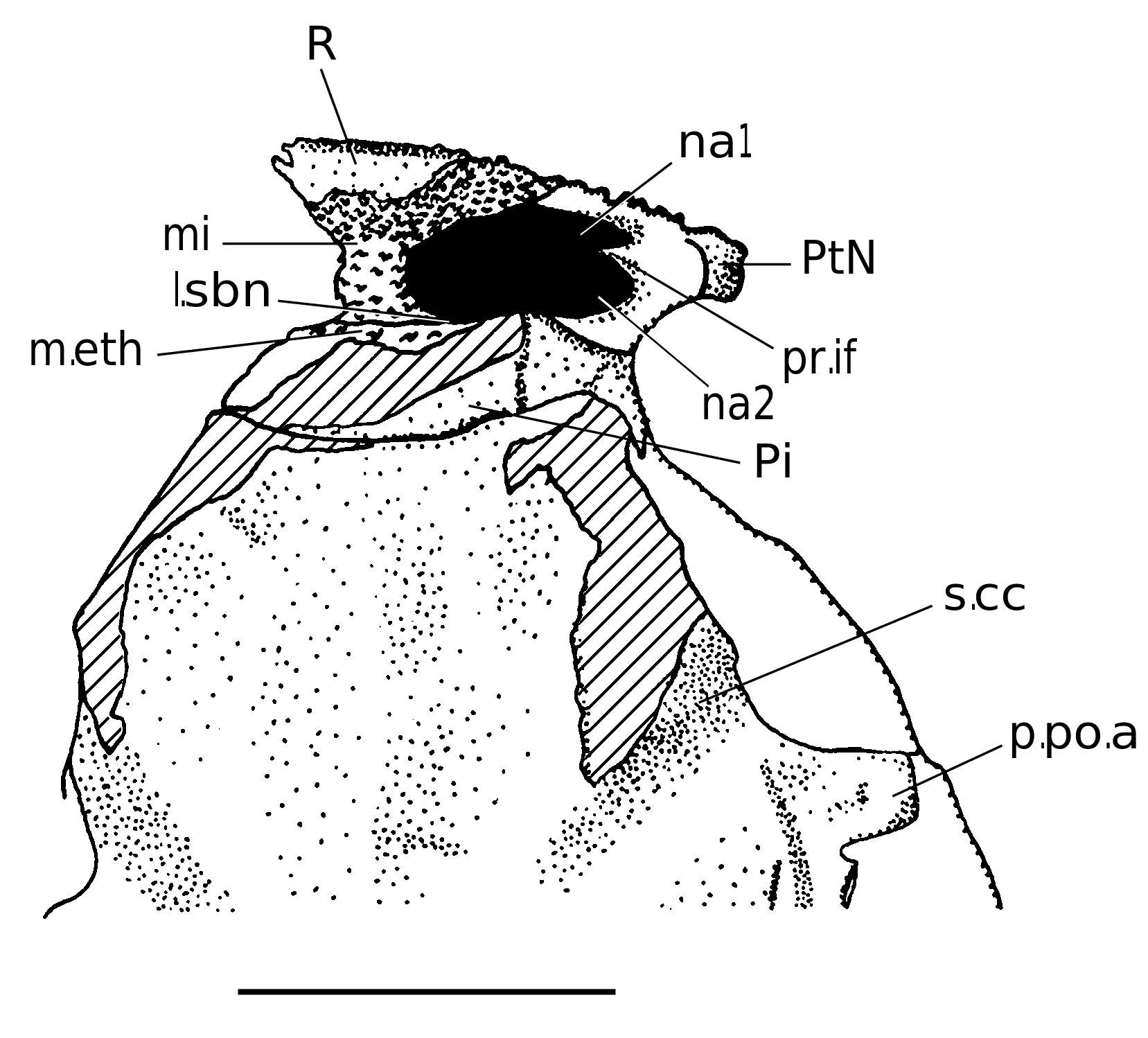
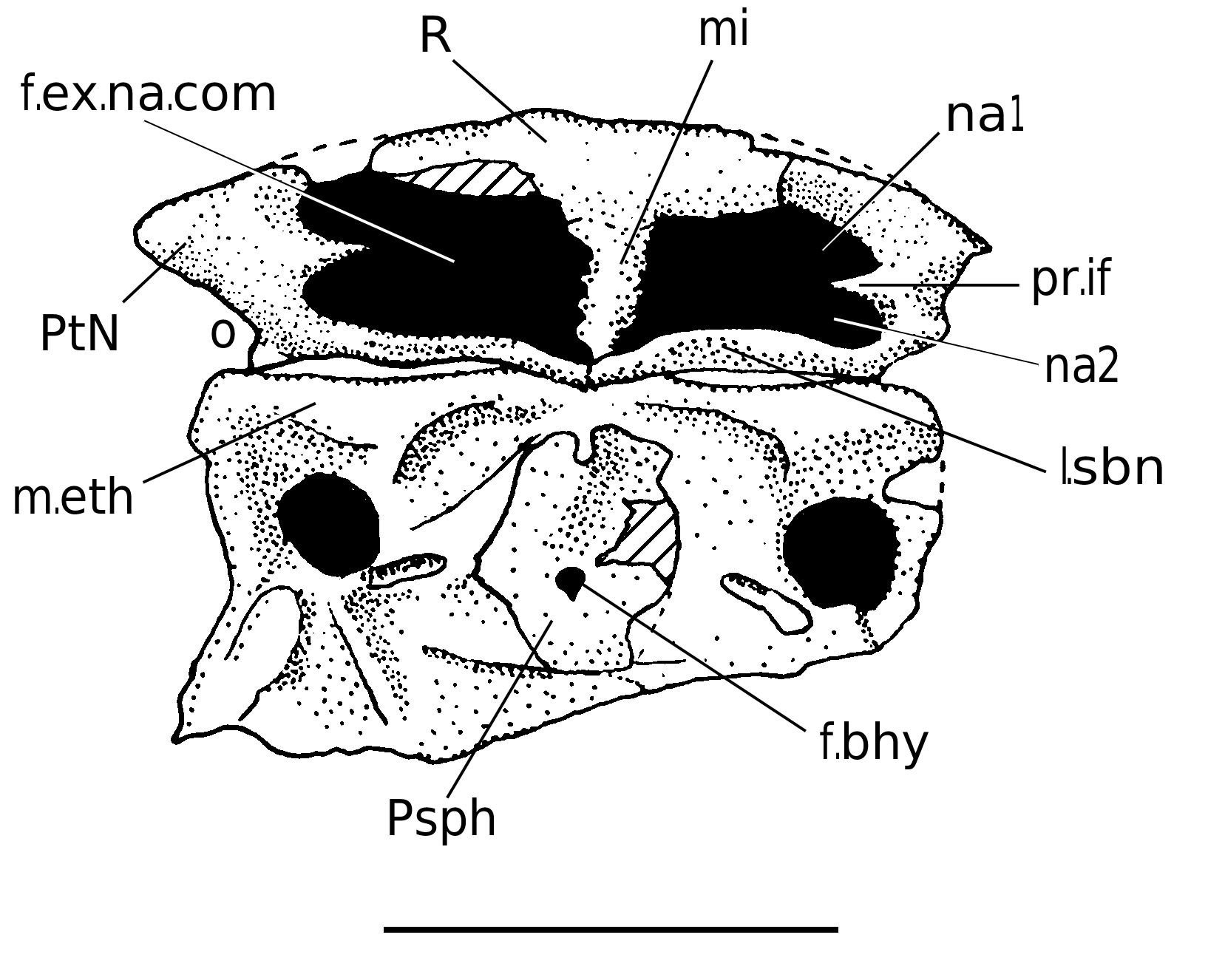
The rostral, pineal and postnasal plates forming the nasal capsule are located above the mouth. The dorsal side of this nasal capsule is clasically composed by the median rostral and pineal plates and the lateral postnasal plates. The lateral sides (“orbital faces” of Stensiö 1942) are posteriorly and slightly laterally directed, and are composed by the postnasal plates alone, forming the anterior edge of the orbit. The anterior side is medially composed by the rostral plate and laterally by the postnasals. The anterior edge of the external inhalant naris (na1, Figs 3 View FIG ; 4 View FIG ) is formed by both the rostral and postnasal plates.
The internal structure shows the anteroventral part of the rostral plate and the postnasal plates anterolaterally. The postnasal plates produce a tiny interfenestral process mesially (pr.if, Figs 3 View FIG ; 4 View FIG ) separating laterally the external inhalant (na1, Figs 3 View FIG ; 4 View FIG ) and exhalant nares (na2, Figs 3 View FIG ; 4 View FIG ). The rostral plate produces a medial process posteriorly (the internasal wall, mi, Figs 3 View FIG ; 4 View FIG ; Ra of Stensiö 1942); Stensiö thought that this medial wall was an independent dermal bone. The ventral side is closed by a pair of subnasal laminae posteriorly (l.sbn, Figs 3 View FIG ; 4 View FIG ) produced by the postnasals, and in mesial contact.
Rostral plate
The rostral plate (R, Figs 3 View FIG ; 4 View FIG ) is trapezoidal dorsally, with a slightly bowed anterior edge. Its anterior side is quite high, flanked by the external inhalant nares laterally. On the ventral side, it protrudes the internasal wall (mi, Figs 3 View FIG ; 4 View FIG ), unless it is an independent ossification (see internasal plate of Coccosteus cuspidatus Miller, 1841 ). Both are tuberculated identically to the adjacent plates. The radiation centre is located on the anterodorsal edge, in the symmetry plane.
Pineal plate The pineal plate (Pi, Figs 3 View FIG ; 4 View FIG ) is pentagonal. Its anterior edge (contacting the rostral plate) is straight, whereas the posterior one is slightly convex. Stensiö (1942: figs 1-3) suggested the presence of a pineal
foramen; nevertheless, this part of specimen NHRM P 5000 was already damaged, and he could not conclude to a true foramen or a closed pit.
Postnasal plate
The postnasal plates (PtN, Figs 3 View FIG ; 4 View FIG ) compose the lateral part of the dermal nasal capsule. They can be divided into three parts ventrally:
– an anterior part constituting the lateral edge of the rostrum, and mesially to it is the external inhalant naris (na1, Figs 3 View FIG ; 4 View FIG );
– a middle part showing the smooth short mesially directed interfenestral process (pr.if, Figs 3 View FIG ; 4 View FIG );
– a posterior part posterolaterally composed by the smooth subnasal laminae (l.sbn, Figs 3 View FIG ; 4 View FIG ), which contact medially behind the internasal wall, contrary to Stensiö’s (1942) interpretation that the internasal wall separated the two laminae. Laterally to the internasal wall are the external exhalant nares (na2, Figs 3 View FIG ; 4 View FIG ).
The external inhalant and exhalant nares form the fenestra exonarina communis (f.ex.na.com, Figs 3 View FIG ; 4 View FIG ). The inhalant naris is more laterally placed than the exhalant one.
Parasphenoid
The parasphenoid (Psph, Figs 3 View FIG ; 4 View FIG ) is more anteroposteriorly lengthened than that of K. podolica . Nevertheless, this element is too poorly represented (only one specimen) to consider it as a diagnostic character for a species (see also Dennis- Bryan 1995).
Postethmoidal part of the skull roof
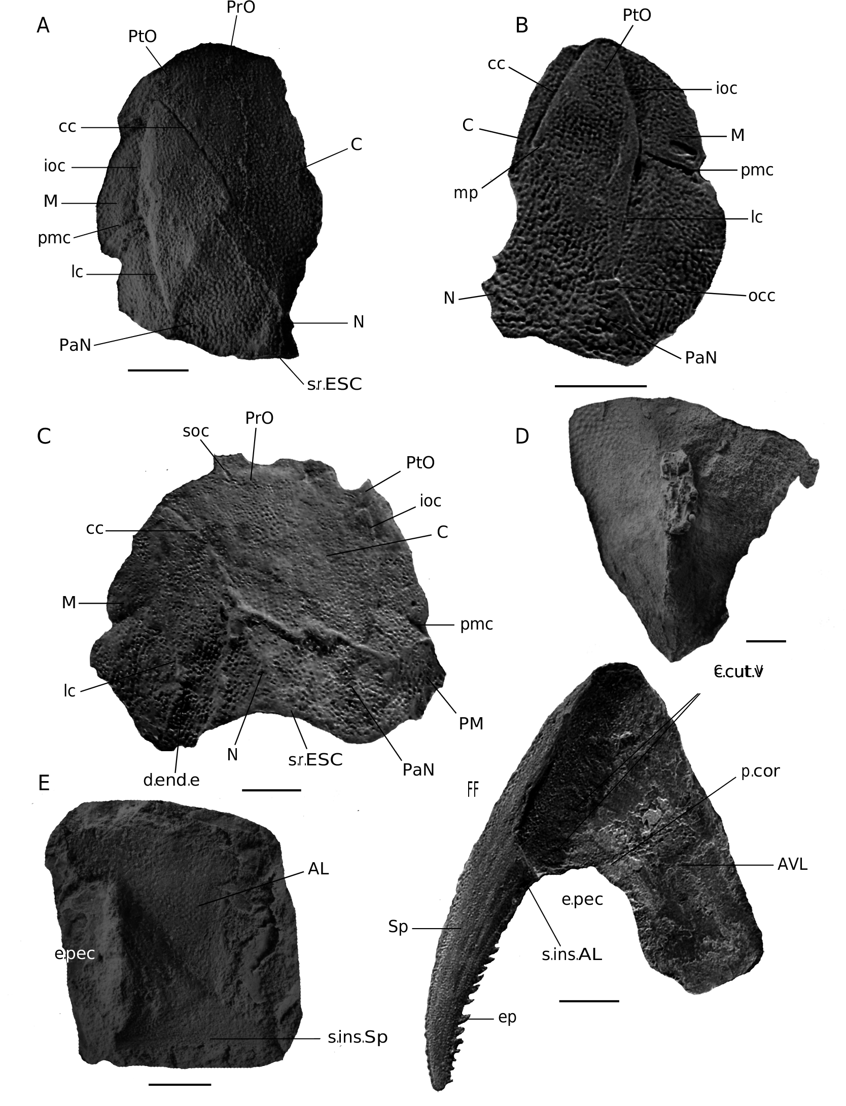
The postethmoidal part of the skull roof ( Fig. 2 View FIG A-C) is covered by very fine tubercles. The plates show the same relationships as in K. podolica , notably the contact between the postorbital and paranuchal plates which separates the central and marginal plates. The nuchal plate is thickened posteromedially, and the posterior edge is smooth and unornamented and possibly for extrascapular elements, as is the case in K. podolica .
Median dorsal plate
The median dorsal plate (MD, Fig. 2D View FIG ) is represented by a single specimen (MB 290a; Brotzen 1934: pl. 9 fig. 2). It is as wide and long as that of K. podolica but the tubercles are very much thinner and more densely distributed. Contrary to K. podolica , it does not show any posterosagittal crest.
The ventral armour ( Fig. 2F View FIG ) is known by left anterior ventrolateral and spinal plates only.
Anterolateral plate
The anterolateral plate (AL, Fig. 2E View FIG ), represented by a single specimen, is higher than long (as is the case in Actinolepis magna Mark-Kurik, 1973 ) and is covered by very fine and dense tubercles.
Anterior ventrolateral plate
The anterior ventrolateral plate (AVL, Fig. 2F View FIG ) is only visible internally. The outline of the cartilagineous scapulocoracoid (sc, Fig. 2F View FIG ) shows the usual trifurcate shape. Vascular canalicles leading to the coracoid process are also visible (c.cut.v, p.cor, Fig. 2F View FIG ).
Spinal plate
The spinal plate is visible in external view. Its distal end extends behind the posterior part of the anterior ventrolateral plate, as is the case in K. podolica . The tubercles are of two types. The first type is the same as that encountered elsewhere on the armour (round, fine and dense); it is located on the dorsal mesial and lateral sides; the lateral side shows slightly bigger tubercles, but still very dense. The second type is visible on the lateroproximal side of the plate, where the tubercles are lozenge in shape, the long axis being anteroposteriorly directed. The reason of this difference is unknown.
The dorsal side of the spinal plate shows the posterior part of the groove for the insertion of the anterolateral plate laterally to the pectoral notch (s.ins.AL, Fig. 2F View FIG ).
No known copyright restrictions apply. See Agosti, D., Egloff, W., 2009. Taxonomic information exchange and copyright: the Plazi approach. BMC Research Notes 2009, 2:53 for further explanation.
|
Kingdom |
|
|
Phylum |
|
|
Class |
|
|
Order |
|
|
Family |
|
|
Genus |
Kujdanowiaspis buczacziensis ( Brotzen, 1934 )
| Dupret, Vincent 2010 |
Phlyctaenaspis buczacziensis
| BROTZEN F. 1934: 118 |