
Saigona latifasciata, Liang, Ai-Ping & Song, Zhi-Shun, 2006
|
publication ID |
https://doi.org/ 10.5281/zenodo.174257 |
|
DOI |
https://doi.org/10.5281/zenodo.6256168 |
|
persistent identifier |
https://treatment.plazi.org/id/03FE879E-C904-1663-FE88-FEB0FE2EB6B9 |
|
treatment provided by |
Plazi |
|
scientific name |
Saigona latifasciata |
| status |
sp. nov. |
Saigona latifasciata View in CoL sp. nov.
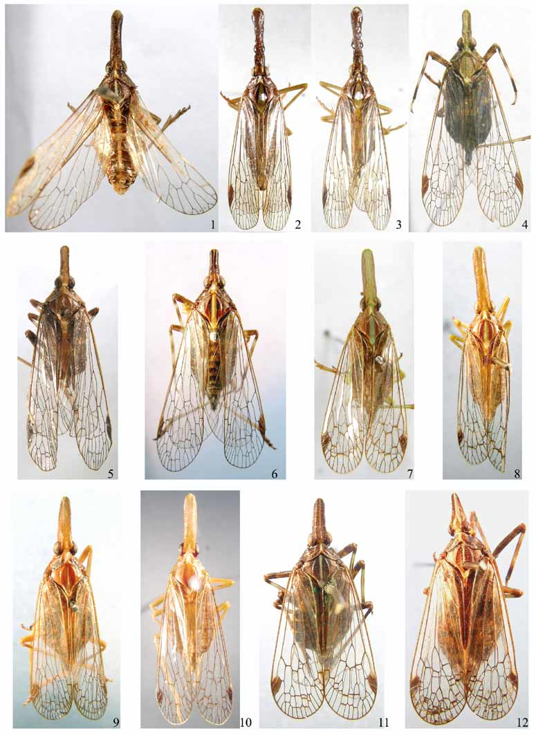
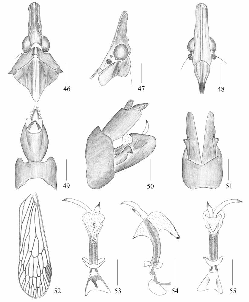
( Figs. 6 View FIGURES 1 – 12 , 46–55 View FIGURES 46 – 55 )
Description
ɗ, BL: 14.7 mm; HL: 4.0 mm; HW: 1.4 mm; FWL: 9.2 mm.
General color dark brown, marked with ochraceous. Vertex fuscous, with median, longitudinal, yellowish stripe. Genae mostly ochraceous, areas surrounding ocellus and antenna beneath eye yellowish brown. Frons and postclypeus yellowish or yellowish brown, anteclypeus and labrum fuscous. Rostrum with basal segment pale ochraceous, apical segment brown with extreme apex black. Pronotum fuscous, suffused with blackish brown; median carina yellowish; lateral, ventrally curved areas yellowish brown. Mesonotum dark brown, with broad, yellow, median, longitudinal stripe; maculae black. Thorax ventrally yellowish, marked with blackish brown. Legs yellowish brown, marked with ochraceous; fore coxae fuscous, middle and hind coxae yellow brown, hind coxae covered with fuscous spots; fore femora dark brown, middle and hind femora yellow brown with fuscous ring at apex; fore and middle tibiae pale brown with two broad fuscous rings basally and medially, respectively, hind tibiae much paler; tarsi and claws brown, hind tarsi and claws much paler; tips of apical spines on hind tibiae and tarsi black. Abdomen with tergites and sternites dark brown, covered with yellowish brown spots; pygofer and parameres fuscous.
Head ( Figs. 6 View FIGURES 1 – 12 , 46–48 View FIGURES 46 – 55 ) short, shorter than pronotum and mesonotum combined. Vertex ( Figs. 6 View FIGURES 1 – 12 , 46 View FIGURES 46 – 55 ) with cephalic process relatively short and robust, somewhat upturned, with median carina only conspicuous at base, lateral carinate margins curved in front of eyes. Frons ( Fig. 48 View FIGURES 46 – 55 ) with lateral carinae reaching to eyes, not to frontoclypeal suture.
Mesonotum ( Figs. 6 View FIGURES 1 – 12 , 46 View FIGURES 46 – 55 ) tricarinate on disc, with median carina faint, not reaching to tip. Fore wing venation as in Fig. 52 View FIGURES 46 – 55 .
Male genitalia with pygofer ( Figs. 49–51 View FIGURES 46 – 55 ) large and broad in lateral aspect ( Fig. 50 View FIGURES 46 – 55 ), posterior margin nearly straight and gently excavated at apical 1/3 to accommodate anal tube, length ratio of upper margin to lower margin about 1: 1.3. Anal tube ( Figs. 49, 50 View FIGURES 46 – 55 ) nearly rectangular, large in lateral view ( Fig. 50 View FIGURES 46 – 55 ) and long oval, large in dorsal view ( Fig. 49 View FIGURES 46 – 55 ) ratio of length to width at middle about 1.6: 1. Anal style ( Figs. 49, 50 View FIGURES 46 – 55 ) large and broad. Parameres ( Figs. 50, 51 View FIGURES 46 – 55 ) relatively small, short in lateral aspect ( Fig. 50 View FIGURES 46 – 55 ), apex bluntly rounded. Aedeagus ( Figs. 53–55 View FIGURES 46 – 55 ) with phallobasal conjunctival processes long, produced lateroposteriorly; phallobase narrow, long, curved dorsally; apical, dorsal, membranous lobe small, directed anterodorsally in lateral view ( Fig. 50 View FIGURES 46 – 55 ); with some spines at apex; apical, ventral, membranous lobe larger, longer, triangular in ventral view ( Fig. 53 View FIGURES 46 – 55 ), converging towards apex, directed anteroventrally in lateral view ( Figs. 50, 54 View FIGURES 46 – 55 ), covered with numerous fine spines on dorsal and ventral surfaces from middle to apex ( Figs. 54, 55 View FIGURES 46 – 55 ).
Material examined
Holotype ɗ, China, Yunnan, Yunlong, 2400 m, 5.vi.1996, L. Y. Zheng (NU).
Remarks
This species can be distinguished from other known Saigona species by its short cephalic process ( Figs. 6 View FIGURES 1 – 12 , 46, 47 View FIGURES 46 – 55 ); mesonotum with a broad yellow longitudinal stripe, and aedeagus with phallobase with apical dorsal and ventral membranous lobes covered with fine spines at apex ( Figs. 53–55 View FIGURES 46 – 55 ).
Distribution
China (Yunnan).
No known copyright restrictions apply. See Agosti, D., Egloff, W., 2009. Taxonomic information exchange and copyright: the Plazi approach. BMC Research Notes 2009, 2:53 for further explanation.