
Leptidea Billberg, 1820
|
publication ID |
https://doi.org/ 10.11646/zootaxa.4402.3.1 |
|
publication LSID |
lsid:zoobank.org:pub:05E1CFBA-B510-4860-AD7F-EA5814F19C0D |
|
DOI |
https://doi.org/10.5281/zenodo.3799942 |
|
persistent identifier |
https://treatment.plazi.org/id/03FEE52B-0A28-9202-88C9-F8FB622DFC45 |
|
treatment provided by |
Plazi |
|
scientific name |
Leptidea Billberg, 1820 |
| status |
|
Leptidea Billberg, 1820 View in CoL View at ENA
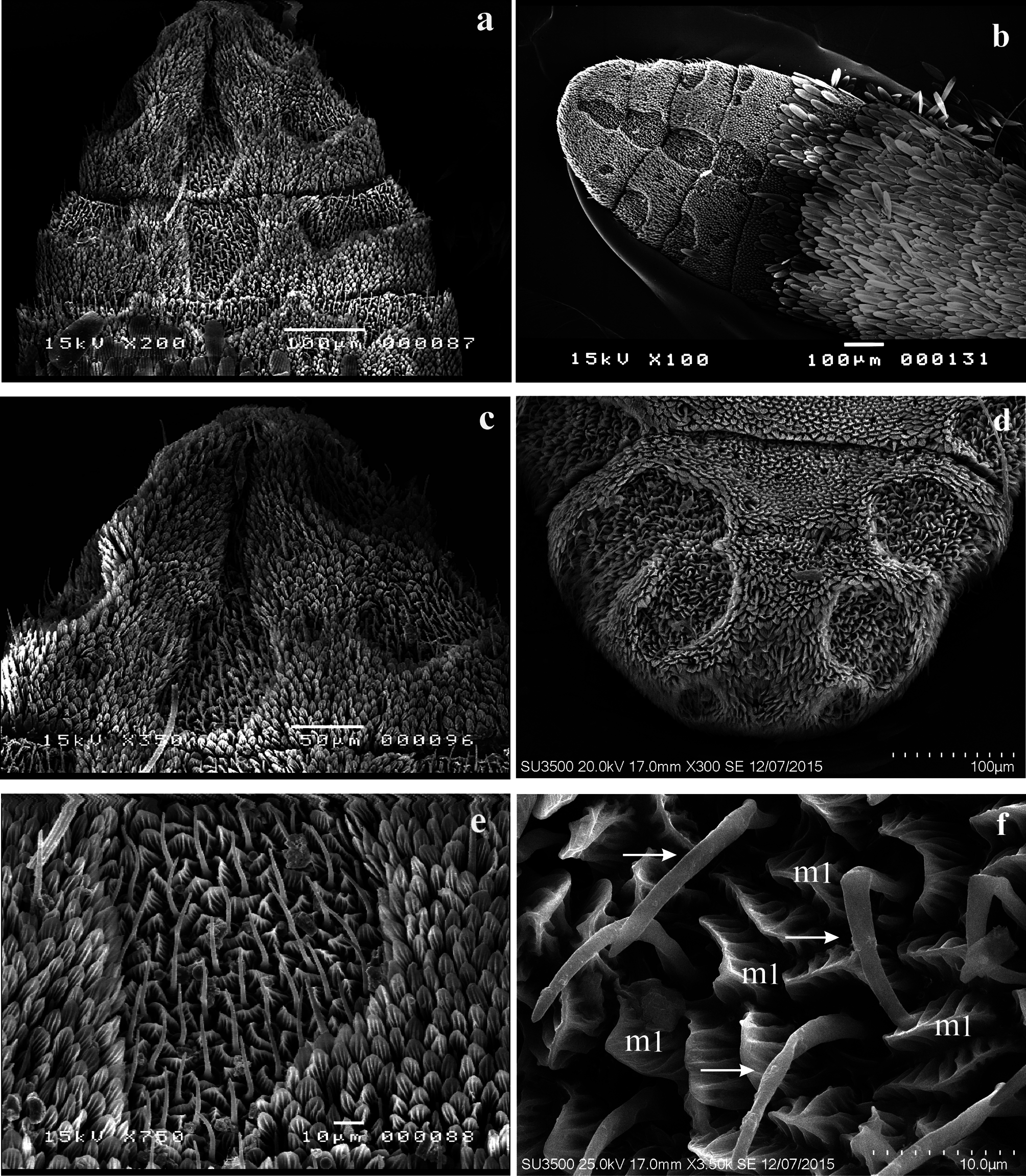
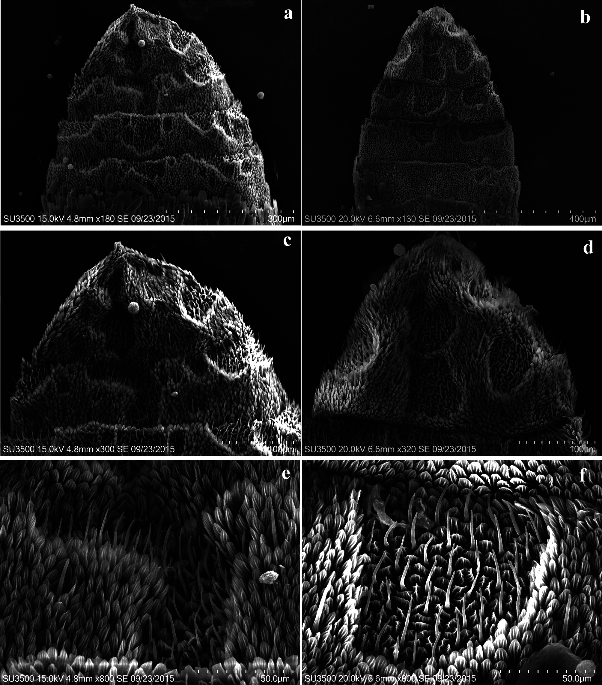
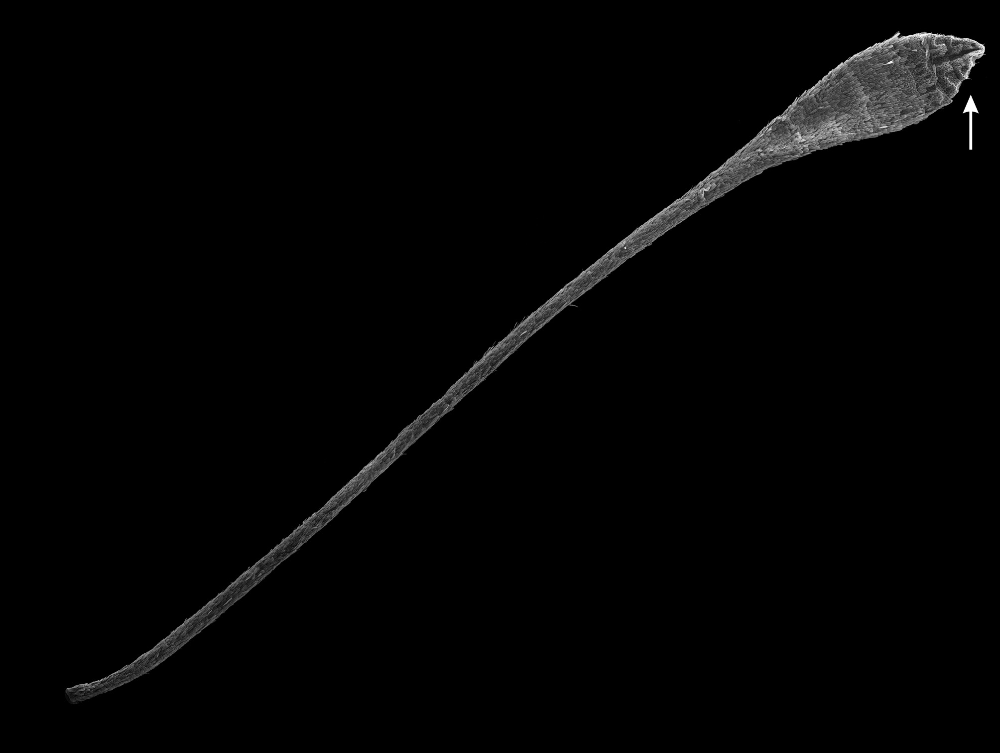
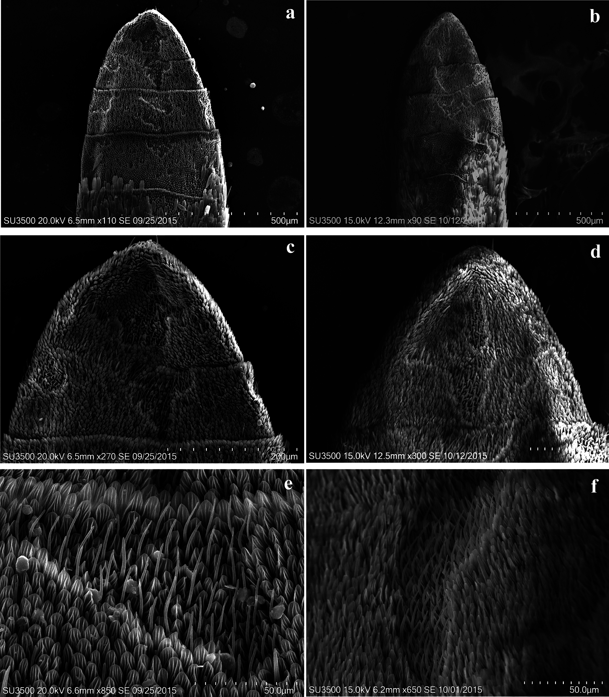
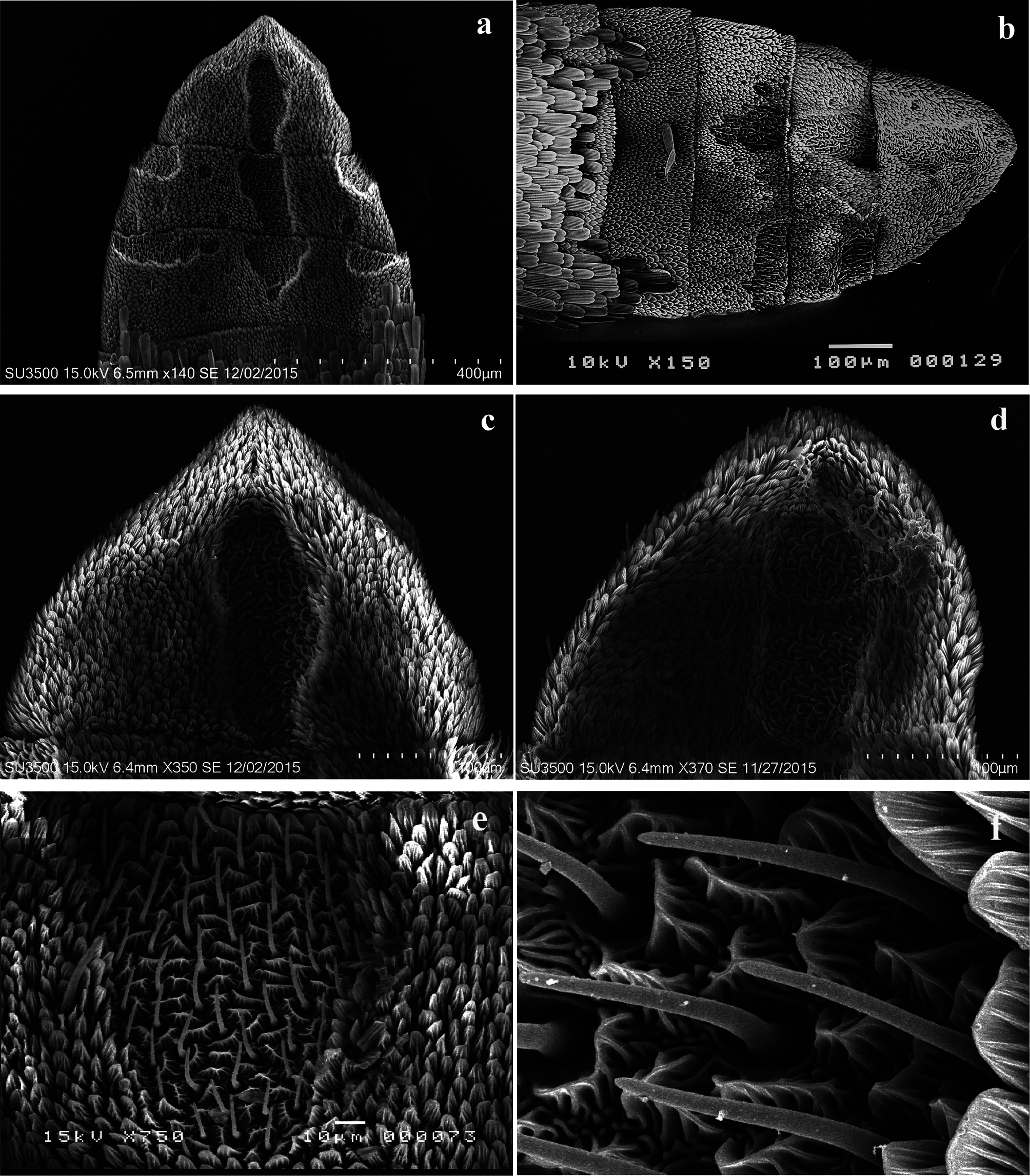
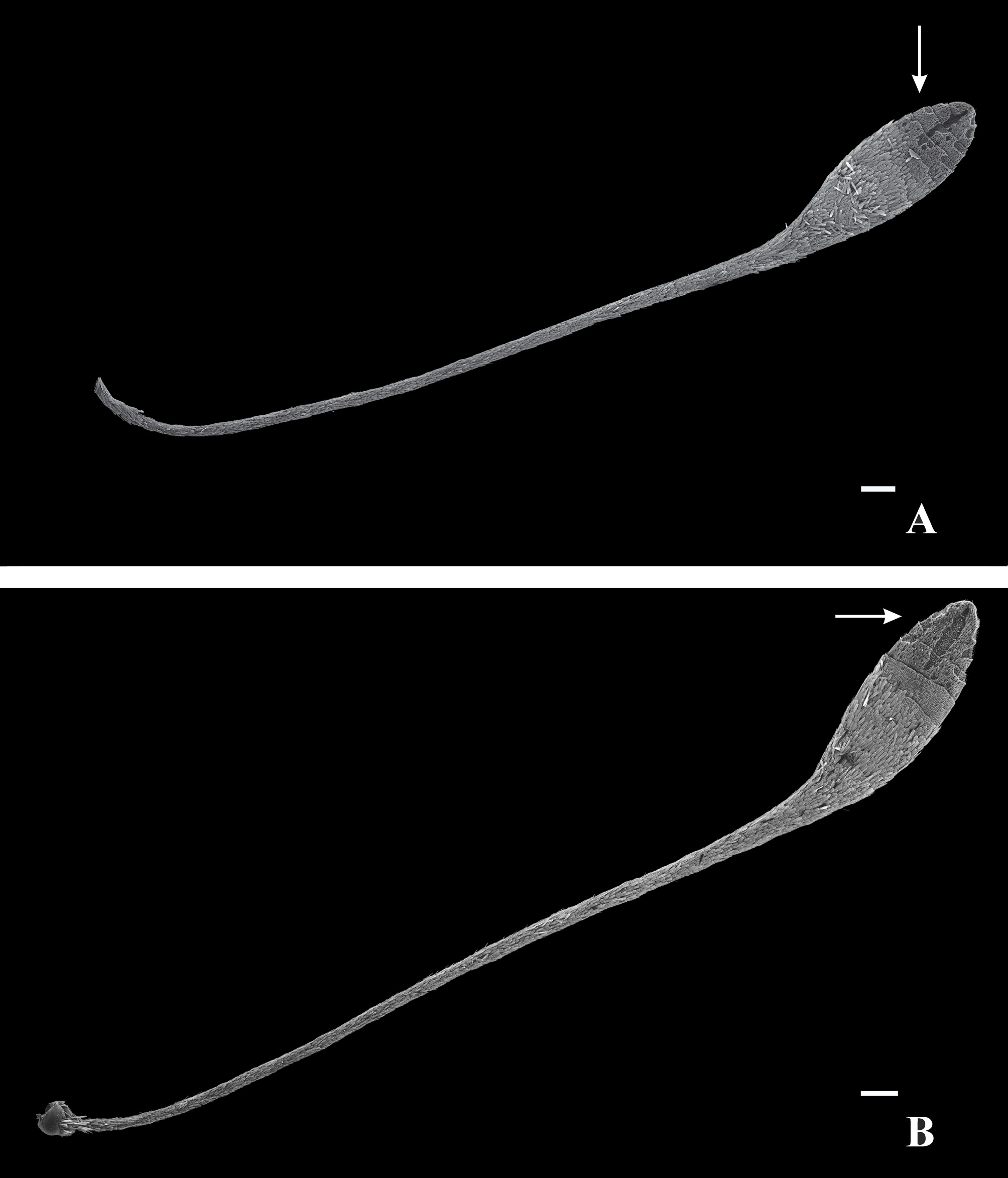
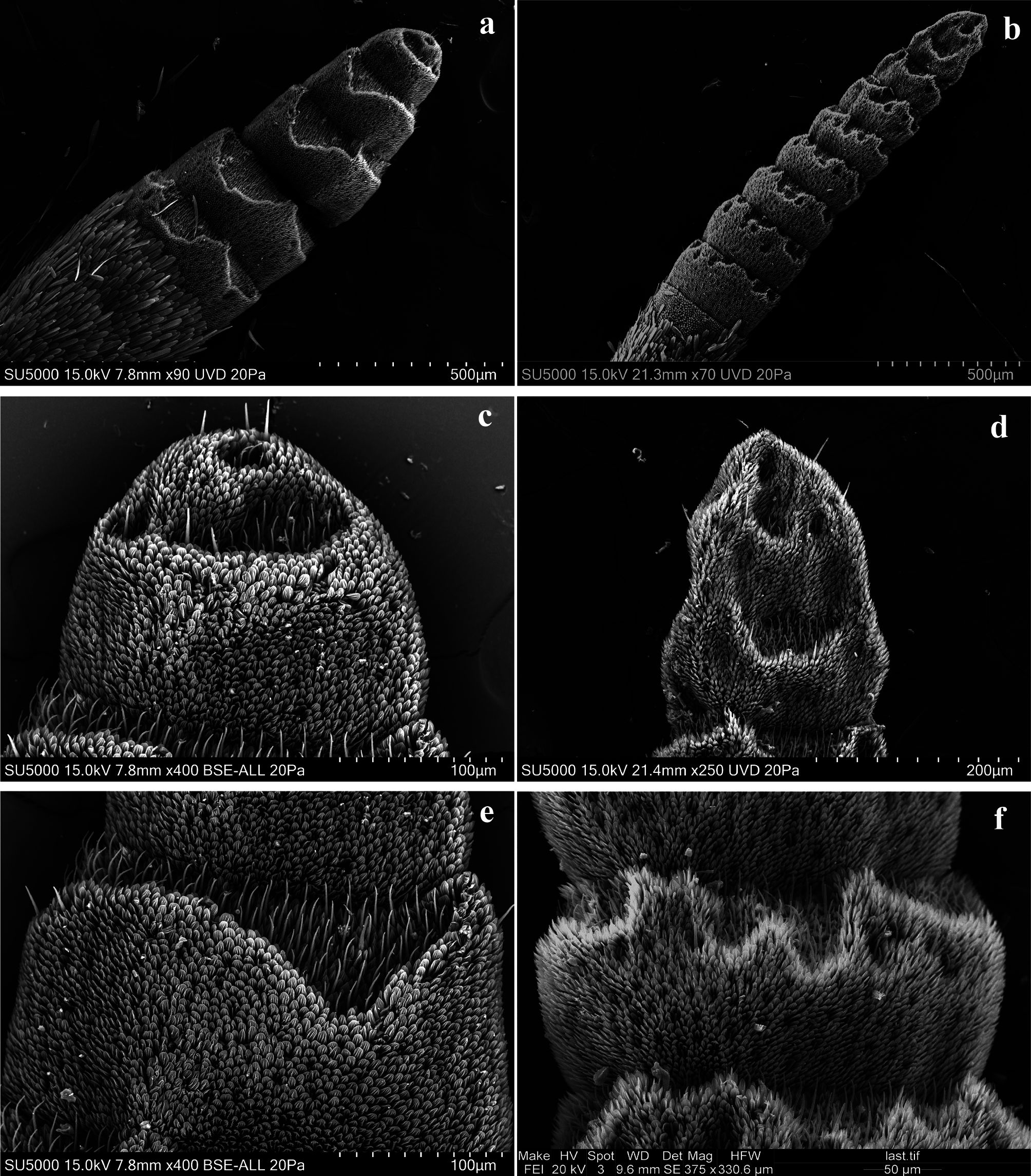
ANTENNAL CLUB ( Figs. 3 View FIGURE 3 , 4–7a, b View FIGURE 4 View FIGURE 5 View FIGURE 6 View FIGURE 7 , 8 View FIGURE 8 , 9–11a, b View FIGURE 9 View FIGURE 10 View FIGURE 11 , 12 View FIGURE 12 , 13–14a, b View FIGURE 13 View FIGURE 14 ). The length of the antenna is less than half the length of the costa of the forewing; the club is subpyriform and flattened ( Higgins 1975). Regardless of the number of antennomeres, the length of the club is greater in females than in males, especially in L. gigantea ( Fig. 14a, b View FIGURE 14 ).
ANTENNOMERES ( Figs. 4–6 View FIGURE 4 View FIGURE 5 View FIGURE 6 a–d, 7a–c, 9–11a–d, 13a–d, 14a–f): The number of scaleless antennomeres is four to ten; both sexes have the same number of segments, or females have one to four more than males; L. gigantea shows the greatest difference. In most cases, the antennomeres are depressed and flattened; they are usually wide doliform (barrel-shaped), and the proximal border is wider than the distal so that the club has a progressively reduced width. The most depressed antennomere has a width up to six times its length. The distal two or three antennomers are fused; occasionally there is an incomplete division or suture. The size of the last antennomere is about twice that of the preceding one; sometimes the two or three distal central sulci make up a single long sulcus. The distal antennomere is cupuliform and is generally slightly flattened. The scaled area dorsally covers the same number of antennomeres (or one or two more) as the ventral surface.
SULCI AND PSEUDOSULCI ( Figs. 4 View FIGURE 4 c–e, 5a–e, 6a–f, 7a–e, 9a–e, 10a–f, 11a–e, 13a–e, 14a–f): Sulci and pseudosulci show the typical trisulcate configuration of Dismorphiinae (one central and two lateral or laterodorsal); in some cases, there are no sulci on the first scaleless antennomere of the club. The number of central (c) and lateral (1) sulci is variable and does not always correspond with the number of scaleless antennomeres. Occasionally, they occupy more than three-fourth or the entire length of the antennomere, forming a continuum with the anterior and posterior sulci (a true groove). Although most are irregular, some are vertically ellipsoidal and are always truncated by the posterior border of the antennomere. Furthermore, they are disaggregated or aggregated with irregular contour and accompanied by some pseudosulci. The lateral sulci are transverse or nearly rectangular semi-elliptical and occupy half or less of the length of the antennomere; these appear disaggregated and occasionally are hidden in the distal antennomere. They usually extend to the dorsal surface from the second or third scaleless antennomere and rarely have a lateral-dorsal location from the first antennomere.
MICROTRICHIA ( Figs. 4 View FIGURE 4 f–h, j, 5f, g, i, 6g–i, 7f–h, 10g, h, 11f, g, 13f, g): Microtrichia m1, m2, and m4 are present. Within the sulci, they are fused with the elliptical cuticular ring of the trichoid sensilla. The sensilla trichoid:microtrichia (st:m1) ratio within sulci, is usually 1:2, 1:3, or 1:4; the last proportion occurs more frequently in the basal sulci. Sometimes where the antennomeres are embedded, the m2 are shorter, almost smooth, and occasionally show a toothed apex.
TRICHOID SENSILLA ( Figs. 4–5f View FIGURE 4 View FIGURE 5 , 6 View FIGURE 6 f–g, 7d–f, 9e, f, 10e–g, 11e, f, 13e, f): These sensilla are usually shorter than the chaetic sensilla and longer than the basiconic; they are 14–29 µm in length. They are surrounded by a cuticular ring and are inclined from their first or last third and pointed toward the apex of the sulcus containing them. The bases of these sensilla may be smooth or have some bumps, or the flagellum may appear to be embedded in a darker socket. In the cuticular wall there are some short stretch marks and near them a few tiny pores. The cuticular rings are partly fused with the m1 microtrichia.
CHAETIC SENSILLA ( Figs. 4g View FIGURE 4 , 6h View FIGURE 6 , 7g View FIGURE 7 , 9h View FIGURE 9 , 11g View FIGURE 11 , 13–14g View FIGURE 13 View FIGURE 14 ): In Leptidea the chaetic sensilla are usually ca. 25 µm long. Some shorter chaetic sensilla are less than 20 µm, whereas others are 20–30 µm long. There are usually six to ten per antennomere on the ventral side, and they are located toward the middle part of the antennomere or toward the distal edge, between the central sulcus and the lateral sulci; there is almost always a chaetic sensilla under each lateral sulcus (typical distribution in the subfamily). On the dorsal side, they are in the middle or distal part of the antennomeres and are often more abundant at the apex of the last antennomere.
BASICONIC SENSILLA ( Figs. 9i View FIGURE 9 , 11h View FIGURE 11 , 13–14h View FIGURE 13 View FIGURE 14 ): These are dispersed outside the sulci and are relatively abundant. They are distributed evenly along the auricular sensilla.
AURICULATE SENSILLA ( Figs. 4h View FIGURE 4 , 5g View FIGURE 5 , 6i View FIGURE 6 , 7h View FIGURE 7 , 9k, l View FIGURE 9 , 10h View FIGURE 10 , 13h View FIGURE 13 , 14i, j View FIGURE 14 ): Like basiconic sensilla, these are distributed outside the sulci in the scaleless antennomeres of the club. In Leptidea they are plentiful.
COELOCONIC SENSILLA ( Figs. 4i, j View FIGURE 4 , 5h, i View FIGURE 5 , 6j View FIGURE 6 , 7i, j View FIGURE 7 , 9j View FIGURE 9 , 10–11i, j View FIGURE 10 View FIGURE 11 , 13i, j View FIGURE 13 , 14k View FIGURE 14 ): These are usually more abundant on the dorsal than the ventral surface of the antennomere; on the ventral one they are near the lateral sulci. There are two types of coeloconic sensilla: type 1 (sc1) and type 2 (sc2), and they occur in pairs in some species.
OTHER SENSILLA ( Figs. 4k, l View FIGURE 4 , 5l View FIGURE 5 , 6–7k, l View FIGURE 6 View FIGURE 7 , 10–11k, l View FIGURE 10 View FIGURE 11 , 13k, l View FIGURE 13 , 14l View FIGURE 14 ): The styloconic sensila without stylus is presented in clusters or there is only one at the apex of the distal antennomere. The campaniform sensilla is most common on the dorsal side of the first scaleless antennomere. The campaniform and styloconic sensilla without stylus are more frequent on the dorsal surface so the same types of sensilla were not always found in all species because sometimes it was not possible to get images of the dorsum of the antennae, where sensilla can be observed that only are in that region .
PORES: Pores are present throughout the antennal club, although they are only visible when the microtrichia are separated to give rise to a sensillum.


No known copyright restrictions apply. See Agosti, D., Egloff, W., 2009. Taxonomic information exchange and copyright: the Plazi approach. BMC Research Notes 2009, 2:53 for further explanation.