
Larsonella pumilus ( Larson & Hoese, 1980 )
|
publication ID |
https://doi.org/ 10.11646/zootaxa.4695.4.4 |
|
publication LSID |
lsid:zoobank.org:pub:38DB2B6D-FA80-40FF-925B-F402787FF7F6 |
|
DOI |
https://doi.org/10.5281/zenodo.5628310 |
|
persistent identifier |
https://treatment.plazi.org/id/03FEE74F-FFC3-FFBA-FF6B-FF2DFBF458E1 |
|
treatment provided by |
Plazi |
|
scientific name |
Larsonella pumilus ( Larson & Hoese, 1980 ) |
| status |
|
Larsonella pumilus ( Larson & Hoese, 1980) View in CoL
[New Japanese name: Yuuna-haze]
( Figs. 1–4 View FIGURE 1 View FIGURE 2 View FIGURE 3 View FIGURE 4 ; Table 2 View TABLE 2 )
Lubricogobius pumilus Larson & Hoese, 1980: 41 View in CoL (type locality: Indian Ocean, 3°25’N 47°14.8’E, 37–38 m depth). Larsonella pumilus ( Larson & Hoese, 1980) View in CoL : Randall & Senou 2001: 11.
Material examined. OCF-P 3808, 21.6 mm SL, East China Sea off Seragaki, Onna Village, Okinawa, Japan, 11 August 2017.
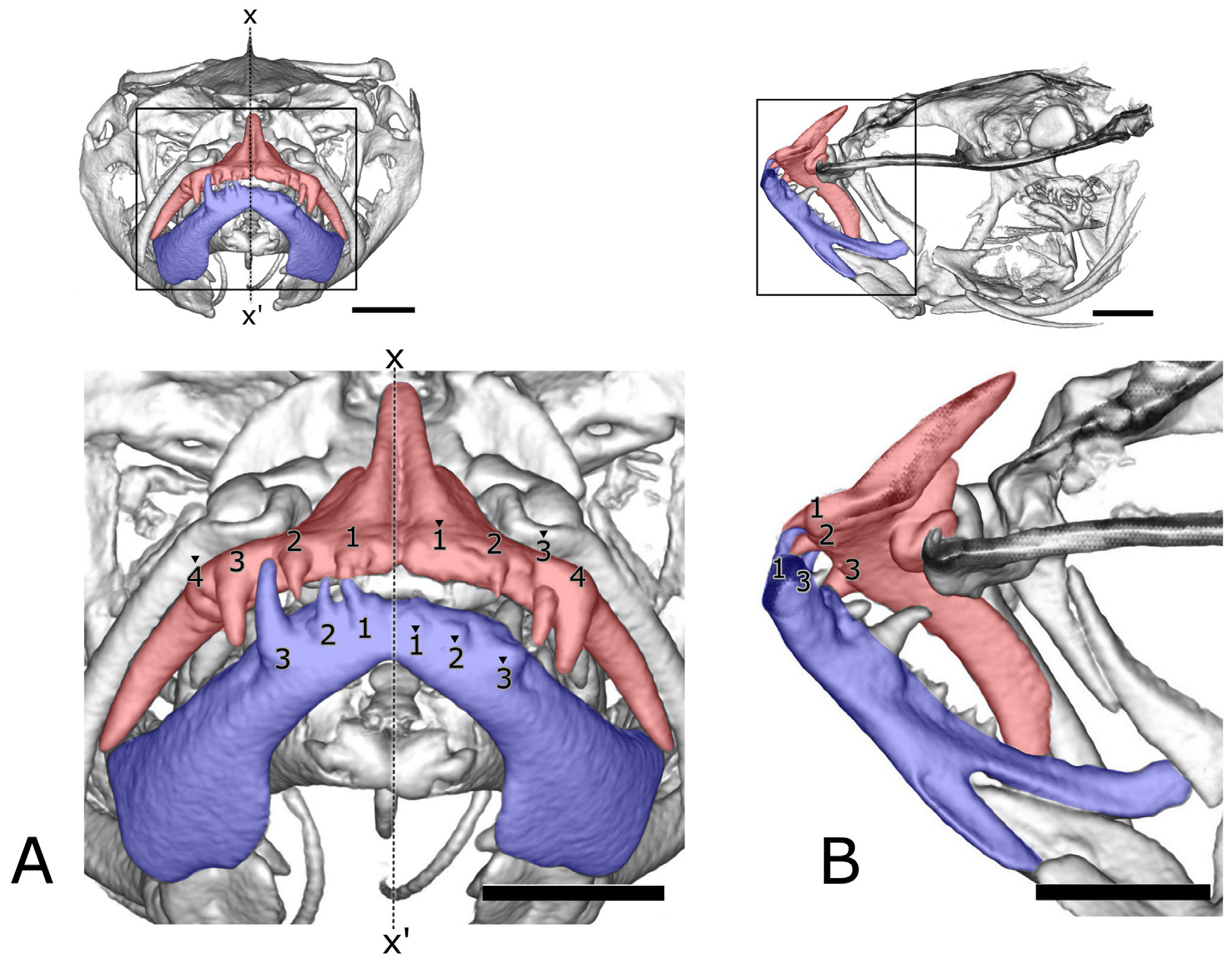
Description. Counts and measurements are shown in Table 2 View TABLE 2 . Head depressed, trunk nearly cylindrical, and tail compressed. Eyes located dorsolaterally. Mouth strongly oblique with angle to body axis about 60 degrees. Lower jaw protruding beyond upper jaw. Posterior end of upper jaw reaching below middle point between anterior margin of iris and anterior margin of pupil. Canine-like teeth aligned on edges of anterior halves of premaxilla and dentary; four and three teeth on one side of premaxilla and dentary, respectively; posterior teeth larger ( Fig. 2A View FIGURE 2 ). An inner row of conical teeth extending from anterior part to more posterior part of dentary than the outer canine-like teeth row ( Fig. 2B View FIGURE 2 ). Additional small conical teeth observed on inner parts of premaxilla and dentary with a stereomicroscope, but no such small teeth with micro-CT.
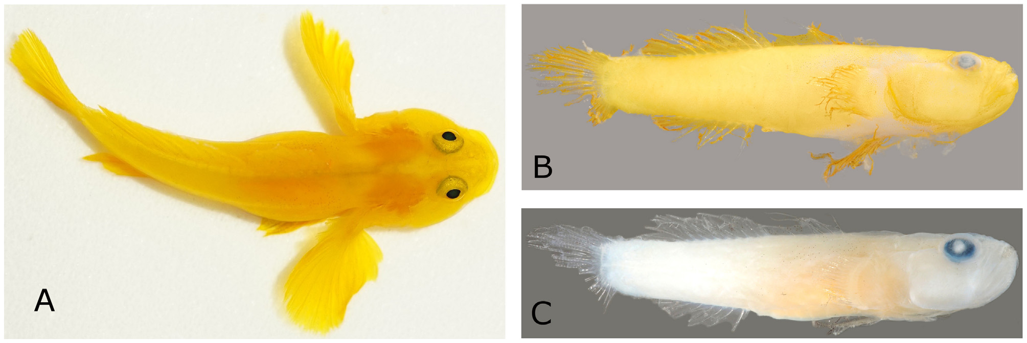
First dorsal fin with six spines. Second dorsal fin with one spine and nine soft rays. First and second dorsal fins connected by a low membrane behind last spine of the first dorsal fin. Anal fin with one spine and eight soft rays. Caudal fin rounded, with 17 segmented rays. Pectoral fin with 18 soft-rays. Pelvic fin with one spine and five soft rays. Posterior tips of pectoral fins reaching or exceeding position of anus, according to a photograph taken in life ( Fig. 1A View FIGURE 1 ), although they are broken in the preserved specimen. Pelvic fins without frenum. Vertebrae 10 + 16 = 26; dorsal-fin pterygiophore formula 3-22110; epural 1; anal-fin pterygiophores anterior to first haemal spine 2.
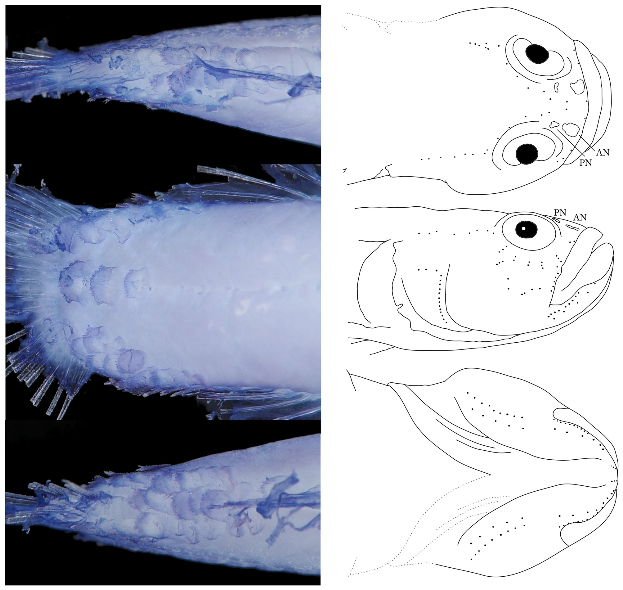
Head and body largely naked except for posterior part of caudal peduncle, involving three rows composed of 7–10 ctenoid scales along dorsal midline. A row of three ctenoid scales along lateral midline, and three rows composed of 6–8 ctenoid scales along ventral midline ( Fig. 3 View FIGURE 3 ). No sensory canals or associated pores on head. Cephalic sensory papillae patterns illustrated in Fig. 3 View FIGURE 3 . Infraorbital area with five transverse rows of sensory papillae.
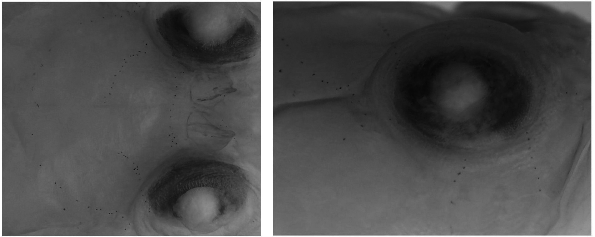
Color in preservative ( Fig. 1C View FIGURE 1 ): Background of head and tail white, trunk yellowish white. All fin membranes transparent. Melanophores scattered on dorsal half of trunk and on membranes of first and second dorsal, anal, and pelvic fins. Pectoral fin also with a few melanophores. Three rows of tiny melanophores arched between right and left eyes ( Fig. 4A View FIGURE 4 ). Infraorbital area with two transverse rows of tiny melanophores ( Fig. 4B View FIGURE 4 ) and another row of tiny melanophores behind eyes ( Fig. 4B View FIGURE 4 ). These melanophore rows arranged radially around eyes.
Color in life ( Fig. 1A View FIGURE 1 ): Body and all fins yellow or yellowish orange. Arrangement of melanophores same as after preservation, described above ( Fig. 1A View FIGURE 1 ).
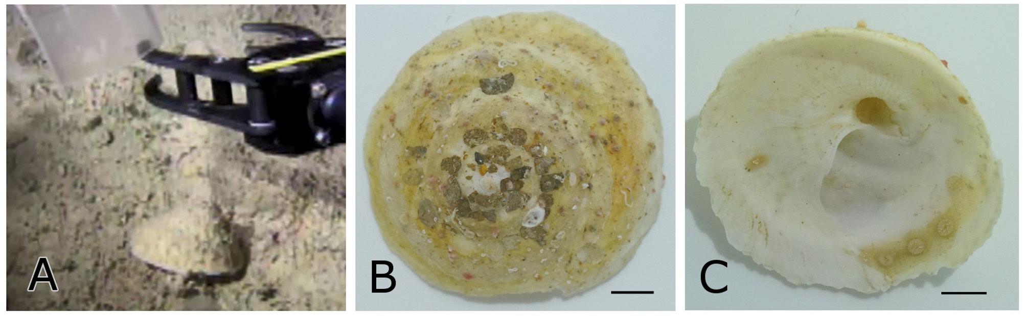
Habitat. We collected ten specimens of five invertebrate species from the muddy bottom at a depth of 209–220 m using the ROV, on 11 August 2017. These included five live comb jellies, Lyrocteis imperatoris , a sea cucumber, Holothuria dura , a starfish, Asterodiscides japonicus , a heart urchin, Pericosmus sp., and an empty shell of Xenophora chinensis ( Fig. 5 View FIGURE 5 ). After these animals were put into a tank on the boat, the L. pumilus specimen was found in the tank. Because Lubricogobius species, close relatives of Larsonella pumilus , often inhabit empty shells, sea urchin tests, tunicate siphons, bottles, etc. ( Randall & Senou 2001; Allen & Erdmann 2016), we believe that the L. pumilus was inside the empty shell of X. chinensis (collected at a depth of 214 m) and was collected with it.
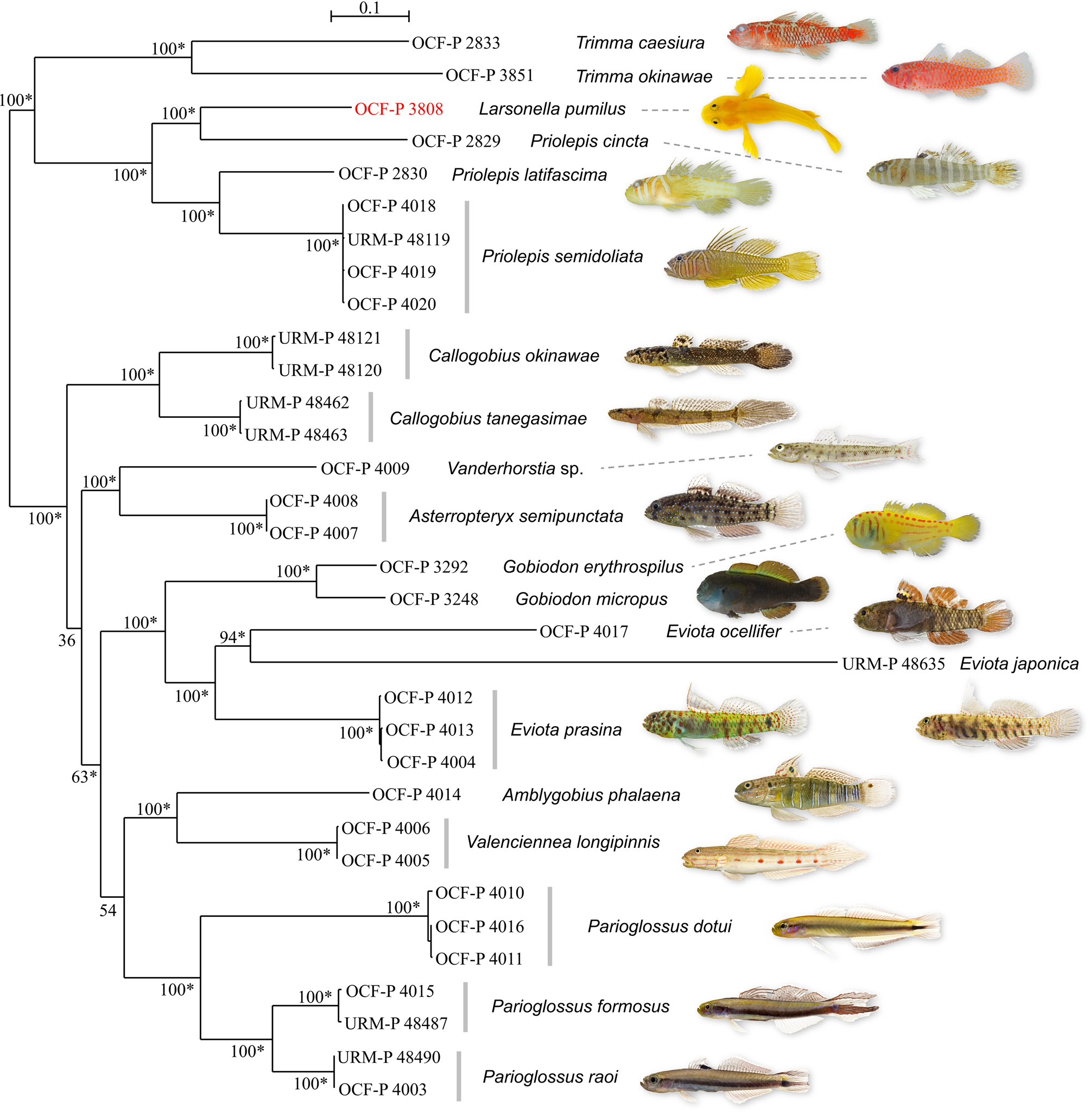
Mitochondrial DNA analysis. We succeeded in assembling the entire mitochondrial genomes of Larsonella pumilus and 19 related species ( Table 1 View TABLE 1 ). In the phylogenetic tree, using 15559 bp of aligned mitochondrial genomes ( Fig. 6 View FIGURE 6 ), most nodes, including L. pumilus , were supported by high bootstrap values (100%) and bayesian posterior probabilities (1), indicating that L. pumilus was placed in a clade including Priolepis spp. and Trimma spp., while Gobiodon spp. was placed in another clade with Callogobius spp., Vanderhorstia sp., Asterropteryx semipunctata , Eviota spp., Amblygobius phalaena , Valenciennea longipinnis , and Parioglossus spp. Larsonella pumilus was paired with Priolepis cincta , and they were placed within the Priolepis lineage.
No known copyright restrictions apply. See Agosti, D., Egloff, W., 2009. Taxonomic information exchange and copyright: the Plazi approach. BMC Research Notes 2009, 2:53 for further explanation.
|
Kingdom |
|
|
Phylum |
|
|
Class |
|
|
Order |
|
|
Family |
|
|
Genus |
Larsonella pumilus ( Larson & Hoese, 1980 )
| Hanahara, Nozomi, Higashiji, Takuo, Shinzato, Chuya, Koyanagi, Ryo & Maeda, Ken 2019 |
Lubricogobius pumilus
| Randall, J. E. & Senou, H. 2001: 11 |
| Larson, H. K. & Hoese, D. F. 1980: 41 |