
Dolichothrombium anatoliae, Mąkol, Joanna & Sevsay, Sevgi, 2011
|
publication ID |
https://doi.org/ 10.5281/zenodo.207445 |
|
DOI |
https://doi.org/10.5281/zenodo.5628237 |
|
persistent identifier |
https://treatment.plazi.org/id/03FF5D19-FFF6-FF97-2A95-72A7DB10FDE7 |
|
treatment provided by |
Plazi |
|
scientific name |
Dolichothrombium anatoliae |
| status |
sp. nov. |
Dolichothrombium anatoliae sp. nov.
Diagnosis. Adult. Anterior process of crista short, formed of two diverging, bulge-like sclerites, not clearly delimited in their anterior parts and not reaching aspidosoma margin. The sclerites encompass the area with two AM setae, located close to aspidosoma termination. Dorsal opisthosomal setae uniform. Setal stem almost parallelsided, with dagger-like or coronate termination, forms relatively wide, fin-like longitudinal ridges on four sides along its axis. Each ridge gradually passes into setulae; the longest setulae present in the basal part of stem. The most distally placed setulae reach the stem termination. Setal bases slightly narrowing apically, smooth, asymmetrically truncated at the top. Subcuticular mesh single-layered, formed of polygonal orifices separated by relatively thin network thread (similar to one present in Andinothrombium , for comparison see: Fig. 95 in Mąkol 2007). Tarsus I elongated, almost parallelsided. For other characters see the diagnosis of the genus.
Deutonymph. Odontus simple. Other characters as in adult (see also Discussion).
Larva. Scutum distinctly narrowing towards anterior margin. Anterior margin of scutellum concave. Index pedibus> 650.
The new species differs from other members of the genus known from active postlarval forms in the length and/or shape of the dorsal opisthosomal setae (for details see the identification key). As far as species known from larvae are concerned, D. anatoliae sp. nov. is most similar to D. telletxeae . It differs from the latter in the shape of scutellum (concave anterior margin in D. anatoliae ), the structure of posterior seta on palp tarsus (nude in D. anatoliae ) and also in the higher value of majority of metric data ( Tab. 2).
Description. Adult. Standard measurements given in Table 1. Colour in life bright red. Body distinctly elongated, densely covered with setae.
Character holotype female [?] DN [?] DN male IL 1952 1313 1156 1078 IW 854 598 510 568 IL /IW 2.3 2.2 2.3 1.9 ChB (L) 145 140 125 137 ChB (W) 45 42 42 47 ChCl (L) 35 30 30 35 PaTr (L) 37 35 32 25
PaTr (W) 42 37 37 35
PaFe (L) 125 125 102 120 PaFe (W) 67 72 62 67
PaGe (L) 42 45 40 40
PaGe (W) 42 47 42 50
PaTi (L) 42 40 40 40
PaTi (W) 30 32 30 30
Odo (L) 42 45 47 40
PaTa (L) 62 57 55 57 continued next page Character holotype female [?] DN [?] DN male ASB—distance between the anterior margin of crista metopica and the level of sensillae, ChB—cheliceral base, ChCl - cheliceral claw (measured along the inner edge of blade), CMW—width of crista metopica, IL—length of idiosoma, IW—width of idiosoma, L—length, Odo—odontus, PSB—distance between the level of sensillae and the posterior margin of crista metopica, W—width.
Character D. anatoliae a sp. nov. D. raphanicum b D. telletxeae c IL 325–370 (x = 347, n = 7) 245–574 240 IW 147–170 (x = 161, n = 8) 106–240 124 ChB (L) 45–51 (x = 48, n = 8) – – ChCl (L) 12–16 (x = 14, n = 8) – – PaFe (L) 24–28 (x = 26, n = 8) – – PaGe (L) 13–14 (x = 13, n = 8) – – PaTi (L) 10–12 (x = 10, n = 8) – – or 7–9 (x = 8, n = 8) 5–8 – bs 32–38 (x = 36, n = 8) 25–32 22 Odo (L) 19–24 (x = 22, n = 8) – – Scutum L 106–115 (x = 111, n = 8) 74–87 78 Scutum W 78–85 (x = 82, n = 8) 74–79 – AM 29–35 (x = 32, n = 8) – 27 AL 23–31 (x = 27, n = 8) 18–25 22 PL 45–50 (x = 47, n = 8) 32–40 34 AA 32–34 (x = 33, n = 8) 32–37 28 AW 52–56 (x = 54, n = 8) 50–57 50 PW 62–71 (x = 66, n = 8) 62–67 56 MA 30–33 (x = 31, n = 8) 24–32 26 AP 21–26 (x = 24, n = 8) 20–25 22 S 48–51 (x = 50, n = 8) 35–47 44 SB 36–39 (x = 37, n = 8) 34–37 32 ASB 70–75 (x = 72, n = 8) 39–57 50 PSB 35–40 (x = 38, n = 8) 25–35 28 HS 30–36 (x = 34, n = 8) 25–42 39 LSS 70–77 (x = 74, n = 8) 67–79 70 SL 45–48 (x = 47, n = 7) 35–42 38 SS 20–27 (x = 24, n = 8) 21–26 20 OL 19–23 (x = 21, n = 8) 19* 18 aO 9–11 (x = 10, n = 7) 8* 9
pO 7–8 (x = 8, n = 7) 9* 9
DS 31–46 (x = 40, n = 8) 27–42 (pDS = 49–62) – h1 66–70 (x = 69, n = 8) – – h2 67–75 (x = 71, n = 8) – – 1a 24–30 (x = 28, n = 7) 14–25 20 1 b 40–48 (x = 43, n = 8) 30– 37 24 2a 35–41 (x = 38, n = 8) 15–30 34 2 b 40–46 (x = 44, n = 8) 25–37 32 3a 30–35 (x = 33, n = 8) 35– 40 30 3b 39–43 (x = 41, n = 8) 22–35 32 VS 35–46 (x = 40, n = 8) – – Cx I 40 –48 (x = 45, n = 8) 32–42 34
continued next page aO—diameter of anterior eye lens, OL—length of ocular sclerite, pO—diameter of posterior eye lens, 1a—medial coxala I,
1b—lateral coxala I, 2a—medial coxala II, 2b—lateral coxala II, 3a—seta located between coxae III, 3b—coxala III.
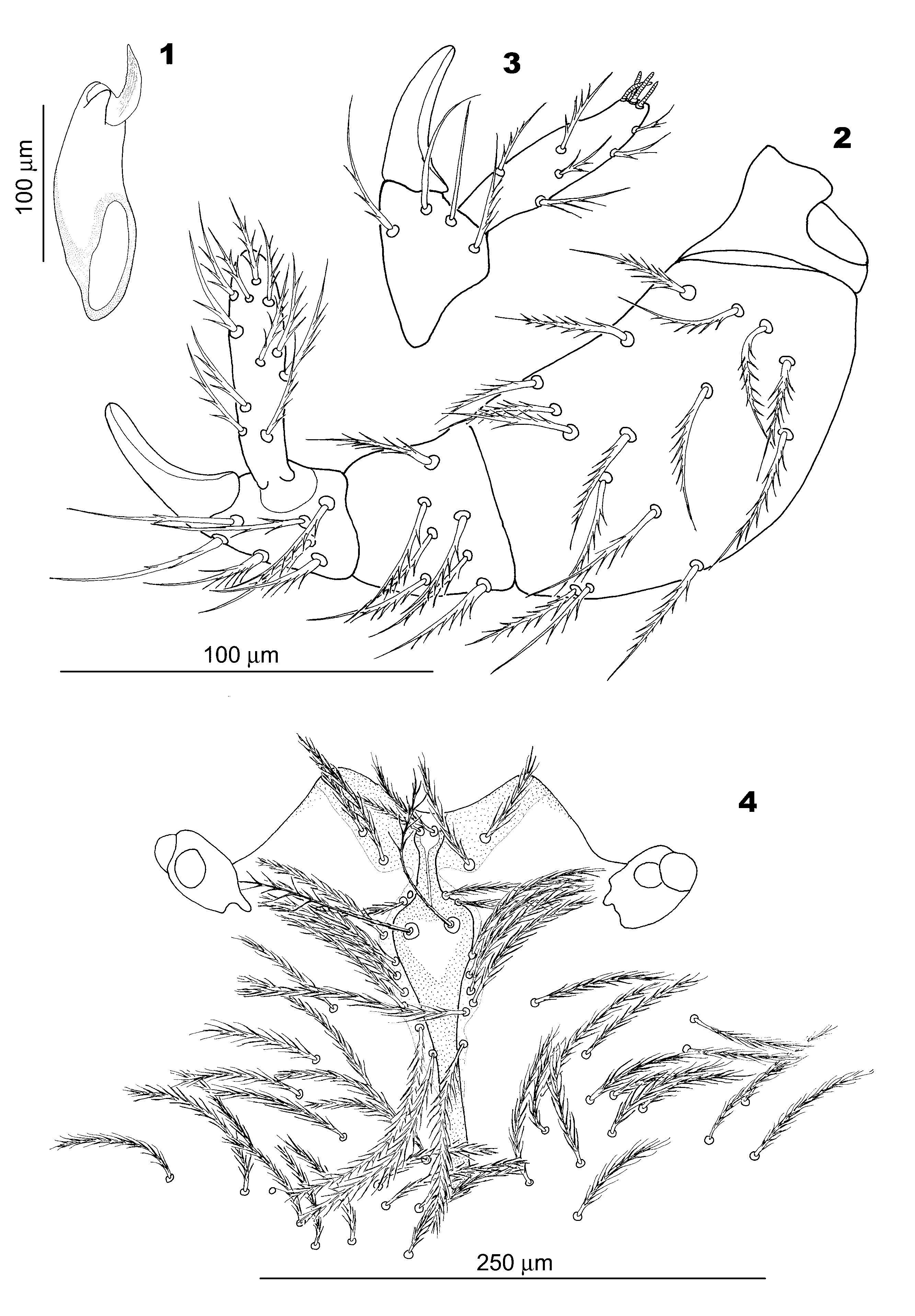
Gnathosoma. Chelicera ( Fig. 1 View FIGURES 1 – 4 ) composed of base, digitus mobilis and short cuticular fold, located at the base of digitus mobilis, in the position of lost digitus fixus. Inner edge of cheliceral blade serrated on almost entire length. Medial face of palp tibia ( Figs 2–3 View FIGURES 1 – 4 ) with c. 1–2 smooth, whip-like setae placed close to odontus base and similar in length to other, setulose setae. On lateral face of palp tibia, in distal position, c. three setae covered with few setulae. The remaining setae on palps setulose. Odontus simple in females. Palp tarsus ( Figs 2–3 View FIGURES 1 – 4 ) elongated, set close to odontus base and extending beyond the termination of palp tibial claw. Circa four solenidia present at the tip of the segment.
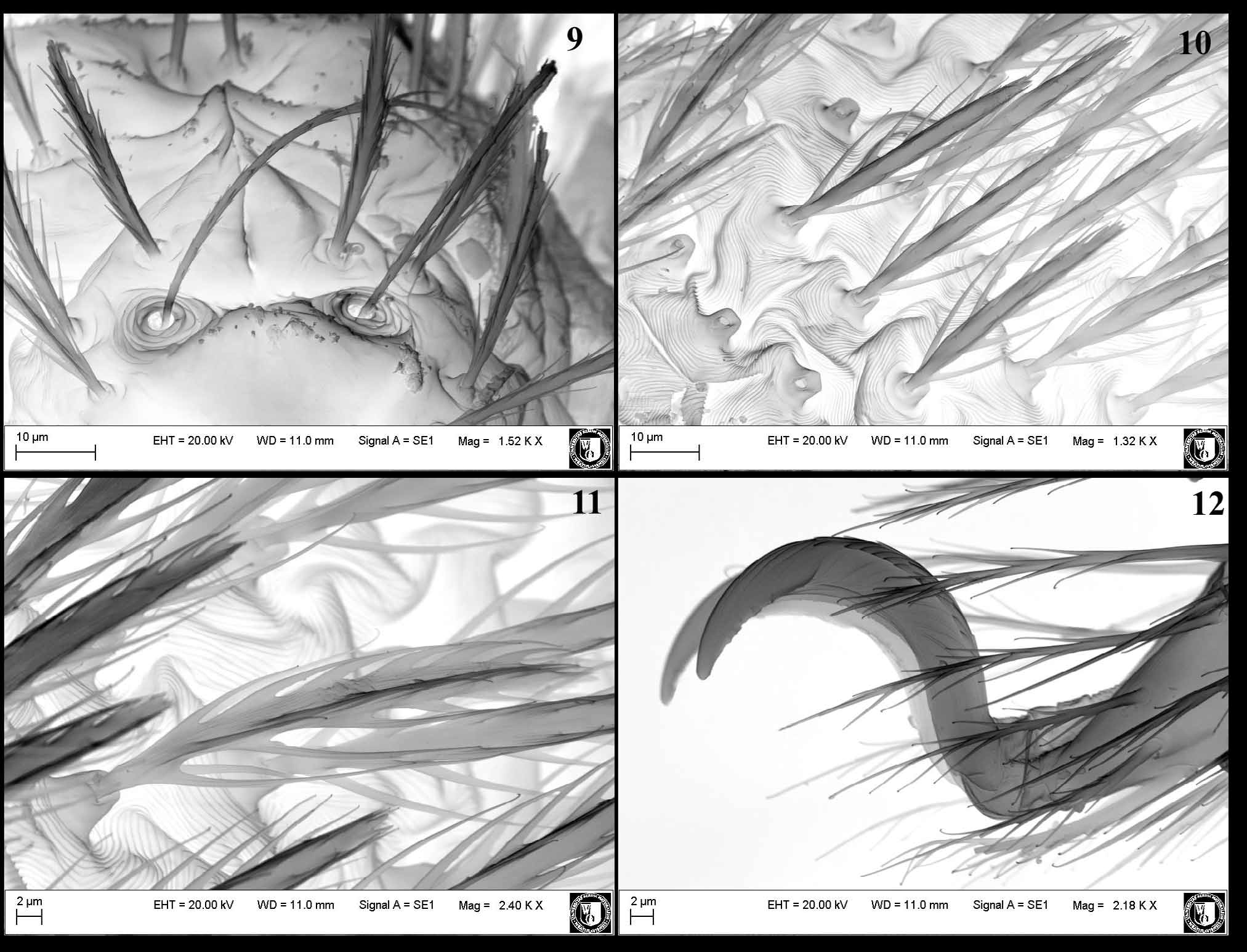
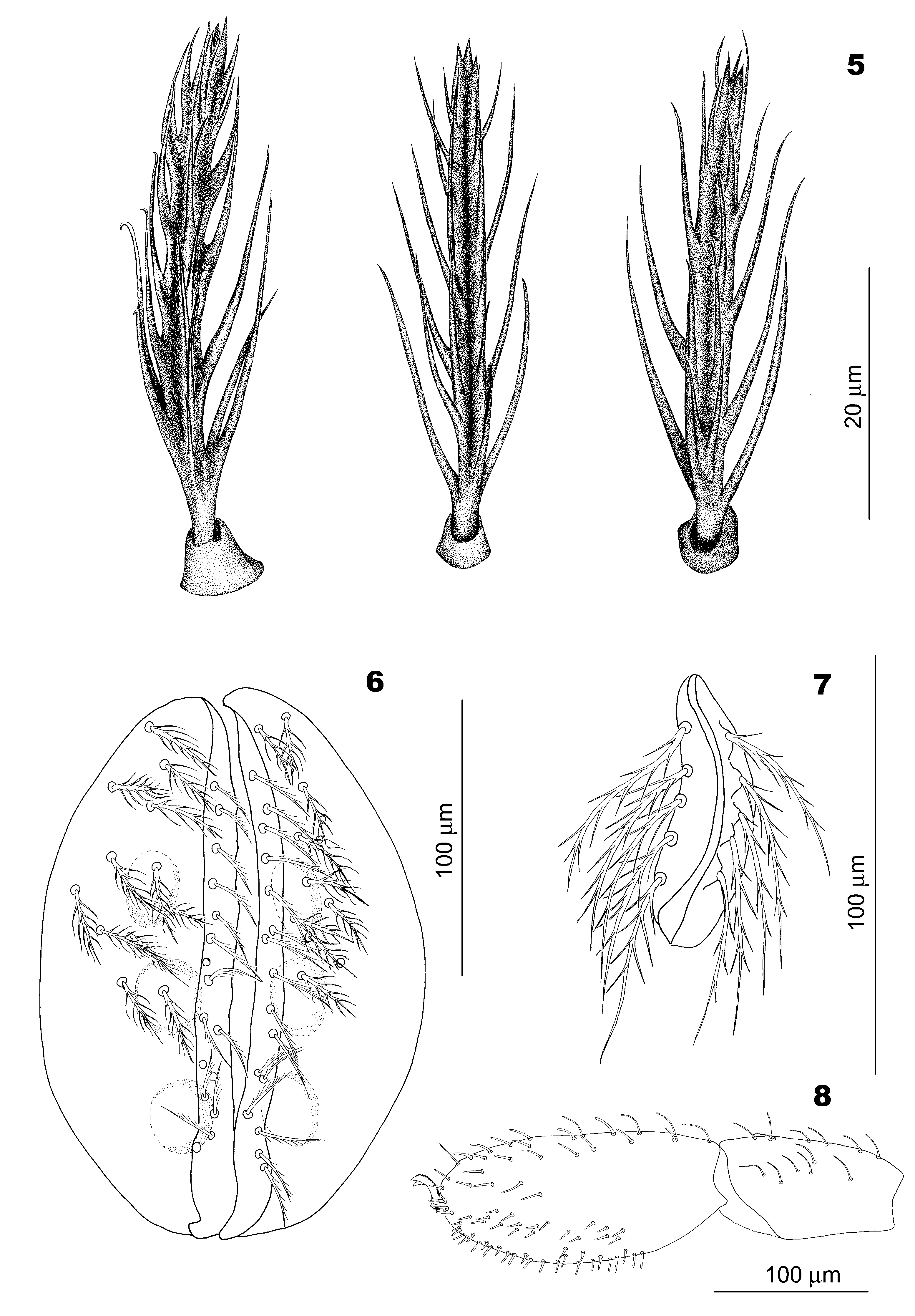
Idiosoma, dorsum. Anterior margin of aspidosoma ( Fig. 4 View FIGURES 1 – 4 ) concave. Sclerotized area, extended laterally, adjoins the anterior margin of aspidosoma. Crista metopica composed of anterior process, sensillary area and posterior process. Anterior process of crista not clearly terminated in its anterior part, does not reach the anterior margin of aspidosoma. The process formed of two diverging, bulge-like sclerites, which encompass the area with two elongated AM setae. Sensillary area ( Figs 4 View FIGURES 1 – 4 , 9 View FIGURES 9 – 12 ) of crista with rounded sides, the widest at the level of sensillae. Sensillary setae covered with tiny barbs in the proximal, and with more distinct setulae in the distal part of stem. Posterior process of crista only slightly narrowing towards the end, rounded terminally. Both sensillary area and posterior process of crista surrounded by weakly sclerotized scutum. Eyes, each composed of a double lens, placed on slightly narrowed at base, robust peduncles and situated at the level of sensillary area of crista. Anterior lens larger than posterior one. Setae at anterior part of aspidosoma narrowing apically, covered with relatively short setulae, then pass into ones with fin-like ridges along the stem, covered with more elongated setulae. Dorsal opisthosomal setae ( Figs 5 View FIGURES 5 – 8 , 10–11 View FIGURES 9 – 12 ) uniform. Setal bases distinctly elevated above idiosoma surface, slightly narrowing apically, smooth, asymmetrically truncated at the top; setal stems, directed towards opisthosoma termination, rest against the longer part of setal base. Setal stems with relatively wide, fin-like, longitudinal ridges on four sides along their axes. Stem termination dagger-like or coronate, formed of several (up to 4) tubercles. Fin-like, longitudinal ridges gradually pass into long setulae. Setulae present at the basal part of stem the longest, with terminations reaching half length of stem. The most distally placed setulae reach the stem termination.
Idiosoma, venter. Ventral setae similar to those covering dorsal side of opisthosoma, but more slender. Genital sclerites of female ( Fig. 6 View FIGURES 5 – 8 ) and male comparable in proportion. Epivalves similar in length to centrovalves; both covered with setae. Setae on epivalves with thicker stem than on centrovalves and with more outstanding setulae. Centrovalval setae more slender, covered with more adhering setulae along the stem. Three pairs of genital papillae. Anus ( Fig. 7 View FIGURES 5 – 8 ) surrounded by membraneous valves. Each valve covered with c. 5 setulose setae.
Legs. Legs relatively short. Legs I longer than IV, legs II, III shorter. Normal setae covering trochanter – tarsus of all legs setulose. Specialized setae present on genua, tibiae and tarsi ( Fig. 8 View FIGURES 5 – 8 ) (Ge I: 9–10, Ti I: 13–20, Ta I: c. 75, Ge II: 2–4, Ti II: 5–7, Ta II: 4–5, Ge III: 4–5, Ti III: 4–6, Ge IV: 4–5, Ti IV: 6–10; specialized setae relatively slender, curved, except for the setae placed along the ventral part of tarsus I, the most distally placed seta on tibia III and IV and the stout solenidion located distally on tarsus II). Tarsus I elongated, oval, in both sexes longer than tibia I. All tarsi terminated with paired claws. The dorsal side of claws with row of fimbriae ( Figs 8 View FIGURES 5 – 8 , 12 View FIGURES 9 – 12 ). Pseudopulvillus absent.
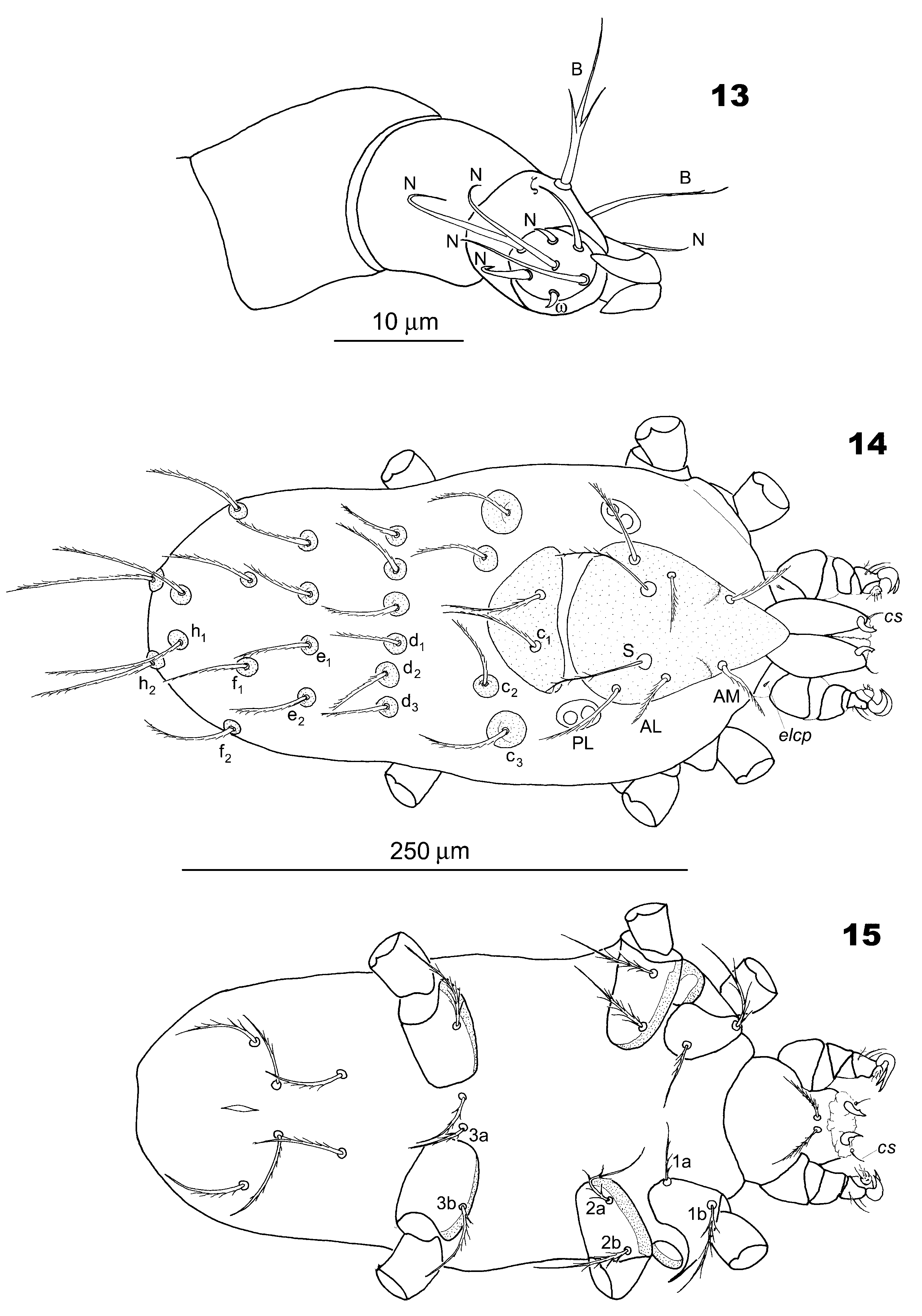
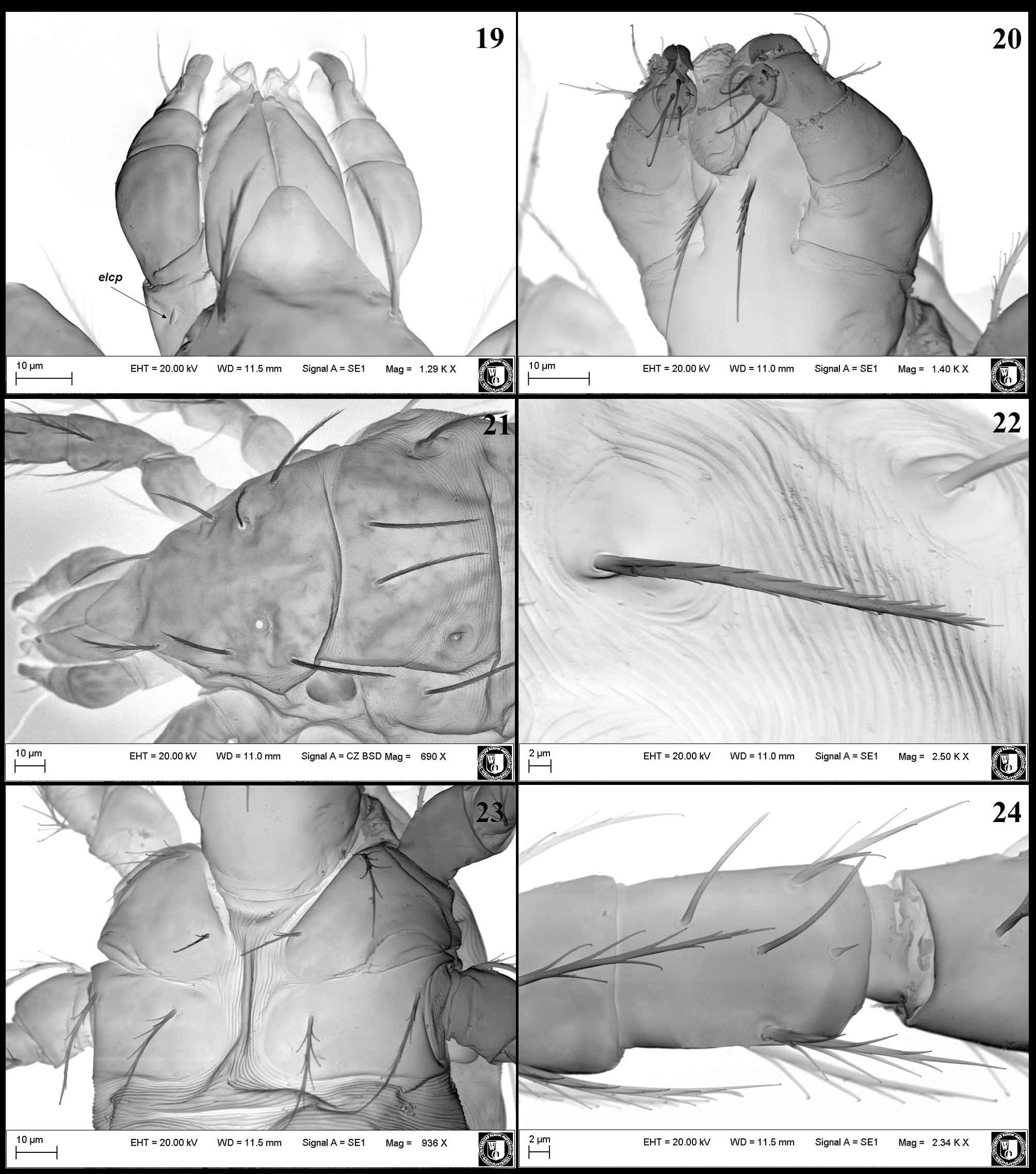
Larva. Gnathosoma. Cheliceral claw ( Figs 14–15 View FIGURES 13 – 15 ) relatively short and curved, with one denticle close to the tip. Adoral seta (cs) ( Figs 14–15 View FIGURES 13 – 15 , 19 View FIGURES 19 – 24 ) spine-like. Dorsally on gnathosoma base a minute (3–4), spine-like supracoxal seta (elcp) ( Figs 14 View FIGURES 13 – 15 , 19 View FIGURES 19 – 24 ), c. three times shorter than adoral seta. At the base of palp femur a collar-like cuticular fold ( Fig. 19 View FIGURES 19 – 24 ), possibly being the vestigial palp trochanter. Palp femur and palp genu ( Figs 13 View FIGURES 13 – 15 , 19 View FIGURES 19 – 24 ) without setae. Palp tibia ( Figs 13 View FIGURES 13 – 15 , 20 View FIGURES 19 – 24 ) with three setae. Posterior seta on palp tibia setulose. Seta placed at c. half length of palp tibia barbed. The most distally placed seta shorter than the preceding setae and nude. Odontus ( Figs 13 View FIGURES 13 – 15 , 19–20 View FIGURES 19 – 24 ) curved, bifid. Palp tarsus ( Fig. 13 View FIGURES 13 – 15 ) with seven setae, including normal setae, eupathidium and [?] solenidion. ƒP p formula: 0-0-0-BBN2-NNNNNζ[?]ω (the actual character of setae on palp tarsus, due to their minute size, difficult to ascertain). All setae on palp tarsus nude. Hypostomalae ( Figs 15 View FIGURES 13 – 15 , 20 View FIGURES 19 – 24 ) elongated, narrowing apically, covered with setulae.
Idiosoma, dorsum. Cuticle covering idiosoma, except for sclerites, folded in lines ( Fig. 22 View FIGURES 19 – 24 ). Scutum and scutellum ( Figs 14 View FIGURES 13 – 15 , 21 View FIGURES 19 – 24 ) smooth. Scutum triangular in outline, distinctly narrowing towards anterior margin. Posterior margin of scutum convex. Setae AM, AL and PL with setulae distributed along the stem. Sensillary setae (S) of scutum located medially between AL and PL, covered with distinct setulae in distal part of stem. Scutellum semicircular in outline, with rounded corners, similar in width to scutum. Anterior margin of scutellum concave. One pair of normal setae (c1) located in anterior half of the sclerite. Eyes, each consisting of a double lens placed on oval sclerite, situated laterally, at the level of scutum/scutellum border. Anterior lens larger and more elevated than the posterior one. ƒD formula: 6 (c1-c3) - 6 (d1-d3) - 4 (e1-e2) - 4 (f1-f2) - 2 (h1-h2). Dorsal setae ( Figs 14 View FIGURES 13 – 15 , 22 View FIGURES 19 – 24 ), except for h1 and h2, similar in length, covered with husk-like setulae and placed on round platelets; c3 plates enlarged. Setae h1 and h2 longer than the remaining dorsal setae.
Idiosoma, venter ( Figs 15 View FIGURES 13 – 15 , 23 View FIGURES 19 – 24 ). A pair of Claparède`s organs located laterally, between coxae I and II. ƒCx formula: BB-BB-B. Medial coxala I (1a) with few barbs, mostly in proximal part of the stem. Lateral coxala I (1b), coxalae II (2a, 2b) and coxala III (3b) with distinct setulae. Setae 3a located between coxal plates III. ƒV formula: 2-2u-2. Ventral setae ( Fig. 15 View FIGURES 13 – 15 ) somewhat thinner than dorsal setae, also covered with setulae on entire length. Anus surrounded by membraneous valves (anal sclerite absent).
Legs ( Figs 16–18 View FIGURES 16 – 18 , 24–28 View FIGURES 19 – 24 View FIGURES 25 – 28 ). Leg segmentation formula 6-6-6. Chaetotaxy of legs: [I] Tr (1n) – Fe (5n) – Ge (4n, 2σ, 1ĸ) – Ti (5n, 2φ, 1ĸ) – Ta (15n, 2ζ, 1ω, 1ε); [II] Tr (1n) – Fe (5n) – Ge (3n, 1σ, 1ĸ) – Ti (5n, 2φ) – Ta (13n, 1ω, 1ε); [III] Tr (1n) – Fe (4n) – Ge (3n, 1σ) – Ti (5n) – Ta (12n). Solenidion (ω) on tarsus I stout, situated at c. half length of segment. All tarsi terminated with two claws and claw-like empodium. Inner (posterior) claw on tarsus III normally developed.
Etymology. The specific epithet refers to the name Anatolia, the geographic and historical area where the new species was collected.
Type material. Holotype ( ZMH Acc. No. A16/11) female (coll. Turkey, Caglayan near Erzincan, N39°35'11.57'', E39°41'51.03'', 1266 m a.s.l., humid meadow with copses, close to the stream, 29.10.2010, leg. S. Sevsay) and paratype ( ZMH Acc. No. A17/11) larva, obtained from holotype female by experimental rearing, deposited in the Zoological Museum in Hamburg, Germany ( ZMH). Other specimens—in author’s collection (Department of Invertebrate Systematics and Ecology, Wrocław University of Environmental and Life Sciences, Poland and the Department of Biology, Science and Literature Faculty, Erzincan University, Turkey).
Distribution. Turkey.
Biology. Oviposition took place under laboratory conditions, at the beginning of November, four days after collecting the females in the field. The eggs were laid in clutches. The prelarvae appeared 20 days after oviposition, and the larvae after the next 11 days. Altogether, four females oviposited and 20– 24 larvae were obtained from each. The total time between oviposition and emergence of larvae was 35 days.

| ZMH |
Zoologisches Museum Hamburg |
No known copyright restrictions apply. See Agosti, D., Egloff, W., 2009. Taxonomic information exchange and copyright: the Plazi approach. BMC Research Notes 2009, 2:53 for further explanation.
|
Kingdom |
|
|
Phylum |
|
|
Class |
|
|
Order |
|
|
Family |
|
|
Genus |