
Echiniscus clavispinosus, Fontoura, Paulo, Pilato, Giovanni & Lisi, Oscar, 2011
|
publication ID |
https://doi.org/10.5281/zenodo.204752 |
|
DOI |
https://doi.org/10.5281/zenodo.5677065 |
|
persistent identifier |
https://treatment.plazi.org/id/051C87C3-B611-FFB5-FF48-2A21FDDCFC7F |
|
treatment provided by |
Plazi |
|
scientific name |
Echiniscus clavispinosus |
| status |
sp. nov. |
Echiniscus clavispinosus sp. nov.
( Figs. 1 View FIGURE 1 , 2 View FIGURE 2 ; Table 1 View TABLE 1 )
Locus typicus. Xôxô, Ribeira da Torre, Municipality of Ribeira Grande, Santo Antão Island, Archipelago of Cape Verde, in a lichen sample growing on rocks.
Material examined. Thirteen specimens, of which four were two-clawed larvae.
Type repository. The holotype (slide N. 5432) and one two-clawed larva (slide N. 5433) are deposited in the collection of Binda & Pilato (Museum of the Department of Animal Biology “Marcello La Greca”, University of Catania, Italy). The other paratypes (slides CIV88 - CIV96) are deposited in the collection of P. Fontoura at the Department of Biology, Faculty of Sciences, University of Porto, Portugal.
Specific diagnosis. Colour green; eye spots absent; dorsal plates well marked. Median plate 3 absent; cuticular ornamentation consisting of slightly raised dark tubercle and fine dots. At a higher focal position very dense, fine dots and small light spots appear. Distance between the dark tubercles shorter than the diameter of the tubercles. Anterior portion of median plates 1 and 2 and area between paired plate III and terminal plate unsculptured. Buccal cirri and cirrus A short, elongated clavae with pointed apices; no other lateral or dorsal appendages present. Spine present on the first pair of legs; papilla and dentate collar on the hind legs; claws well developed; internal claws with well developed spur.
Description of the holotype. Body length 220 µm; plate colour green in specimens observed with transmitted light, after mounting on a slide, but reddish brown when observed with reflected light; eye spots not observed in either living nor slide mounted specimens. A narrow finely dotted transverse band was present anterior to the cephalic plate ( Fig. 1 View FIGURE 1 A, D); this plate with an anterior notch; a true neck plate was absent but there was a thickened and faint ornamented transverse area between the cephalic plate and the scapular plate ( Fig. 1 View FIGURE 1 A, D); the scapular plate, 43.6 µm long in the medium line, was subdivided into a wide central portion and two small lateral, almost triangular, portions ( Fig. 1 View FIGURE 1 A, D). Median plate 2 with unsculptured anterior portion ( Fig. 1 View FIGURE 1 B); median plate 3 absent and the area between paired plate III and terminal plate unsculptured ( Fig. 1 View FIGURE 1 B, C); terminal plate unfaceted and with two indentations ( Fig. 1 View FIGURE 1 B). Plate sculpture of the adults comprised of slightly raised dark tubercles and very fine, dense dots ( Fig. 1 View FIGURE 1 A–D); at a higher focal position small light spots appear ( Fig. 1 View FIGURE 1 D). Distance between the dark tubercles shorter than the diameter of the tubercles; largest tubercles (diameter c. 2.3 µm) are present in the central portion of the scapular plate. Cephalic plate sculptured with only fine dots ( Fig. 1 View FIGURE 1 D). In the anterior portion of the paired plates dark tubercles ( Fig. 1 View FIGURE 1 B, C) were present (and a transverse area with almost invisible dots). Leg plates sculptured with only fine dots. Ventral body surface had an extremely fine granulation more visible in the cephalic region. Small spine (2.4 µm long) was present on the first pair of legs ( Fig. 2 View FIGURE 2 A); small papilla 3.3 µm long and dentate collar with about 10 unequal, sharp teeth (the longest c. 2.2 µm long) were present in the fourth pair of legs ( Fig. 2 View FIGURE 2 C).
Internal buccal cirri 9.6 µm long; external cephalic cirri slightly longer (11.9 µm); cephalic papillae 6.8 µm long ( Fig. 2 View FIGURE 2 A); clavae, 7.2 µm long, had a distinctive sharp-tipped, conical shape rather than the rounded apices ( Fig. 2 View FIGURE 2 B). Cirri A, a short filament 26.0 µm long ( Fig. 2 View FIGURE 2 A, B); no other trunk appendages present.
Claws well developed ( Fig. 2 View FIGURE 2 A, C); internal and external claws on the first pair of legs 13.3 and 11.8 µm long respectively; they were slightly longer on the fourth pair of legs (14.3 µm and 13.3 µm respectively). Internal claws with a well developed spur directed downwards ( Fig. 2 View FIGURE 2 C).
Eggs unknown.
Variability. The paratypes (body length 133–241 µm) were similar to the holotype in both qualitative and metric characters ( Table 1 View TABLE 1 ). However, we would like to emphasise the curiosity found in two of the paratypes. One had a deeply forked buccal cirrus, and the other a deeply forked cirrusi A. The cause of this anomaly was not found.
We consider it important to stress the differences between the two-clawed larvae (body lengths c. 142–144 µm) and the adults. In the larvae the cuticular ornamentation lacked the dark tubercles and was instead formed by small, dense dots, sometimes joined together, and by sparse light spots ( Fig. 2 View FIGURE 2 D); these light spots, more evident in the posterior margins of the scapular, median and paired plates, were larger than those observed on the adults. We were sure that the two-clawed larvae belong to Echiniscus clavispinosus sp. nov. due to the distinctive (pointed) clavae shape and because they have the smooth unornamented anterior portion of the median plate 2 and the unornamented area between the paired plates III and the terminal plate. The differences of the cuticular ornamentation seemed to be in relation to the developmental stage and not body length because the four two-clawed larvae were slightly larger (142–144 µm long) than a four-clawed paratype (133 µm) which had typical adult cuticular ornamentation.
Characters Smallest specimen Holotype Largest specimen
Etymology. The name clavispinosus refers to the pointed apex shape of the clava.
Differential diagnosis. Echiniscus clavispinosus sp. nov. belongs to the viridis group, a small group of species within the genus Echiniscus characterised by the green colour, by plate ornamentation comprised of dark tubercles, fine dots and light spots, by the absence of dorsal and lateral trunk appendages (cirrus A excluded), and well developed claws. Traditionally, on the basis of the colour, five species were attributed to this group: E. viridis , E. rufoviridis , E.viridissimus , E. perviridis and E. viridianus . However, two of these species, E. rufoviridis and E. viridissimus , have distinctly different cuticular ornamentation ( Fig. 3 View FIGURE 3 ) (Pilato et al., 2007; 2008a) and should probably be excluded from the group. Nevertheless, to avoid misunderstandings and to make the species diagnosis easier, we incorporated all the green coloured Echiniscus species lacking dorsal and lateral trunk appendages (including E. viridissimus and E. rufoviridis ) into the key below.
Echiniscus clavispinosus sp. nov. differed from the other three true ‘ viridis group’ species by the unsculptured area between paired plate III and terminal plate; pointed clavae, instead of with rounded apices; and a stronger spur on internal claws. Differences among these species regarding quantitative characters are illustrated in Table 2. It is important to stress that the values relative to the external buccal cirrus, clava and spine on the first pair of legs of E. viridis and the values relative to the leg IV claws of E. viridianus are different from those indicated by Pilato et al. (2008a; tables 1 and 2) in which they, for a misunderstanding, are wrong.
The new species differed from E. viridis in the absence of dark tubercles on the cephalic and neck plates; the smooth anterior portion of the median plate 2; the distance between the dark tubercles much shorter than the diameter of the tubercles ( Fig. 3 View FIGURE 3 , A and B); more visible fine dots and light spots; and slightly longer cirrus A ( Table 2).
Echiniscus clavispinosus sp. nov. also differed from E. perviridis in lacking tubercles in the cephalic and neck plates, the smooth anterior portion of median plate 2; finer and denser dots and light spots on the plates ( Fig. 3 View FIGURE 3 , A and D), less visible green area bordering the trunk plates; dentate collar with smaller teeth; and much shorter cirrus A and shorter claws ( Table 2).
The new species differed from E. viridianus , the most similar species, by the absence of dark tubercles on the cephalic and neck plates; less visible and more dense fine dots and light spots on the plates ( Fig. 3 View FIGURE 3 , A and C); smooth anterior portion of the median plate 2; less visible green area bordering the trunk plates.
Characters E. clavispinosus sp. nov. E. viridis * E. perviridis E. viridianus *
µm % bo % sc µm % bo % sc µm % bo % sc µm % bo % sc
Body length 241 - - 190 290 231
Scapular plate length 48.8 20.2 - 41.8 22.0 56.5 19.5 43.1 18.7
Internal buccal cirrus 12.8 5.3 26.2 9.0 4.7 21.5 - - - 13.9 6.0 32.3 External buccal cirrus 15.8 6.6 32.4 10.3 5.4 24.6 - - - 14.7 6.4 34.1 Cephalic papilla 7.3 3.0 15.0 6.4 3.4 15.3 6.0 2.1 10.6 5.8 2.5 13.5 Clava 8.1 3.4 16.6 6.0 3.2 14.4 - - - 5.5 2.4 12.8 Cirrus A 32.4 13.4 66.4 18.2 9.6 43.5 114.0 39.3 201.8 29.0 12.6 67.3 Spine on leg I 3.4 1.4 7.0 3.3 1.7 7.9 - - - 2.6 1.1 6.0 Internal claw leg I 15.4 6.4 31.6 12.6 6.6 30.1 - - - 15.1 6.5 35.0 External claw leg I 14.4 6.0 29.5 11.9 6.3 28.5 21.4 7.4 37.9 14.5 6.3 33.6 Internal claw leg IV 17.6 7.3 36.1 15.5 8.2 37.1 30.0 10.3 53.1 16.9 7.3 39.2 External claw leg IV 15.5 6.4 31.8 14.7 7.7 35.2 28.5 9.8 50.4 15.2 6.6 35.3 Papilla leg IV - - - 3.0 1.6 7.2 - - - 4.8 2.1 11.1
* As above mentioned in the text, the %bo and %sc values relative to the external buccal cirrus, clava and spine on the first pair of legs of E. viridis and the values regarding the leg IV claws of E. viridianus differ from those indicated by Pilato et al. (2008a, tables 1 and 2), which were incorrect.
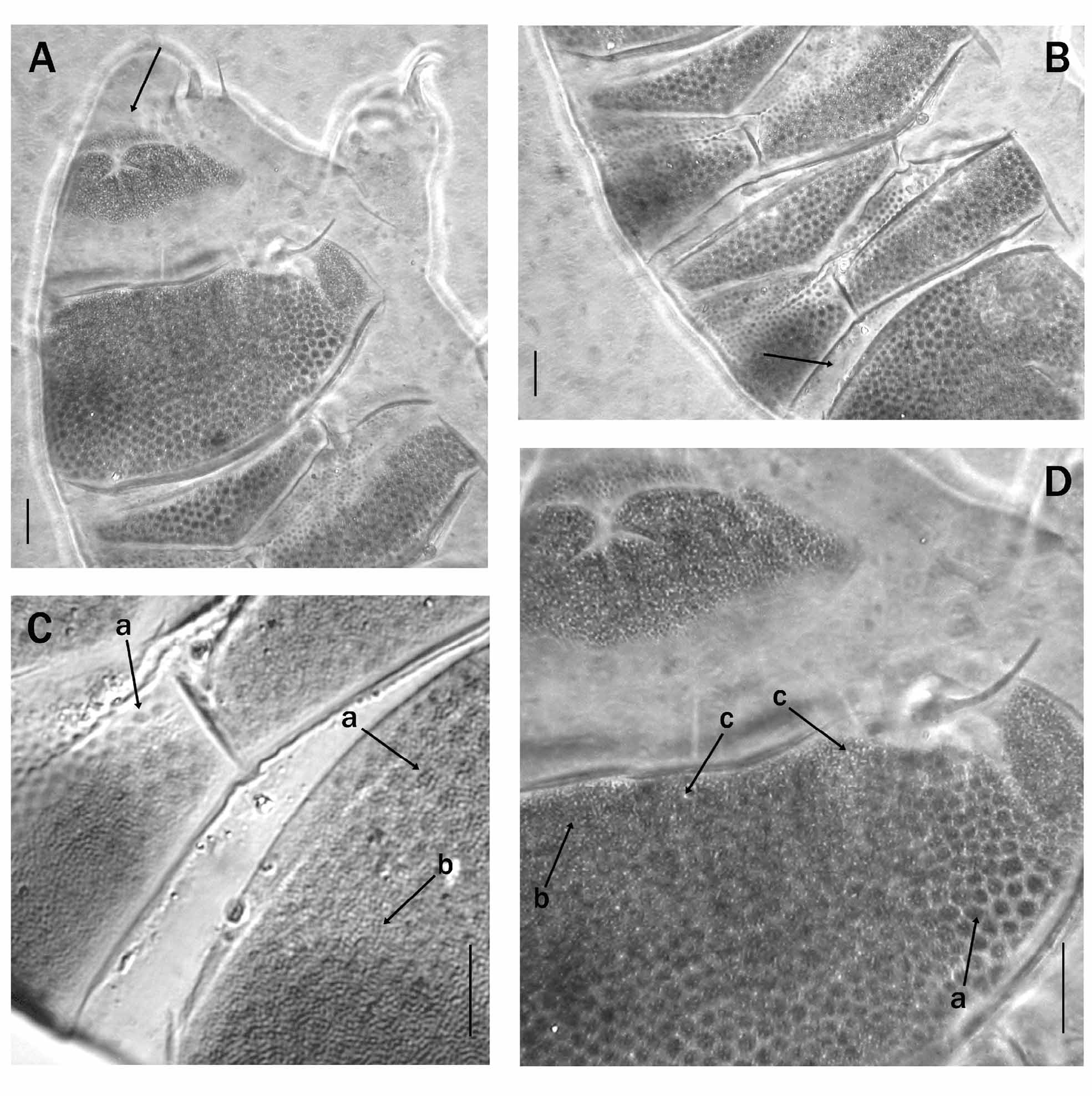


TABLE 1. Measurements of the holotype, the smallest and the largest specimens of Echiniscus clavispinosus sp. nov. (% bo = percentage of body length; % sc = percentage of scapular plate length).
| Body length Scapular plate length | µm 133 26.6 | % bo - 20.0 | % sc - - | µm 220 43.6 | % bo % sc - - 19.8 - | µm 241 48.8 | % bo - 20.2 | % sc - - |
|---|---|---|---|---|---|---|---|---|
| Internal buccal cirrus External buccal cirrus Cephalic papilla Clava | 7.1 8.6 4.5 4.8 | 5.3 6.5 3.4 3.6 | 26.7 32.3 16.9 18.1 | 9.6 11.9 6.8 7.2 | 4.4 22.0 5.4 27.3 3.1 15.6 3.3 16.5 | 12.8 15.8 7.3 8.1 | 5.3 6.6 3.0 3.4 | 26.2 32.4 15.0 16.6 |
| Cirrus A Spine on leg I | 14.5? | 10.9 - | 54.5 - | 26.0 2.4 | 11.8 59.6 1.1 5.5 | 32.4 3.4 | 13.4 1.4 | 66.4 7.0 |
| Internal claw leg I External claw leg I Internal claw leg IV External claw leg IV Papilla leg IV | 9.2 8.1 9.3 8.1? | 6.9 6.1 7.0 6.1 - | 34.6 30.5 35.0 30.5 - | 13.3 11.8 14.3 13.3 3.3 | 6.1 30.5 5.4 27.1 6.5 32.8 6.1 30.5 1.5 7.6 | 15.4 14.4 17.6 15.5? | 6.4 6.0 7.3 6.4 - | 31.6 29.5 36.1 31.8 - |
No known copyright restrictions apply. See Agosti, D., Egloff, W., 2009. Taxonomic information exchange and copyright: the Plazi approach. BMC Research Notes 2009, 2:53 for further explanation.
|
Kingdom |
|
|
Phylum |
|
|
Class |
|
|
Order |
|
|
Family |
|
|
Genus |