
Othelosoma impensum Sluys & Neumann
|
publication ID |
https://doi.org/ 10.5281/zenodo.250240 |
|
publication LSID |
lsid:zoobank.org:pub:B0B97269-AE03-4A41-811A-5F135988055E |
|
DOI |
https://doi.org/10.5281/zenodo.6022396 |
|
persistent identifier |
https://treatment.plazi.org/id/06116D51-FFC5-8440-FF02-FC7AFE84FF45 |
|
treatment provided by |
Plazi |
|
scientific name |
Othelosoma impensum Sluys & Neumann |
| status |
sp. nov. |
Othelosoma impensum Sluys & Neumann , sp. nov.
Material examined. Holotype: ZMA V.Pl. 7250.1, São Tomé Island, Nova Moka , Monte Café, Mé Zóchi district, 30 October 2014, coll. Leonel Viegas, sagittal sections of the anterior part on 13 slides; transverse sections of the prepharyngeal part on 6 slides; sagittal sections of the pharynx region on 8 slides; sagittal sections of the copulatory apparatus on 11 slides; sagittal sections of the tail on 10 slides.
Other material: ZMA V.Pl. 7251.1, São Tomé Island, Nova Moka , Monte Café, Mé Zóchi district, 9 November 2014, coll. Leonel Viegas, sagittal sections of the anterior part of the specimen on 10 slides; horizontal sections of the pharynx portion on 7 slides; sagittal sections of the copulatory apparatus on 13 slides; horizontal sections of the tail end on 4 slides.
Etymology. The specific epithet is derived from the Latin adjective impensus, great, ample or strong, and alludes to strong muscular sphincter on the vaginal duct.
Diagnosis. Dark green Othelosoma species with two dirty-white or light greenish dorsal bands separated by a narrow dark green (or dull black) stripe. Anterior tip orange. Ventral surface white or dirty white and light greenish. Ventral testes, extending posteriorly under the anterior end of the seminal vesicle. Dorsal retractor extending from the anterior margin to between one-fourth and one-fifth of the total body length. Non-constricted, elongated seminal vesicle, running parallel to the body surface. Short penis papilla with a dorsally shifted ejaculatory duct. Copulatory bursa receives the single opening of the vaginal duct, the latter receiving the opening of the bursal canal near its communication with the bursa or slightly ventrally to the copulatory bursa. Large, bean-shaped muscular sphincter around the vaginal duct.
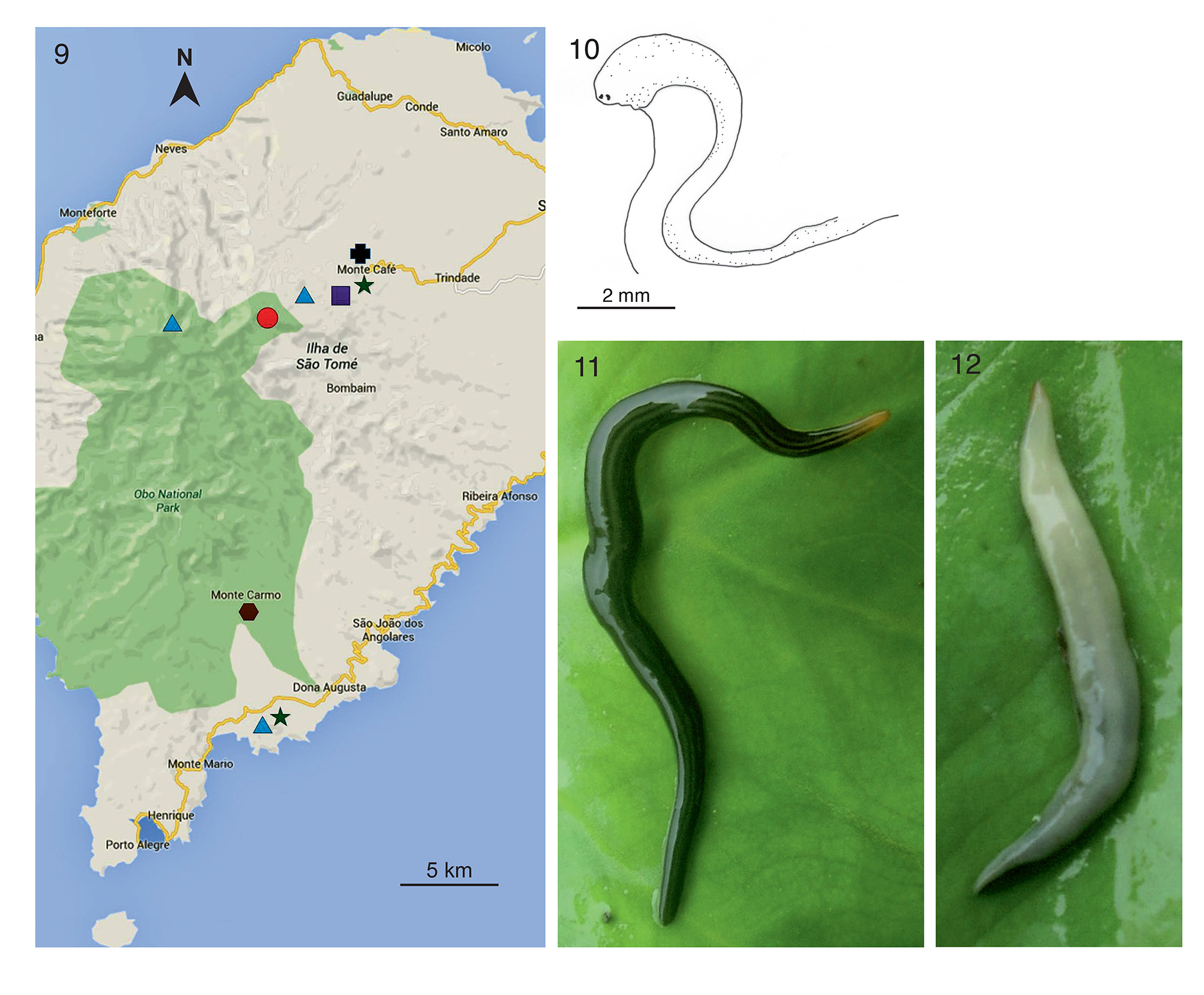
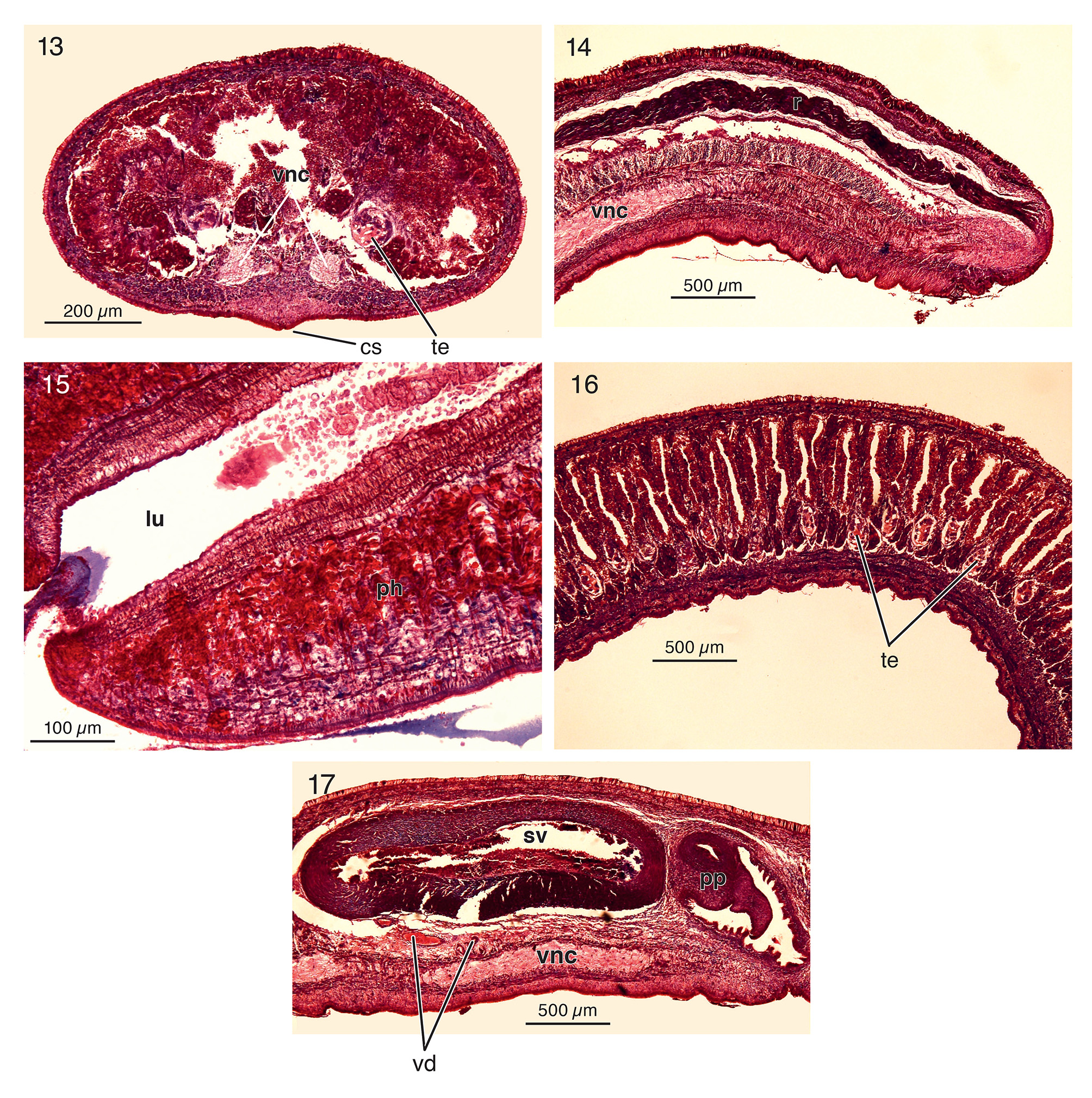
Description. Dorsal surface pale yellow (ZMA V. Pl. 7251.1; Fig. 10 View FIGURES 9 – 12 ), or very dark green (ZMA V.Pl. 7250.1; dull black in preserved specimen) with two dirty-white or light greenish bands, separated by a narrow dark green (or dull black) stripe ( Fig. 11 View FIGURES 9 – 12 ). Ventral surface white (V.Pl. 7251.1) or dirty white and light greenish (V.Pl. 7250.1) ( Fig. 12 View FIGURES 9 – 12 ). Anterior tip of holotype V.Pl. 7250.1 pale orange and devoid of any bands or stripe; bands and stripe do extend to the very tail end. A pair of well developed eyes is situated at the very anterior end of the body. The preserved specimen V.Pl. 7251.1 measured 24mm in length and had a body width of 1.8mm. The preserved holotype measured 34mm in length and had a body width of 2.2mm. Narrow creeping sole, comprising around onefourth of the total body width of the preserved specimens ( Fig. 13 View FIGURES 13 – 17 ).
Subepidermal musculature consisting of a thin layer of circular muscle, followed by a thin layer of longitudinal muscle. Parenchymal longitudinal muscles well developed, particularly on the ventral side, where longitudinal muscles are present also dorsally to the ventral nerve cords. Strong retractor muscle developed in the anterior end of the body, extending throughout the anterior fifth of the body length in the preserved specimens ( Fig. 14 View FIGURES 13 – 17 ).
The root of the cylindrical pharynx is placed at halfway the distance between the anterior tip and the posterior end of specimen ZMA V.Pl. 7251.1. Pharynx length is approximately 1/15th of the total body length in preserved specimen ZMA V.Pl. 7251.1. The position of the mouth in relation to the pharyngeal cavity is almost halfway between the posterior wall of the pharyngeal pouch and the root of the pharynx in V.Pl. 7251.1 and at 7/8th in the holotype. Pharyngeal pouch musculature composed of few layers of subepithelial circular muscles, followed by a weak and simple layer of longitudinal muscles.
Outer pharynx epithelium underlain by a simple layer of longitudinal muscles, followed by stronger layers of circular muscles, while the inner pharynx epithelium is underlain by intermingled muscles ( Fig. 15 View FIGURES 13 – 17 ).
In the holotype the first testes are located 1.5mm behind the ovaries and 3.5mm behind the centre of the brain. The testes extend posteriorly to the anterior section of the muscular seminal vesicle. In other words, the testes extend posteriorly to well behind the mouth. The testes are located ventrally on either side of the body between the intestinal branches and the parenchymal muscle band which is located dorsally to the nerve cords. The shape of the testes follicles is oblong, the follicles occupying between one-fourth and one-fifth of the dorso-ventral diameter in the pre-pharyngeal part of the body ( Fig. 16 View FIGURES 13 – 17 ).
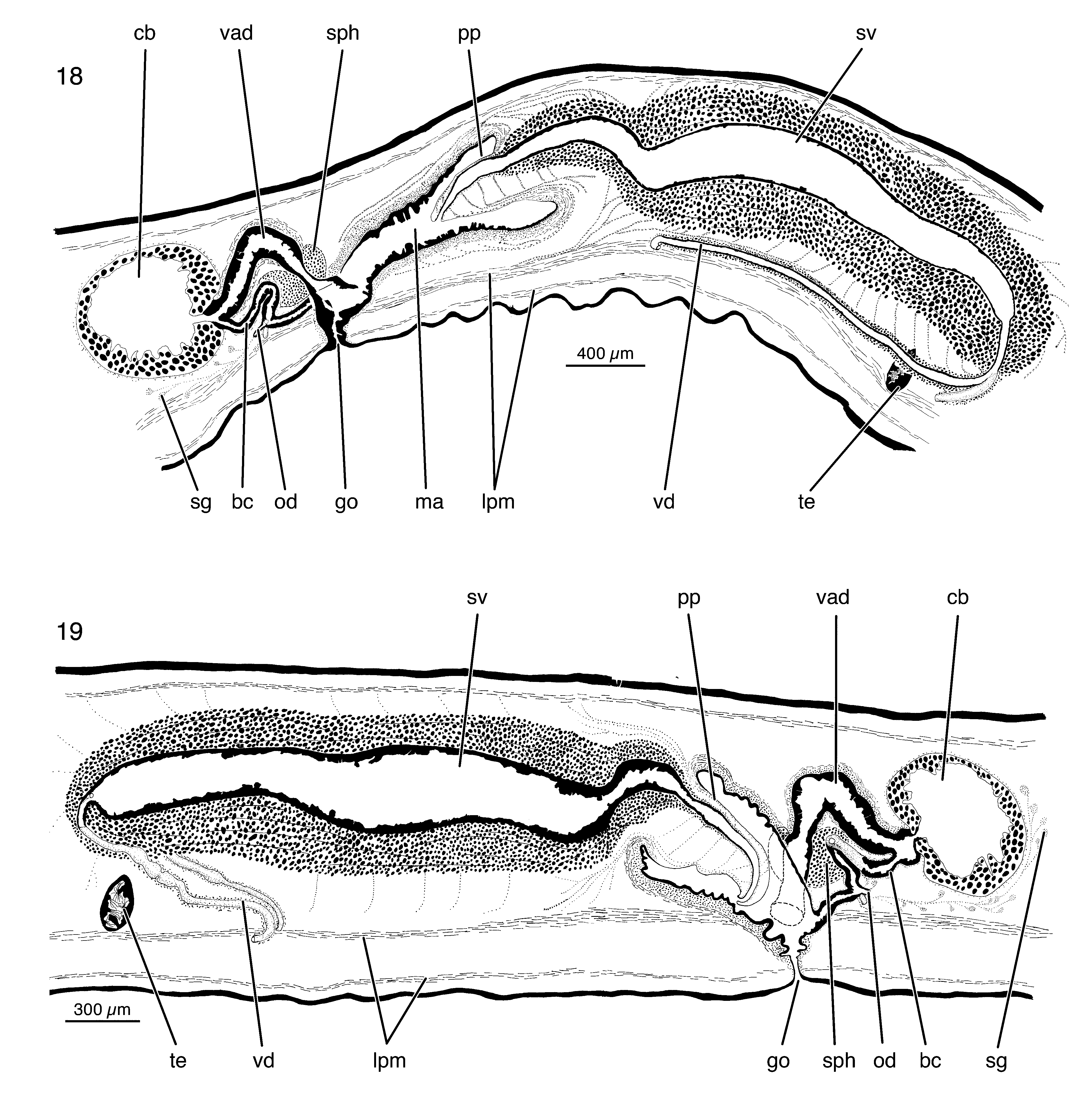
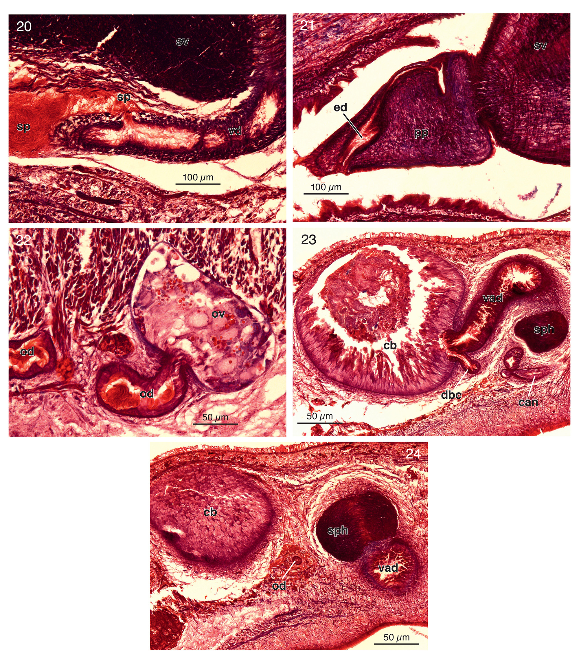
The sperm ducts run amongst the fibers of the layer of longitudinal parenchymal muscle located dorsally to the ventral nerve cords. The distribution of muscle fibers around the ducts is irregular, being more abundant on the dorsal than on the ventral side. Before the sperm ducts form spermiducal vesicles they run an erratic course, in ZMA V.Pl. 7251.1 giving rise to Z-shaped bends to anterior, posterior, ventral and dorsal direction ( Fig. 17 View FIGURES 13 – 17 ). However, in the holotype the sperm ducts do not follow an erratic course. The sperm ducts of the holotype run posteriorly to about three-quarters of the length of the seminal vesicle, after which the ducts recurve and run towards the anterior end of the vesicle ( Fig. 18 View FIGURES 18 – 19 ). In both specimens examined the sperm ducts expand slightly after curving and running towards the anterior section of the seminal vesicle. In ZMA V.Pl. 7251.1 the sperm ducts, after recurving, form narrow spermiducal vesicles ( Fig. 19 View FIGURES 18 – 19 ). Along the portion where the sperm ducts recurve, sperm has burst out at various places of the ducts ( Fig. 20 View FIGURES 20 – 24 ).The sperm ducts of both specimens are relatively muscular, being surrounded by decussate fibres.
Having approached the very antero-ventral surface of the coat of muscles surrounding the seminal vesicle, the sperm ducts open separately, albeit very closely together, into the rather narrow proximal section of the seminal vesicle. This narrow proximal section widens to become the broad lumen that occupies the major part of the seminal vesicle and which gradually narrows before communicating with the ejaculatory duct. The elongated cylindrical seminal vesicle runs parallel to the body surface and is surrounded by a very strong zone of circular muscles, interspersed with longitudinal muscles ( Figs 18, 19 View FIGURES 18 – 19 ).
The ejaculatory duct runs a dorsally displaced course through the penis and opens at the tip of the papilla ( Fig. 21 View FIGURES 20 – 24 ). The lining epithelium of the ejaculatory duct is nucleated and stratified, while the epithelium of the seminal vesicle is nucleated and pseudostratified.
The short, cone-shaped penis papilla sits in a spacious atrium and is covered with a thin, nucleated epithelium. In ZMA V.Pl. 7251.1 the penis papilla proper, i.e. from its point of insertion to the tip, measures 510µm in length, while the seminal vesicle measures 2450µm in length. In the holotype the penis papilla measures 580µm in length, while the entire seminal vesicle measures 2700µm in length ( Fig. 19 View FIGURES 18 – 19 ).
The penis glands are mainly distributed dorsally and ventrally of the root of the penis papilla and ventrally to the posterior fifth of the seminal vesicle; furthermore, many glands are located anteriorly to the seminal vesicle. These erythrophil glands discharge their secretion into the ejaculatory duct and the seminal vesicle, or prostatic vesicle, perpendicular to their longitudinal axes.
The genital atrium is lined with a thin, nucleated epithelium, underlain with a subepithelial circular muscle layer, followed by intermingled muscles. The gonopore is located at two-thirds of the distance between the anterior and posterior end of the specimen. Distance between mouth and gonopore between 1/4th and 1/5th of the total body length in the preserved specimens.
The slightly dorso-ventrally elongated ovaries are situated above the ventral nerve cords, occupying about 1/8th of dorso-ventral diameter of the anterior part of the body. They are positioned at approximately 1/6th of the distance between the brain and the root of the pharynx. The oviducts arise from the ventro-lateral sides of the ovaries ( Fig. 22 View FIGURES 20 – 24 ). Oviducts run in posterior direction between the fibers of the parenchymal muscle band, dorsally to the nerve cords. Dispersion of these muscle fibers around the duct is irregular, more fibres being present to the dorsal than to the ventral side. After passing the gonoduct, the oviducts curve dorso-medially to open separately, but very closely together, into the bursal canal. The openings of the oviducts are located at about 1/4th of the distance between the atrial opening of the bursal canal and its communication with the vaginal duct, close to the copulatory bursa ( Figs 18, 19 View FIGURES 18 – 19 ). The oviducts receive the secretion of shell glands, which are located in the area ventrally to the bursa.
From the point where it receives the oviducal openings, the bursal canal of ZMA V.Pl.7251.1 runs for about 200µm obliquely into antero-ventral direction to open into the atrium, about 220µm dorsally to the ventral surface (this section of the bursal canal corresponds to the so-called canalis anonymus). From the point of the opening of the common oviduct, a dorsal branch of the bursal canal (corresponding to Beauchamp's canal) rises anterodorsally for about 220µm, thereafter abruptly changing its course in postero-ventral direction for the next 200µm, after which it opens after 130µm into the vaginal duct ( Fig. 19 View FIGURES 18 – 19 ). The latter opens through the antero-lateral wall of the copulatory bursa; the lining epithelium of the vaginal duct extends for a short distance into the bursa, although the height of the epithelium in ZMA V.Pl. 7251.1 is here highly reduced. Very similar sinuous courses and conditions of the female ducts and canals are also present in the holotype ( Figs 19 View FIGURES 18 – 19 , 23 View FIGURES 20 – 24 ).
In ZMA V.Pl. 7251.1, the vaginal duct is surrounded by a very large, bean-shaped sphincter, consisting of strong circular muscle fibres, at approximately 120µm dorsally to its opening into the atrium ( Fig. 24 View FIGURES 20 – 24 ). This sphincter surrounds a section of about 190µm of the vaginal duct and forms a structure measuring about 305µm in anterior-posterior direction and 220µm in dorso-ventral direction. A similar condition is present in the holotype ( Fig. 23 View FIGURES 20 – 24 ).
Vaginal duct and bursal canal are lined with a nucleated, ciliated and pseudostratified epithelium. The musculature around the vaginal duct consists of a well developed layer of circular muscles, followed by a layer of intermingled muscles. The musculature around Beauchamp's canal is composed of a thin layer of circular muscles, followed by weak layer of longitudinal muscles. The canalis anonymus is surrounded by weak circular muscles ( Figs 18, 19 View FIGURES 18 – 19 ).
The obliquely oriented sac-shaped bursa measures in the holotype about 600µm in anterior-posterior direction and about 410µm in dorso-ventral direction, thus occupying less than 50% of the dorso-ventral diameter of the body in this part of the specimen. The bursa is lined with a well developed, pseudostratified, nucleated epithelium. The bursal musculature is composed of a weak layer of decussate fibres ( Figs 18, 19 View FIGURES 18 – 19 ).
Distribution. The species is known only from type locality ( Fig. 9 View FIGURES 9 – 12 ).
Discussion. Othelosoma impensum seems to be a species with a variable external appearance, at least judging by the two specimens available to us, viz. ZMA V.Pl. 7251.1 and the holotype ZMA V.Pl. 7250.1, which show conspicuous external differences. While the holotype is a dark green flatworm with two light greenish bands, separated by a narrow dark green stripe, V.Pl. 7251.1 is pale yellow dorsally, while it did not show the pale orange tip. In contrast to their external differences, the two specimens show only very few anatomical differences, mostly in the course of their sperm ducts. In the holotype the ducts runs posteriorly to about three-quarters of the length of the seminal vesicle before each recurves, whereas the sperm ducts of V.Pl. 7251.1 run not so far into posterior direction, while they follow a much more erratic, sometimes Z-shaped, course. Another difference concerns the location of the mouth in the pharyngeal cavity, which is located almost halfway between the anterior and the posterior wall in V.Pl. 7251.1, whereas in the holotype it is located at 7/8th between the anterior and the posterior wall of the pharyngeal pocket. However, these anatomical differences in the course of the sperm ducts and the location of the mouth opening, may be due to preservation artefacts, giving rise to various degrees of contraction. Other anatomical features, especially of the copulatory apparatus, suggest, with very little doubt, that the specimens V.Pl. 7250.1 and V.Pl. 7251.1 belong to one and the same species, viz. O. impensum (compare Figs 18, 19 View FIGURES 18 – 19 ).
The colour pattern of the holotype specimen V.Pl. 7250.1 of Othelosoma impensum resembles O. simile Sluys & Neumann , sp. nov., described below. Both have two dark dorso-lateral stripes, a dorsal narrow dark line, and an orange tip. But in O. simile the bands are made up of black pigment, whereas the dorsal bands of O. impensum (V.Pl. 7250.1) are dark green, at least in the live animal. Thus, the only shared similarity in the external appearance of V.Pl. 7251.1 of O. impensum and O. simile is that both specimens have an orange tip.
In addition to these external similarities between the holotype of Othelosoma impensum and O. simile , anatomical similarities between all specimens of the two species O. impensum and O. simile are present. Both species have (a) a well developed antero-dorsal retractor, extending from the anterior margin to the anterior fifth of the specimen, (b) a dorsally shifted ejaculatory duct running through a small penis papilla and (c) a long male atrium.
However, O. impensum can be distinguished from O. simile in the presence of a muscular sphincter on the vaginal duct. In contrast, O. simile has no sphincter around the vaginal duct, albeit that strong intermingled muscles are present on that part of the vaginal duct of O. simile where a sphincter is present in O. impensum . Further differences between O. impensum and O. simile can be found in the shape and musculature of the seminal vesicle. While the seminal vesicle of O. impensum is long, very muscular and orientated parallel to the body surface, the seminal vesicle of O. simile (a) is much smaller, in comparison to the dorsoventral diameter of the preserved specimen in that part of the body, (b) is slightly curved, and (c) has a relatively thin muscular coat. Other anatomical differences of the male reproductive systems between these two species can be found in (a) the course of the sperm ducts, and (b) in the location of the testes in the body. While the sperm ducts of Othelosoma impensum extend posteriorly to well behind the anterior end of the seminal vesicle, the ducts of Othelosoma simile recurve immediately after reaching the anterior end of the seminal vesicle. The testes of Othelosoma impensum reach the proximal section of the muscular seminal vesicle, while extending posteriorly, whereas the testes of Othelosoma simile extend posteriorly to about 1.5mm anterior to the seminal vesicle.
Further conspicuous differences in the female reproductive systems of O. impensum and O. simile are as follows. The large copulatory bursa of O. impensum has just one opening where the vaginal duct enters, in contrast to the small bursa of O. simile that receives the two separate entries of the vaginal duct and the bursal canal. In other words, the bursal canal and the vaginal duct of O. simile stay separate, whereas in O. impensum the bursal canal opens into the vaginal duct shortly before the latter enters the bursa. The bursal canals in O. impensum and O. simile follow a similar course but only in O. simile it receives the opening of a short common oviduct, whereas in O. impensum the oviducts open separately into the bursal canal. The course of the vaginal duct of O. impensum is characterised by an abrupt change, in that after leaving the bursa it first rises antero-dorsally and then turns posteroventrally, while reaching the level of the dorsal margin of the bursa. In contrast, the vaginal duct of O. simile does not show any abrupt changes during its course.
Othelosoma impensum shares the following combination of anatomical characteristics with O. conyum Marcus, 1953 , O. pugum Marcus, 1953 , O. rudebecki Marcus, 1955 , and O. saegeri Marcus, 1955 : (1) presence of a dorsal retractor, (2) presence of a muscular sphincter around the vaginal duct, (3) vaginal duct and bursal canal communicating shortly before the vaginal duct opens into the copulatory bursa.
However, the seminal vesicle of both O. rudebecki and O. saegeri is characterised by the presence of constrictions of the lumen, thus dividing the vesicle into several parts ( Marcus 1955a; Marcus 1955b). Further, the penis papillae of O. rudebecki and O. saegeri have similar sizes as their seminal vesicles, whereas the seminal vesicle of Othelosoma impensum is between four and five times longer than the penis papilla.
The proportions between the penis papilla and the seminal vesicle of O. pugum and O. conyum are similar to those of O. impensum and the sperm ducts also run posteriorly well behind the proximal section of the seminal vesicle. But the shape of the seminal vesicle is different. While in O. pugum and O. conyum the anterior end of the seminal vesicle is curved, in O. impensum the whole seminal vesicle runs more or less parallel to the body surface.
The course of the seminal vesicle in O. polecatum Marcus, 1953 resembles that of O. impensum in which the vesicle runs parallel to the body surface. Another anatomical similarity of O. polecatum is that a dorsal retractor in the anterior part of the body is present. But in comparison to the long dorsal retractor of O. impensum , extending throughout the anterior fifth of the whole preserved specimen, the dorsal retractor of O. polecatum is much shorter, measuring around 2mm in the 18mm long specimen ( Marcus 1953). Furthermore, the copulatory bursa of O. polecatum receives two distinct openings, whereas the bursa of O. impensum receives only the single entry of the vaginal duct. That means that there is no communication between the vaginal duct and the bursal canal of O. polecatum , while the bursal canal and the vaginal duct of O. impensum communicate shortly before the latter enters into the bursa.
| ZMA |
Universiteit van Amsterdam, Zoologisch Museum |
No known copyright restrictions apply. See Agosti, D., Egloff, W., 2009. Taxonomic information exchange and copyright: the Plazi approach. BMC Research Notes 2009, 2:53 for further explanation.
|
Kingdom |
|
|
Phylum |
|
|
Class |
|
|
Order |
|
|
Family |
|
|
SubFamily |
Microplaninae |
|
Genus |
Othelosoma impensum Sluys & Neumann
| Sluys, Ronald, Neumann, Matthias, De Lima, Ricardo F. & Drewes, Robert C. 2017 |
O. rudebecki
| Marcus 1955 |
O. saegeri
| Marcus 1955 |
O. conyum
| Marcus 1953 |
O. pugum
| Marcus 1953 |