
Losbanosia taivaniae, Szwedo, Jacek & Adamczewska, Malgorzata, 2004
|
publication ID |
https://doi.org/ 10.5281/zenodo.158482 |
|
publication LSID |
lsid:zoobank.org:pub:F9DB5503-EA81-4EBE-8796-719BF88F6A9F |
|
DOI |
https://doi.org/10.5281/zenodo.6271304 |
|
persistent identifier |
https://treatment.plazi.org/id/06176719-5452-FFE4-FEA3-F9F6FEEDF8CF |
|
treatment provided by |
Plazi |
|
scientific name |
Losbanosia taivaniae |
| status |
sp. nov. |
Losbanosia taivaniae View in CoL sp. nov.
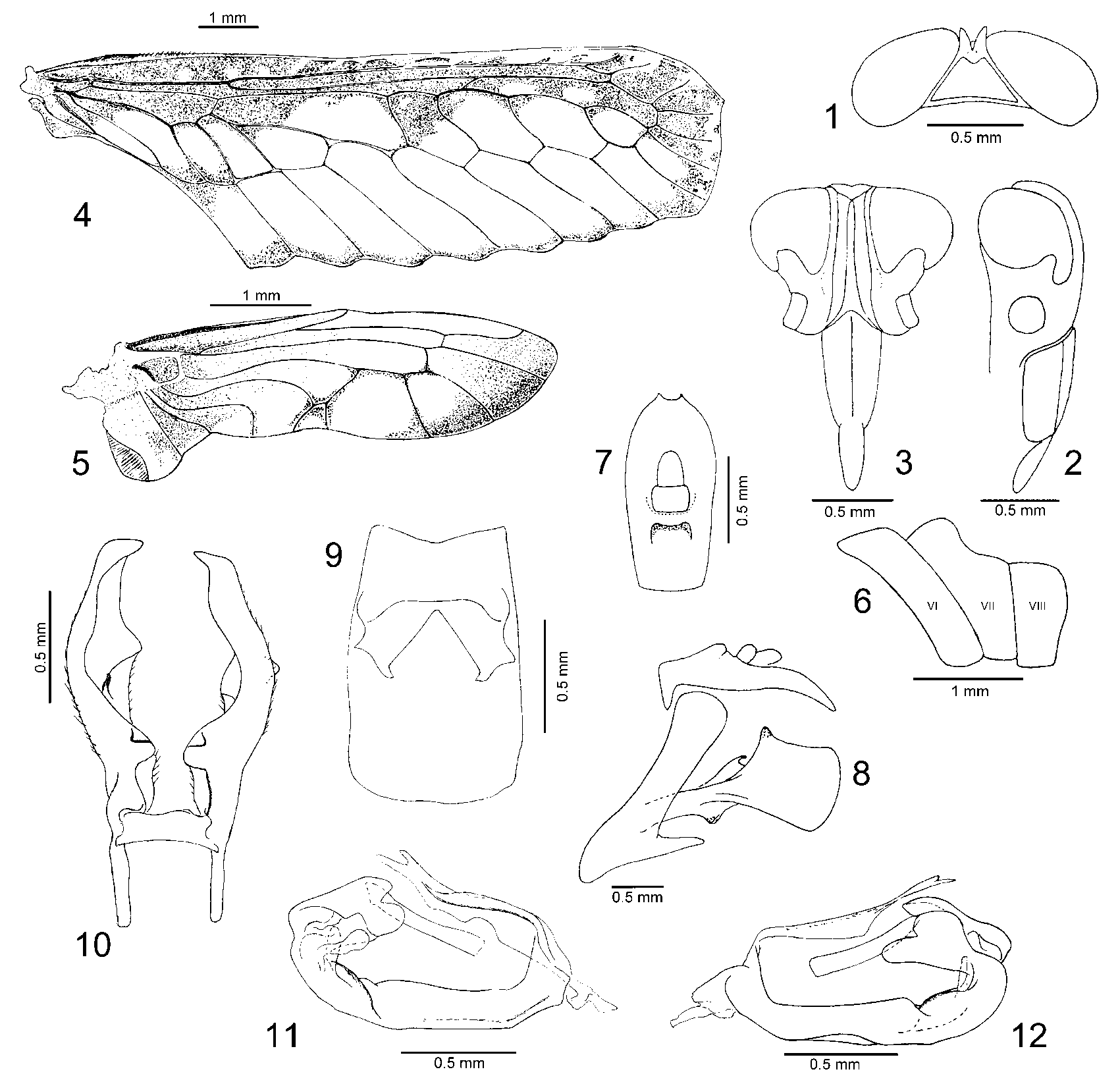
( Figs. 1–12 View FIGURES 1 – 12 )
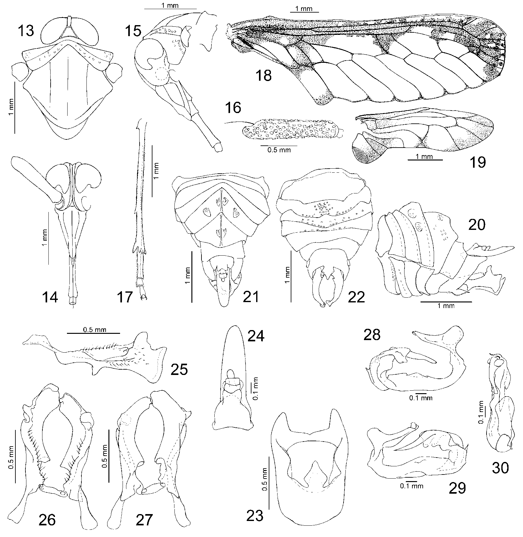
Losbanosia hiberensis [sic!] ( Matsumura, 1935): Yang & Wu 1993: 37, Fig.14 View FIGURES 13 – 30 A–J.
Losbanosia hiberensis [sic!] (Muir) [sic!]: Yang & Chang 2000: 545, 546, Fig. 444A–F.
DIAGNOSIS: It differs from Losbanosia hibarensis (Mats.) by the almost subtriangular, only slightly diamondlike shape of the ventral median process of the pygofer (distinctly diamondshaped in L. hibarensis ); the posterolateral margin of pygofer lacking a process, angulately rounded (angulate process in L. hibarensis ); the anal tube about three times as long as wide at the base, and not widened at the base (shorter and widened at base in L. hibarensis ); the apical portion of the anal tube with two short teeth (lack of such teeth in L. hibarensis , apical portion rounded); curved ventrad (straight in L. hibarensis ); the aedeagus with the endosomal process blunt at apex (acute in L. hibarensis ); VIIIth abdominal segment a without distinct dorsal crest (small crest in L. hibarensis ).
DESCRIPTION (Based also on data provided by Yang & Wu 1993): Length of body (including tegmina) 12.8 mm. General colour brown; rostrum and legs pale yellowish; tegmina white with brown to black markings as in Fig. 4 View FIGURES 1 – 12 , veins on markings red.
Vertex ( Fig. 1 View FIGURES 1 – 12 ) triangular with bifurcate apex, disc shallowly excavate; lateral carinae of frons distinctly separated throughout; compound eyes deeply incised ventrally; ocelli absent ( Figs. 2 & 3 View FIGURES 1 – 12 ).
Tegmina ( Fig. 4 View FIGURES 1 – 12 ) longer than widest part about 3.5:1, longer than wings about 1:0.35, with anterior and posterior margins nearly parallel; vein RA with two terminals, RP with three terminals, M with 5 terminals, CuA + CuP with 4 terminals; second transverse veinlet mcu short.
Wings ( Fig. 5 View FIGURES 1 – 12 ) with anterior margin arched anteriad in median portion, ScRA reaching margin in about half of wing length, M simple, CuA with three terminals, CuP distinctly curved and deflected basad in terminal portion, at the level of oblique veinlet uniting CuA2 with CuP, PCu distinctly curved, stridulatory area on anal field distinct.
Tibiometatarsal formula 5:(5–6):(5–6).
Abdominal tergite VII ( Fig. 6 View FIGURES 1 – 12 ) in profile distinctly longer than ventrally, dorsal margin with basal half produced dorsad, in form of rounded crest.
Anal segment ( Fig. 7 View FIGURES 1 – 12 ) slender, apical third slightly curved, in dorsal view evenly widened at apical third, then narrowed, apical margin distinctly incised medially, each side with a small process; pygofer ( Figs. 8 & 9 View FIGURES 1 – 12 ) with ventral margin sharply oblique, dorsal margin rounded, in ventral view medioventral process subtriangular (diamondshaped), lateral incisions very small; aedeagus ( Figs. 11 & 12 View FIGURES 1 – 12 ) in left lateral view with membranous flagellar lobe, in right lateral view small process arising from phallotheca and directed dorsomediad, endosomal process rodlike, blunt at apex; genital styles with basal portion rather narrow, apical portion quadrate, with dorsobasal angle produced dorsad roundly, dorsal margin at middle with lower portion hooked at apex, same position at ventral margin produced ventrad roundly.
ETYMOLOGY: Species named after Latin form of the name of Taiwan—Taivania.
REMARKS: The description and figures in Yang & Wu (1993) are based on a single male, collected in Chihsingshan, Taipei Hsien, 7IX1985, by S.C. Tsaur. Yang & Yeh (1994) described a nymph identified as Losbanosia hiberensis [sic!] ( Matsumura, 1935), collected in Hualien Hsien, Tailuko, 14II1990, by W.B. Yeh, under rotten wood, and identified by Yeh on the basis of two males. These data should probably also be referred to the species described above.
MATERIAL EXAMINED: Holotype, male, dissected specimen. Labelled: [ Losbanosia taivaniae Szw. et Ada. ɗ]; red label [HOLOTYPE]; [ TAIWAN: Taipei / Chihsingshan / 7IX1985 / col. S.C. Tsaur]; [ Losbanosia hiber / ensis (Mats.)]; [1]; tegmina and wings on separate microscopic slides with the same labels and annotations: [2]–tegmina and [3]– wings.
No known copyright restrictions apply. See Agosti, D., Egloff, W., 2009. Taxonomic information exchange and copyright: the Plazi approach. BMC Research Notes 2009, 2:53 for further explanation.
|
Kingdom |
|
|
Phylum |
|
|
Class |
|
|
Order |
|
|
Family |
|
|
Genus |
Losbanosia taivaniae
| Szwedo, Jacek & Adamczewska, Malgorzata 2004 |
Losbanosia hiberensis
| Yang 2000: 545 |
Losbanosia hiberensis
| Yang 1993: 37 |