
Scelimena marta Skejo & Tan, 2018
|
publication ID |
https://doi.org/ 10.11646/zootaxa.4485.1.1 |
|
publication LSID |
lsid:zoobank.org:pub:EDC89718-2F45-494A-80F1-A187DA926CC4 |
|
DOI |
https://doi.org/10.5281/zenodo.5959388 |
|
persistent identifier |
https://treatment.plazi.org/id/0636885F-FFBB-7274-FF74-560BFE3987B5 |
|
treatment provided by |
Plazi |
|
scientific name |
Scelimena marta Skejo & Tan |
| status |
sp. nov. |
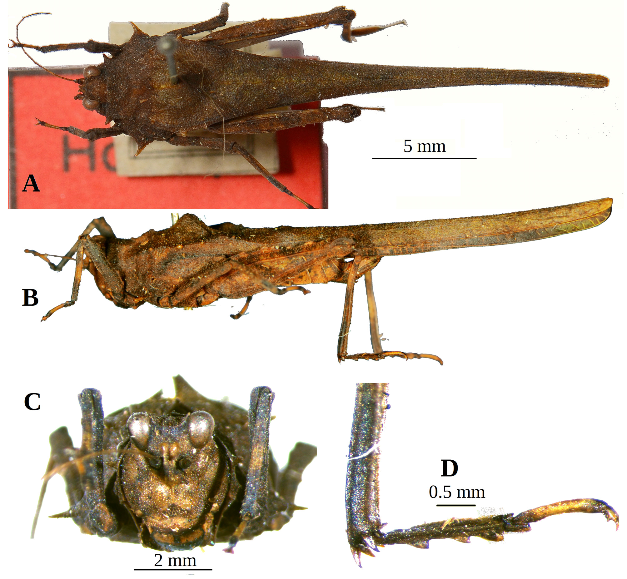
Scelimena marta Skejo & Tan View in CoL , sp. nov.
( Figs 3–5 View FIGURE 3 View FIGURE 4 View FIGURE 5 )
Material examined. Holotype (female, MNCN _ Ent 219686, abbreviated HT, Fig. 3 View FIGURE 3 ): Indonesia: Sumatra: mid Sumatra [locality not precise on the labels] leg. Sumatra Expedition 1877–1878, collected in VIII.1878. det. [as Hexocera marta sp. nov.] J. Skejo 12.V.2017. ( MNCN) . Paratypes: 2 males ( MNCN _Ent 219688, abbreviated PT1— Fig. 4 View FIGURE 4 ; and MNCN _Ent 219687, PT2— Fig. 5 View FIGURE 5 ). Probably from the very same locality and environments as the holotype. leg . Sumatra Expedition 1877–1878, collected in VIII.1878. det. [as Hexocera marta sp. nov.] J. Skejo 12.V.2017. ( MNCN)
Diagnosis. This new species most resembles Scelimena hexodon (de Haan, 1843) comb. resurr. and is assigned to the Scelimena hexodon species group. The species can be easily separated from Bornean S. hexodon by the following states of the characters: (I) second frontolateral projections (FL2) are more produced and more spiky in S. hexodon , (II) metalateral projection (ML) is weaker in S. marta , (III) first metamedial projection (MM1) is much higher in S. hexodon than in S. marta , (IV) ventrolateral spines are shorter in S. marta , (V) S. hexodon has wider shoulders than S. marta , and (VI) is generally larger species (body length including pronotum about 24 mm in males, while about 28 mm in females). In other characters, the two species are very close and are thus both assigned to this species group.
Description. Holotype female ( Fig. 3 View FIGURE 3 ).
Antennae. Very long and thin, 13 segmented: 1 st massive scapus (width of the frontal ridge between eyes about 2 times width of scapus), 2nd stout pedicel, 3rd–6th elongated basal segments, 7–8th extremely elongated mid segments (about 14 times as long as wide), elongated 9–10th subapical segments, 11–13th reduced, small apical segments. Coloration. Scapus and pedicel dark, flagellum yellowish greenish or brown.
Head. In frontal view . Transverse and lateral carinae of the vertex slightly elevated. Frontal costa long, bifurcates below the mid of the compound eyes height, just between the paired lateral ocelli. Scutellum in its widest part as wide as an antennal groove. Scutellum forms shallow concavity, becoming wider ventrad. Upper margin of the antennal grooves visibly below (about 0.1 mm) the lower margins of the compound eyes. Vertex width (1.02 mm) 1.6 times a compound eye width (0.63 mm). Compound eyes ovoid. Coloration. Vertex dark, frons, upper half of the facial carinae yellowish green, frons yellowish green, genae with dark stripes, labium dark with two light spots (yellowish green). In dorsal view . Frontal costa cut in the fastigium, and its anterior margin is concave. Medial carina of the vertex is short and low, present only in the upper fifth of the vertex length. Fossulae deep. Lateral and transverse carinae of the vertex forming obtuse angle. Compound eyes oval. Vertex wider than a compound eye 1.6 times. Coloration. Greenish yellow with dark patches. In lateral view . Eyes subglobular in shape, exerted above the vertex. Frontal costa slightly projected in front. Paired lateral ocelli large, visible just below the anterior lower margin of the compound eyes. Palpi without widened apical segments. Vertex, fastigium, and frons not visible. Coloration. Dark, after the eyes there is yellowish green stripe. Palpi yellowish green with dark patches.
Pronotum. Pronotum very long (macropronotal state), surpassing abdominal apex for 1.6 length of the hind femur. In frontal view . Ventrolateral projection of the lateral lobes (VL), metalateral projections of the shoulders (ML) and first metamedial projection (MM) highly raised as spines. Second frontolateral projection (FL2) and second prolateral (PL2) produced forwards as tooth. In dorsal view . Anterior margin of the pronotum truncated. FM small, FL1 unrecognizable, FL2 strong, tooth-like and projected in the front, FL3 weak, prozonal carinae straight and parallel, PM rounded (larger than FM, smaller than MM1), PL1 distinct and small, while PL2 larger and tooth-like. PML1 and PML2 small, wart-like. Ventrolateral (VL) projection of the lateral lobes strongly projected outwards as acute spine, directed forwards. Interhumeral carinae unrecognizable (barely seen between PML2 and MML1). Angle between humeral (also known as humero-apical) and external lateral carina obtuse, shoulders bearing strong metalateral (ML) tubercles raised as spines. After ML, there are two more tubercles on the external lateral carina of the pronotum, much smaller than ML: MM1 is the largest of medial unpaired projections, while size of MM2 to MM4 decreases caudad. MML1 to MML3 small, decreasing in size towards the apex. Median carina of the pronotum continuous from the anterior margin to the tip of the pronotum, low and granulated. Internal lateral carina decurved and slightly visible only in the level between the hind knees. Pronotum slightly depressed after MM1, and after the mentioned depression flat. Pronotal apex truncated, slightly concave. Pronotal surface granulated with fine and medium sized tubercles. In lateral view . Prozona directed slightly upwards, not straight. FM small and wart like, prozonal carinae granulated with medium sized tubercles, PML1 small, PML2 larger and wart-like. FL1 unrecognizable, FL2 projected forwards as a tooth, PL1 small, PL2 larger tubercle. Ventral margin of the lateral lobes straight, not saw-like, ventrolateral spine sharp, ventral sinus (triangular) slightly deeper than tegminal (rectangular) sinus. Infrascapular area large and triangular, not as wide as tegmina. PM is the second largest projection on the dorsum, just after large rounded MM1. MM2 and MM3 low. MML1,
MML2, MML3 and MML4 low, decreasing in size caudad. Infrascapular area fused with lateral area, which runs to the apex of the pronotum. Before ML, one tubercle present in humeral carina, and after strong ML, two more tubercles present in the external lateral carina. Pronotum with depression after MM1, then flat to the apex. Coloration. Smaller projections, spines, tubercles, area around and after MM1, and pronotal dorsum after the level of the hind knees yellowish green, rest of the pronotum (including MM1) dark.
Wings. Tegmenula elongated, oval, and smooth (finely granulated), with apical margin subacute; three times longer than wide; visible part of tegmen 0.6 times width of the mid femur. Hind wings long, not surpassing apex of pronotum. Coloration. Tegmenula yellowish green, and alae dark.
Legs. Fore legs. Femur with carinated and undulated dorsal and ventral margins, dorsal more than ventral, dark with weak greenish patches. Femur about 5.3 times as long as wide. Tibia rectangular in cross-section, having fine denticles in its margins, bearing two light rings, one in proximal part and one in the middle. Proximal tarsal segment much shorter than the prolonged distal. Two thirds of the distal segment yellowish green in color, while proximal segment and the distal part of the distal segment black. Claws pale colored with dark apices. Mid legs. Femur with carinated dorsal margin. Besides the genicular tooth, three weak teeth present in the dorsal margin, while the ventral margin has a weaker undulation. Mid femur 5.6 times as long as wide. Tibia rectangular in crosssection, dark in color, and with three pale rings (in the beginning, in the middle and distally). Proximal tarsal segment much shorter than the prolonged distal. Two thirds of the distal segment yellowish green in color, while proximal segment and the distal part of the distal segment black. Hind femora with alternating yellowish green and dark parts, lappets (spines) being bright in color. Hind legs. Femur slender, 3.8 times as long as wide. Dorsal margin with three dentiform lappets before the antigenicular tooth, while ventral margin bearing four strong spines (lappets transformed into strong denticles). Four transverse ridges hardly recognizable in the external lateral area of the hind femur. Genicular and antigenicular teeth small and blunt, incision clearly visible before the knee. Tibia widened towards the apex, in the distal part lamellate. Upper side of hind tibia a few (3–5) larger outer and (3–5) inner spines and minute denticles between those spines. Hind tibia dark, with a light band in the proximal third of its length. First tarsal segment widened (adapted for swimming), 1.4 times longer than the third. First pulvillus of the proximal segment of the hind tarsus angular (without apical tooth) with right-angled apex, second angular with obtuse angle, and the third one rounded, and blunt. First and second tarsal segments are dark in color, almost black, while the distal (third) is yellowish green. Claws pale colored with dark apices.
Abdominal apex. Subgenital plate 1.13 times longer than broad; posterior margin of plate lobed. Cerci hairy and stout basally, with pointed apex. Valves of ovipositor narrow, dentate. Upper valve of ovipositor 5 times longer than its maximum width. Lower valve of ovipositor 4.5 times longer than its maximum width; apical tooth curved downwards. Ovipositor dark with yellowish patches, with apices of denticles blackish. Tergites and sternites yellowish brown with dark markings.
Male paratypes. In morphology correspond to the female description, except for smaller size (consult Table 2. and Figs. 3 View FIGURE 3 and 4 View FIGURE 4 for comparative measurements) and different shape of the abdominal apex, due to sexual dimorphism.
Measurements. Table 2.
Etymology. This species is named after María Marta Cigliano (currently working at the División Entomología in the Museo de La Plata, and National Scientific and Technical Research Council, Argentina). María Marta is a famous orthopterist specialized in taxonomy of Tristiridae and Acrididae : Melanoplinae, especially in America. She has described one tribe, 15 genera and 70 species. The specific epithet is noun in apposition, (Marta, ae, f.) and is made of her mid name.


| MNCN |
Museo Nacional de Ciencias Naturales |
No known copyright restrictions apply. See Agosti, D., Egloff, W., 2009. Taxonomic information exchange and copyright: the Plazi approach. BMC Research Notes 2009, 2:53 for further explanation.