
Paramacroxiphus C.Willemse, 1961
|
publication ID |
https://doi.org/10.11646/zootaxa.1755.1.1 |
|
persistent identifier |
https://treatment.plazi.org/id/0638878C-FFC4-FFED-19EC-FDA0FED8ABE1 |
|
treatment provided by |
Felipe |
|
scientific name |
Paramacroxiphus C.Willemse, 1961 |
| status |
|
Paramacroxiphus C.Willemse, 1961 View in CoL
Type species: Paramacroxiphus aberrans Willemse, 1961 , by original designation
Paramacroxiphus C. Willemse 1961a View in CoL , Publ. natuurh. Gen. Limburg 12: 32.
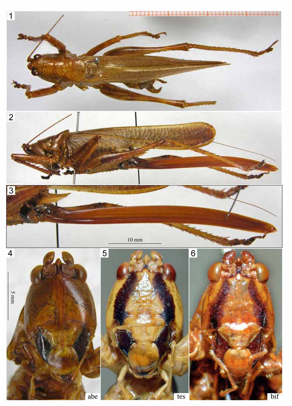
Description. Fastigium verticis conical, shorter than scapus; ventral margin separated by a shallow sinuosity from fastigium frontis. Frons subsmooth, with shallow, scattered or more dense impressed dots, more distinct towards genae ( Figs. 4–6 View FIGURES 1–6 ). Pronotum shining, faintly rugose towards margins; disc broadly rounded into paranota, posterior area often faintly raised, flat and shouldered; anterior margin broadly rounded but faintly concave or truncate in middle; posterior margin rounded (often almost straight in middle); transverse sulcus little impressed but entire; second transverse sulcus deeply cut on paranota, widening to a shallow furrow on disc; paranota with ventral margin slightly concave, strongly descending posteriorly; posterior angle rounded; auditory swelling distinct, ovoid; humeral sinus little indicated. Fully winged, tegmen surpassing hind knees, with approaching margins in basal half, subparallel margins in apical half, in some species slightly widening again towards apex, apex rounded ( Fig. 2 View FIGURES 1–6 ). Prosternal spines medium to long (somewhat variable, but always shorter than coxa), apex acute. Mesosternal lobes conical, metasternal lobes short-conical; medial plate with a spine at both posterior angles. Anterior tibia with dorsal angles broadly rounded, only near apex little angular. All femora with spines on both ventral margins; on ventro-internal margin of postfemur numerous and distributed over the whole femur length. Knee lobes of profemur triangular or rarely obtuse on external, spinose on internal side; of mesofemur spinose on both sides; of postfemur bi-spinose on both sides.
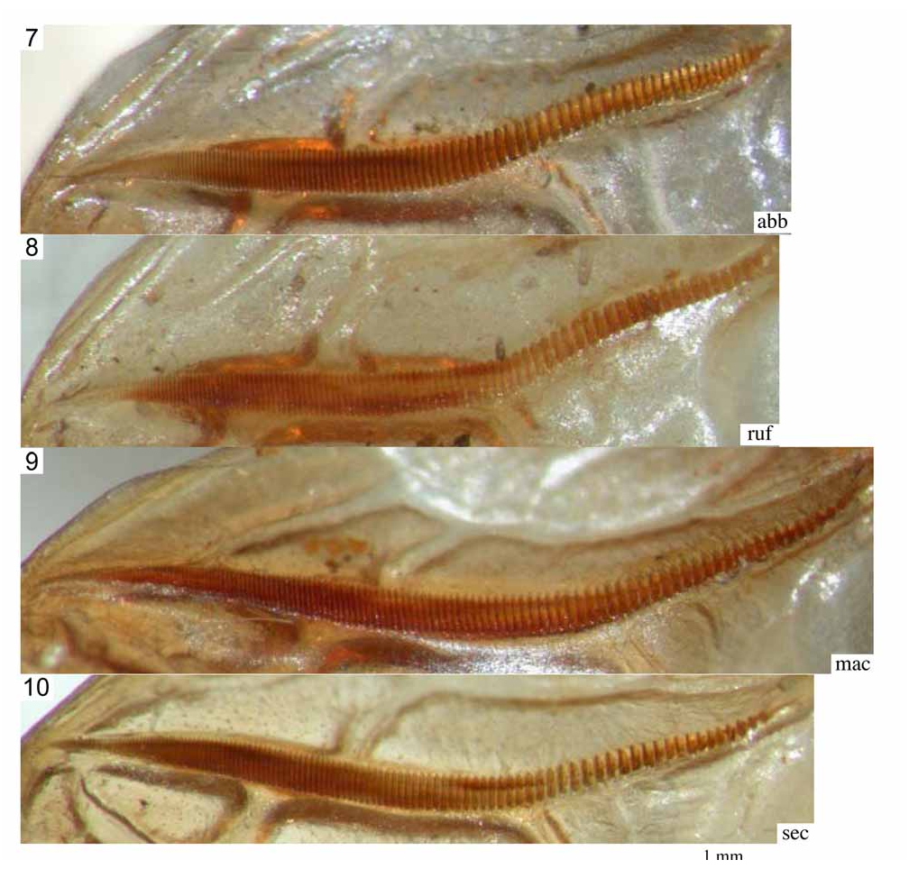
Male. Stridulatory file curved in about middle; basal half with large teeth, gradually turned into narrow teeth in apical half ( Figs. 7–15 View FIGURES 7–10 View FIGURES 11–15 ). Dorsal area of right tegmen behind mirror with a field of minute spinules sitting on veins and veinlets ( Fig. 44 View FIGURES 35–44 ).
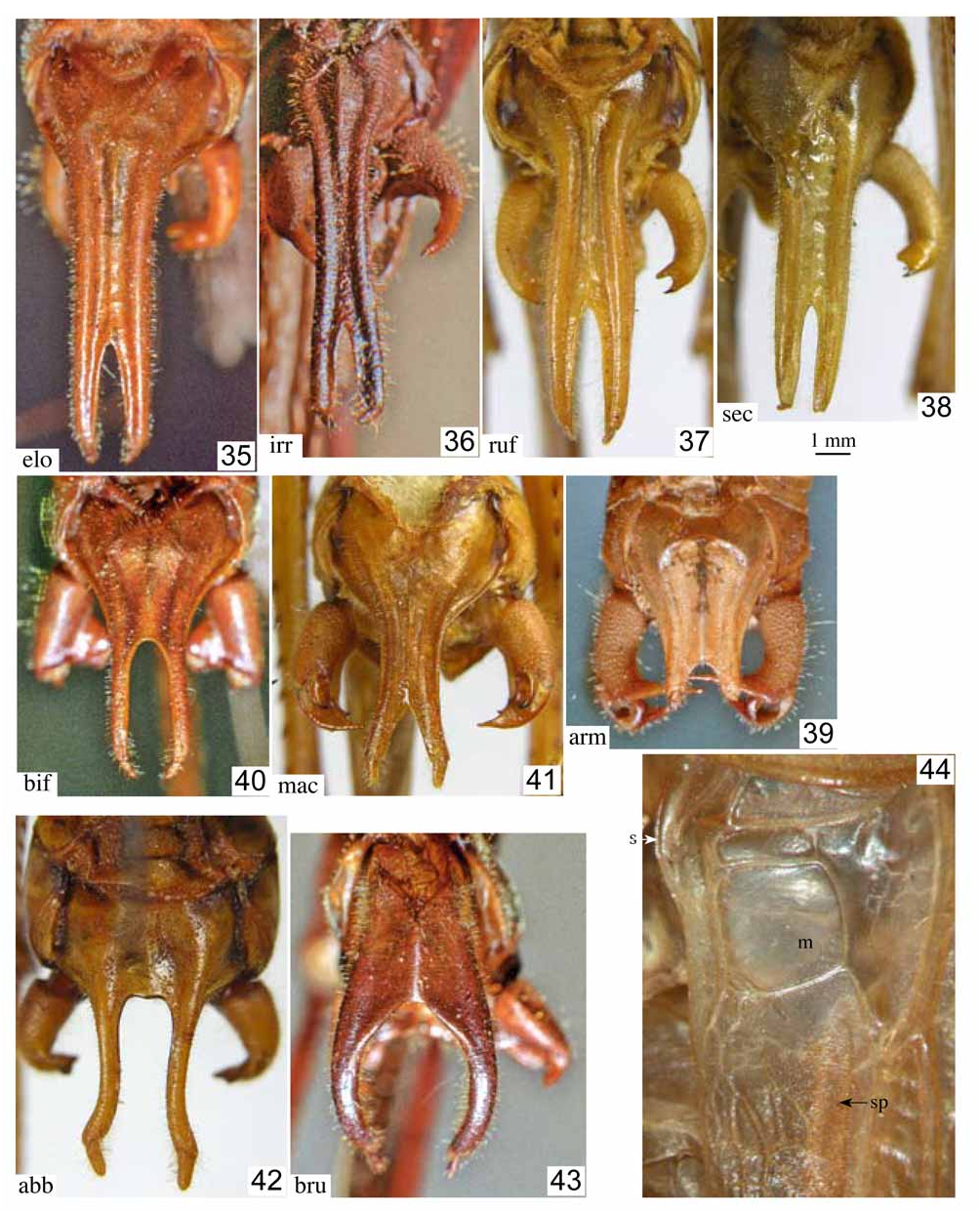
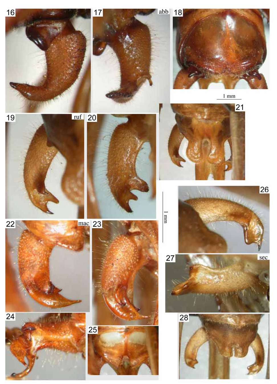
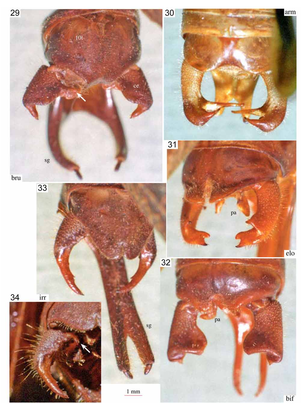
Tenth abdominal tergite varying in size and shape between species, short or prolonged, globular or almost flat; apical margin excised or with projections of various shape ( Figs. 18, 21, 25, 28 View FIGURES 16–28 , 29–33 View FIGURES 29–34 ). Subgenital plate with convex and ascending lateral areas; with obtuse lateral carinae; central area between and including carinae or carinae alone prolonged behind, in some species very long; resulting shapes strongly differing between species ( Figs. 35–43 View FIGURES 35–44 ). Epiproct and cerci species specific.
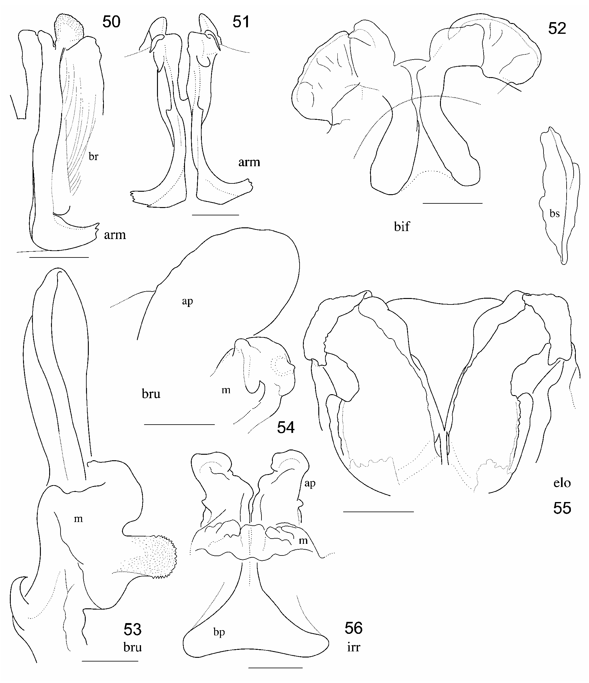
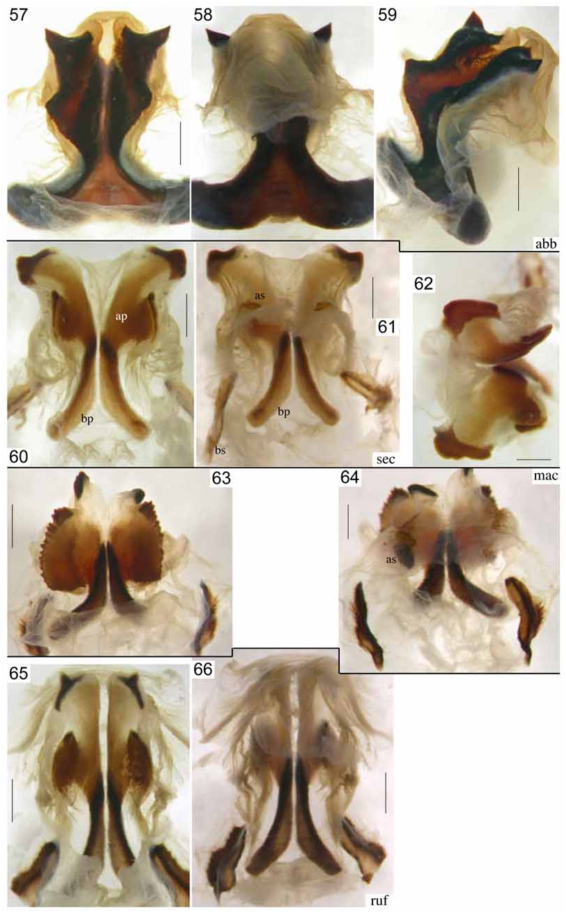
Titillators with basal parts simple; apical parts widened and to varying degree divided into two branches ( Figs. 45–66 View FIGURES 45–49 View FIGURES 50–56 View FIGURES 57–66 ). With large separate basal sclerites.
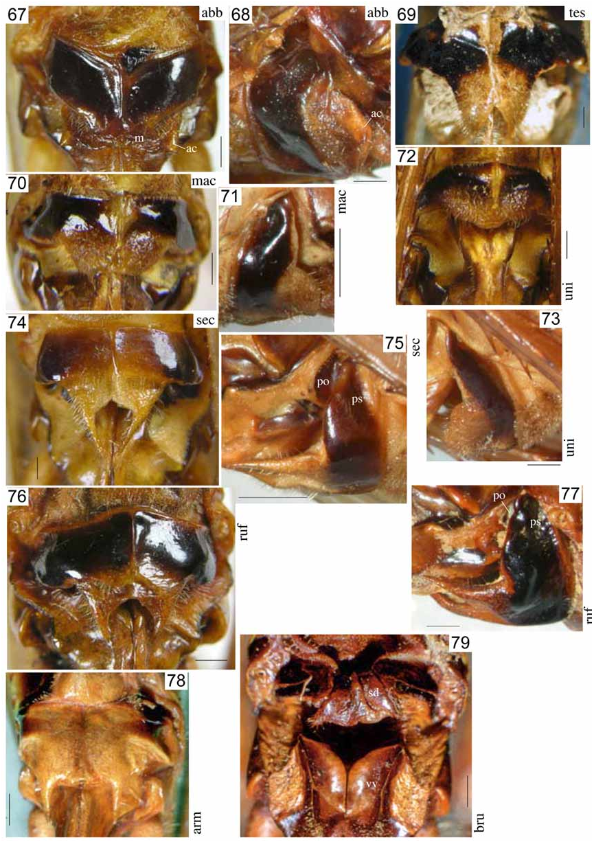
Female. Tenth abdominal tergite transverse. Epiproct rounded or triangular, surface shallowly depressed. Cerci long, conical, slightly curved, apex acute. Ovipositor long, compressed, blade-shaped, highest in or slightly before or behind middle, faintly curved dorsad in basal half, and ventrad in apical half ( Fig. 3 View FIGURES 1–6 ). Subgenital plate transverse, divided in midline by a narrow membranous suture; apex with a membranous lobe of varies shape and often stiffened apical projections.
Coloration. Often uniformly ochre, some species tessellate (dark brown with ochre). Frons often concolorous, clypeus often yellow, mandibles dark brown or black; some species with black vertical bands or other dark pattern. Tegmen light brown with little conspicuous brown spots, or strikingly tessellate in the dark colour form. Ovipositor brown. Female subgenital plate black, suture and membranous apical area light.
Discussion. The genus contains large, robust species. It is characterised by the pronotum with deep paranota, weak humeral sinus and moderately prolonged apical area, covering not more than the base of the stridulatory area in male ( Fig. 1 View FIGURES 1–6 ). Characteristic for the male stridulatory apparatus is the sinuate stridulatory file with rather large teeth ( Figs. 7–10 View FIGURES 7–10 ) and the dorsal field of the right tegmen, which carries a field of minute spinules behind the mirror area ( Fig. 44 View FIGURES 35–44 ). The male abdominal terminalia and internal genitalia show species specific modifications. The cerci are short, conical or cylindrical with internal teeth of variable number and shape. The titillators have the apical parts often divided into two branches, the surface with a very fine striation or granulation and the margins crenulated. The baso-lateral sclerites are large and elongate; the apico-lateral sclerites usually small or reduced. In some species the titillators are provided with bunches of hairs, bristles or short spines. The female subgenital plate has the apical area membranous or prolonged into a median process or a pair of spines; the basal sclerite is divided or furrowed in midline. The ovipositor varies from little longer to twice as long as the hind femur, is slightly curved down, and highest in middle of length. All known species are fully winged with the tegmen surpassing the abdomen but do not reach the apex of the stretched hind tibia.
The male tenth abdominal tergite and male and female subgenital plates show evolutionary trends within the genus. The male tenth abdominal tergite is in the simplest form little longer than the preceding sclerites, and with moderately emarginated hind margin as in P. armatus ( Fig. 30 View FIGURES 29–34 ). There are two main trends of modification: the tergite is prolonged (most expressed in P. irregularius , Fig. 33 View FIGURES 29–34 ) or it becomes globular (most expressed in P. aberrans , Fig. 18 View FIGURES 16–28 ). Additionally, the lobes resulting from the apical emargination can become separately prolonged and modified (e.g. in P. rufus , Fig. 21 View FIGURES 16–28 ). With regard to the male subgenital plate, the most primitive form also occurs in P. armatus ( Fig. 39 View FIGURES 35–44 ) with only little prolonged medial area and moderately excised apex. The main trend goes via intermediate forms ( Figs. 40–41 View FIGURES 35–44 ) to a strong prolongation of the central area of the subgenital plate ( Figs. 35–38 View FIGURES 35–44 ). Additionally, the apical excision can become longer to the extreme that the prolongation is only formed by the lateral carinae ( Figs. 42–43 View FIGURES 35–44 ). The female subgenital plate shows in several species a membranous apical area without (e.g. Fig. 72 View FIGURES 67–79 ) or with (e.g. Figs. 67-68 View FIGURES 67–79 ) sclerotised cones or horns at the apical angles. Two evolutionary hypotheses are plausible, (1) a reduction: apical lobes of the subgenital plate (as in Fig. 69 View FIGURES 67–79 ) developed into acute cones (as in Fig. 78 View FIGURES 67–79 ), sclerotisation of the area between the cones and the wider basal area got lost (e.g. Figs. 67, 74 View FIGURES 67–79 ), and finally also the cones were reduced ( Figs. 70, 72 View FIGURES 67–79 ); (2) the membranous apical area is a newly evolved structure that might have resulted from an outgrowth of the dorsal surface of the subgenital plate behind the apical margin. In the latter case, subgenital plates with simple membranous apical area would be the most primitive, those fully sclerotised the most advanced.
The titillators are very complex. However, the titillators proper are simple clips that form the basal parts. They are fused with formerly probably membranous lobes of the phallus that became sclerotised to a varying degree forming the complex apical parts. Basal and apical parts can be readily recognised as separate units when sclerotisation of the apical parts is less pronounced ( Figs. 60–66 View FIGURES 57–66 ), while in other species both parts form a single strongly sclerotised sclerite ( Figs. 57–59 View FIGURES 57–66 ). The surface of the apical parts shows a very fine striation or granulation while the margins are coarsely granular. Similar titillators can also be found in other Agraeciini genera of the Indo-Australian region, e.g. Axylus Stål, 1877 or Pseudonicsara Karny, 1912 . Those of Paramacroxiphus are characterised by the two-branched apical parts.
The male stridulatory file is rather uniform between species apart from differences in size. The modified male internal genitalia show characteristic differences between species. However, all species known so far can also be differentiated by external characters as the male tenth abdominal tergite and cerci and male and female subgenital plates.


No known copyright restrictions apply. See Agosti, D., Egloff, W., 2009. Taxonomic information exchange and copyright: the Plazi approach. BMC Research Notes 2009, 2:53 for further explanation.
|
Kingdom |
|
|
Phylum |
|
|
Class |
|
|
Order |
|
|
Family |
Paramacroxiphus C.Willemse, 1961
| Ingrisch, Sigfrid 2008 |
Paramacroxiphus
| C. Willemse 1961 |