
Bursaphelenchus singaporensis, GU, JIANFENG, ZHANG, JIANCHENG, BRAASCH, HELEN & BURGERMEISTER, WOLFGANG, 2005
|
publication ID |
https://doi.org/ 10.11646/zootaxa.988.1.1 |
|
persistent identifier |
https://treatment.plazi.org/id/06738799-8C0D-6A0A-FECB-FBB9FB992168 |
|
treatment provided by |
Felipe |
|
scientific name |
Bursaphelenchus singaporensis |
| status |
|
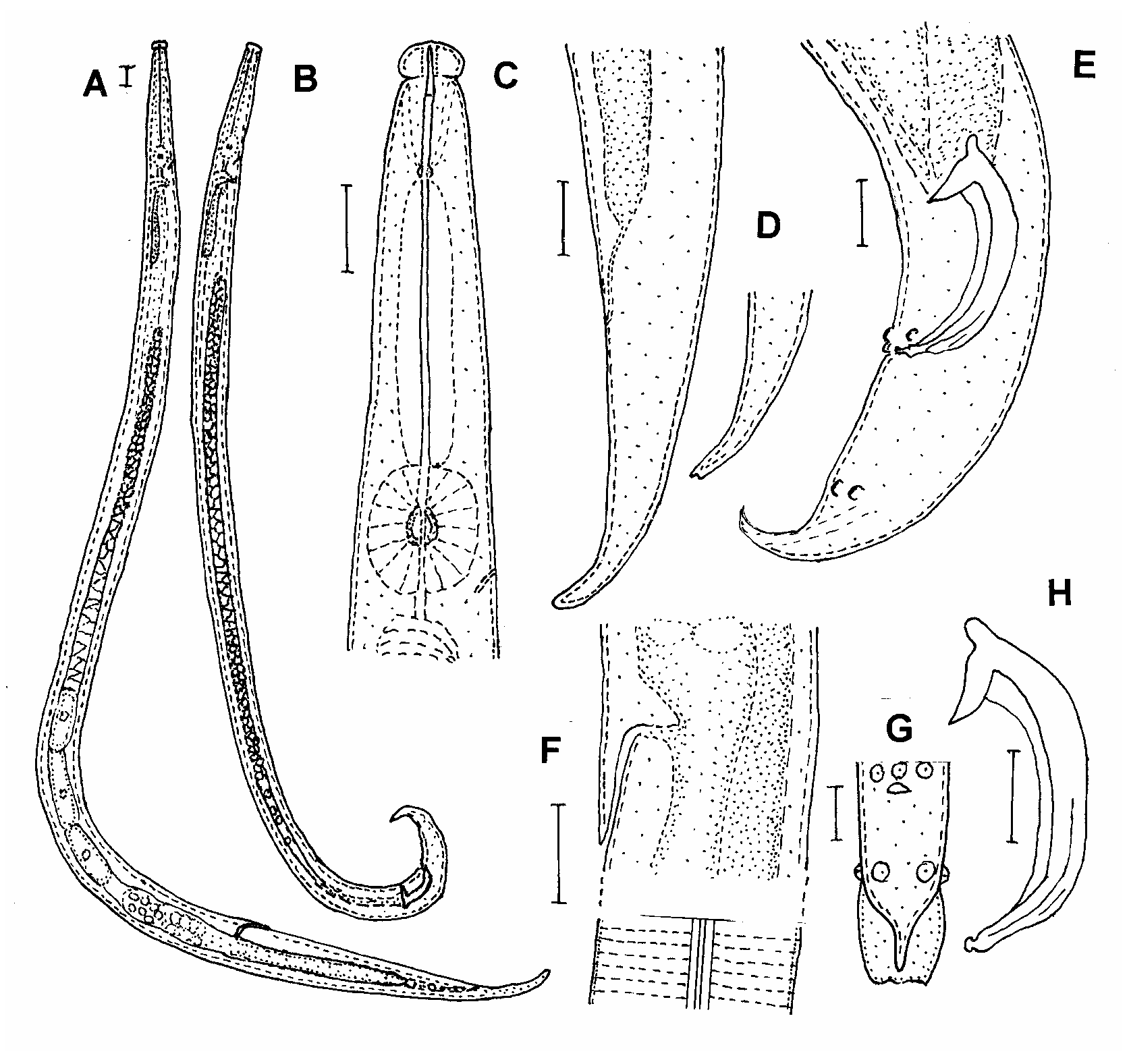
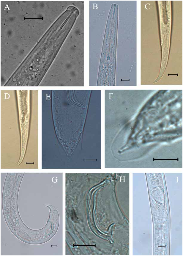
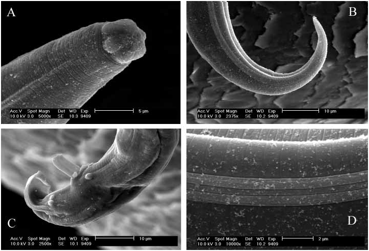
Description of Bursaphelenchus singaporensis sp.n. ( Figs 1–3 View FIGURE 1 View FIGURE 2 View FIGURE 3 )
MEASUREMENTS See Table 1.
MORPHOLOGICAL DESCRIPTION
Female
Displaying all the features of the Aphelenchoidoidea according to Hunt (1993), heatrelaxed form slim and ventrally arcuate. Cuticle marked by fine annules. Lateral field 5 µm wide with four lines. Lip region convex, 4.2 µm high, 9.4 µm wide, and offset by a distinct constriction. Stylet slender with small basal swellings, shaft forming about two thirds of total stylet length. Procorpus cylindrical. Median bulb oval, sometimes almost rectangular. Valve plates slightly posterior to the middle of the median bulb. Oesophageal gland lobe extending dorsally for a considerable distance (two to three body widths long) down the body. Nerve ring located at ca. one quarter of body diameter posterior to the median bulb. Excretory pore position at the level of median bulb or slightly posterior to it. Hemizonid at the level of intestine, ca. 100 µm from anterior end. Reproductive system prodelphic, gonad outstretched, occupying about half of the body length. Developing oocytes arranged in multiple rows. Anterior vulval lip prolonged posteriorly forming a ca. 8 µmlong vulval flap. Vulva situated in a slightly sunken area, about as long as the vulval flap and alongside strengthened by two small ribs. Postuterine branch extending over two thirds of vulva to anus distance, acting as a seminal receptacle. Tail long and slender, tapering to a slightly ventrally bent and finely rounded terminus, sometimes with a very small mucro.
Male
Anterior body region and cuticle similar to that of female. Body bent ventrally when killed by heat. Testis outstretched, occupying about half of the body length. Spermatocytes arranged in multiple rows. Spicules paired, very large and arcuate with pointed rostrum, sometimes with slightly dorsally hooked condylus. Distal ends of spicules with small cucullus. Tail ventrally arcuate with a pointed, talonlike terminus bearing a distinct, ovalshaped terminal bursa, which can be seen in dorsoventral position. There are three pairs of caudal papillae (one preanal and two pairs close to each other just anterior to the bursa) and a single midventral papilla anterior to the anus. Their position is shown in Figures 1 View FIGURE 1 and 3 View FIGURE 3 .
DIAGNOSIS AND RELATIONSHIPS
Bursaphelenchus singaporensis sp. n. clearly belongs to the B. xylophilus group having males with the typically shaped spicules with a cucullus at their distal extremity, the typical number and position of caudal papillae (a pair and a single one preanal, two adjacent pairs postanal just before the bursa) and the anterior vulval lip of the females developed as a distinct flap. The new species is characterized by a body length of 792 (553– 950) µm and 850 (690–961) µm of males and females, respectively, robust body (a= 34 for males and 31 for females), 15–16 µmlong stylet, lateral field with four lines, long postu terine branch (mean 102 µm) occupying about two thirds of vulva to anus distance, and a strongly conoid female tail (c= 20) with a finely rounded, only slightly ventrally bent terminus, male with very strong spicules (41–48 µm long), distinct rostrum and small cucullus, and a dorsoventral visible terminal bursa.
Having the typical characters of the B. xylophilus group, Bursaphelenchus singaporensis sp. n. is easily distinguishable from almost all other Bursaphelenchus species except species of this group. It is morphologically similar to B. abruptus GiblinDavis , Mundo Ocampo, Baldwin, Norden & Batra, 1993 (not in the B. xylophilus group), B. xylophilus , B. mucronatus Mamiya & Enda, 1979 , B. kolymensis Korenchenko, 1980 , B. fraudulentus Rühm, 1956 , B. conicaudatus Kanzaki, Tsuda & Futai, 2003 , B. baujardi Walia, Negi, Bajaj & Kalia, 2003 and B. luxuriosae Kanzaki & Futai, 2003 . The differentiation within the B. xylophilus group is mainly based on the shape of the female tail, whereas the spicules of males in the group widely resemble each other. However, B. singaporensis sp. n. differs from all other species of the B. xylophilus group by having larger spicules.
B. singaporensis sp. n. differs from B. abruptus by the degree of ventral curvature of the spicules (less bent in B. abruptus ), the larger spicules (41–48 vs. 23–30 µm) and the longer postuterine sac of females averaging 28% of the vulvaanus distance in B. abruptus (mean 57 µm) compared to 67% in B. singaporensis sp. n. (mean 102 µm).
B. singaporensis sp. n. differs from B. xylophilus mainly by the tapering female tail compared to the cylindrical tail with rounded tail terminus in B. xylophilus . It is more robust than B. xylophilus , B. mucronatus , B. kolymensis (a = 34/ 31 for males/ females vs. a averaging more than 40 in both males and females of the other three species). B. singaporensis sp. n. differs from the mucronate form of B. xylophilus and from B. mucronatus , B. kolymensis , B. fraudulentus , B. conicaudatus and B. baujardi by the lack of a distinct mucro at the female tail terminus. B. fraudulentus may have an indistinct mucro, but is reported as having spicules of 21–22 µm in length ( Rühm, 1956) vs. 41–48 µm in B. singaporensis sp. n. and a differently shaped tail. B. conicaudatus and B. baujardi , possessing a short mucro, also have shorter spicules of 23–28 µm and 25–28 µm in length, respectively.
B. singaporensis sp. n. is most similar to B. luxuriosae . The female tails of both species resemble each other in shape. B. singaporensis sp. n. differs morphologically from B. luxuriosae by the size of the spicules (41–48 vs. 27–30 µm), the vulva position (76 % vs. 71% of body length) and the position of the excretory pore, which is usually more anterior in B. singaporensis sp. n. (position at the level of median bulb or slightly posterior to it) than in B. luxuriosae (position ranging between centre of median bulb and hemizonid level).
MOLECULAR DIFFERENTIATION OF B. SINGAPORENSIS SP. N. FROM SIMILAR
SPECIES
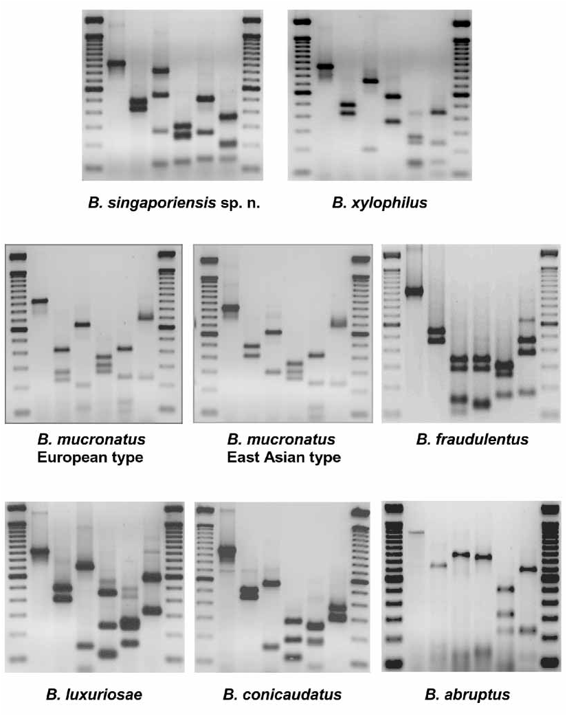
The pattern of DNA restriction fragments obtained in the ITSRFLP analysis of B. singaporensis sp. n. is different from the patterns of five other species of the B. xylophilus group and of the morphologically similar B. abruptus ( Fig. 4 View FIGURE 4 and Table 2). It is also distinct from the ITSRFLP patterns of 20 other Bursaphelenchus species summarized by Burgermeister et al. (2005).
As shown in the ITSRFLP pattern of B. singaporensis , digestion of the amplified rDNA sequence (950 bp) with Hae III resulted in four restriction fragments of approximately 840, 550, 290 and 120 bp, respectively. With some DNA extracts of the nematode, only the restriction fragments of 840 and 120 bp were obtained which together correspond to the size of the undigested rDNA sequence. The appearance of the additional restriction fragments of 550 and 290 bp may be explained by the presence of a Hae III restriction site within the 840 bp fragment in part of the amplified DNA. This may be caused by microheterogeneity of rDNA either between different specimens or in the same specimen, as has been observed, for instance, in Meloidogyne spp. ( Zijlstra et al., 1995). The situation may be clarified by sequencing of amplified and cloned rDNA of B. singaporensis (experiments in progress).
TYPE LOCALITY AND HABITAT
Packaging wood of nonconiferous hardwood exported from Singapore and inspected in Ningbo Entryexit Inspection and Quarantine Bureau, China, in 2003.
TYPES
Collected from a culture on Botrytis cinerea on malt agar, developed from a sample taken in China from packaging wood arriving from Singapore with other commodities. Slides in the nematode collection of Helen Braasch (holotype, allotype, paratypes 6 males and 6 females) , in the USDA Nematode Collection , Beltsville, Maryland, USA (paratypes 3 males and 3 females) and in the nematode collection of Ningbo Entryexit Inspection and Quarantine Bureau, China (paratypes 6 males and 6 females) .
| USDA |
United States Department of Agriculture |
No known copyright restrictions apply. See Agosti, D., Egloff, W., 2009. Taxonomic information exchange and copyright: the Plazi approach. BMC Research Notes 2009, 2:53 for further explanation.
|
Kingdom |
|
|
Phylum |
|
|
Class |
|
|
Order |
|
|
Family |
|
|
Genus |