
Dilemma frumarkernorum, Leal, Jos Ẽ H., 2008
|
publication ID |
https://doi.org/10.5281/zenodo.181996 |
|
DOI |
https://doi.org/10.5281/zenodo.6228963 |
|
persistent identifier |
https://treatment.plazi.org/id/081987A1-A218-BB56-FF5D-DA84FE49891A |
|
treatment provided by |
Plazi |
|
scientific name |
Dilemma frumarkernorum |
| status |
sp. nov. |
Dilemma frumarkernorum View in CoL new species
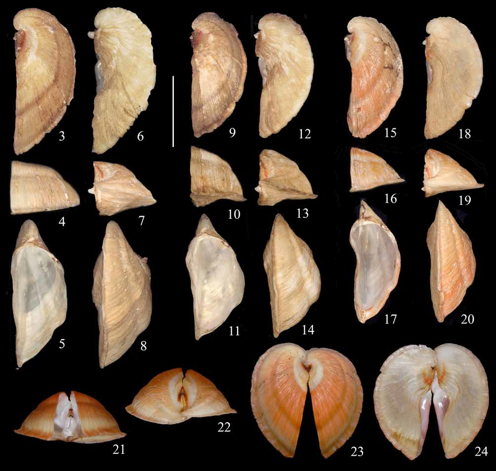
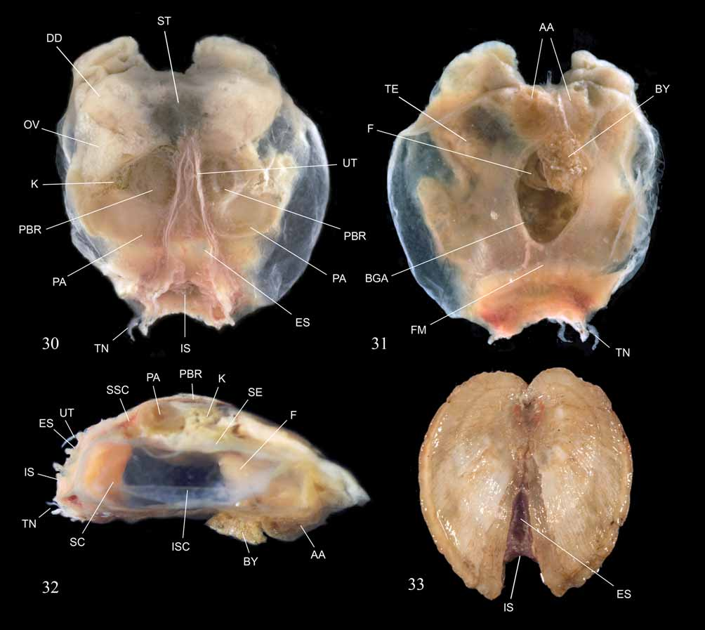
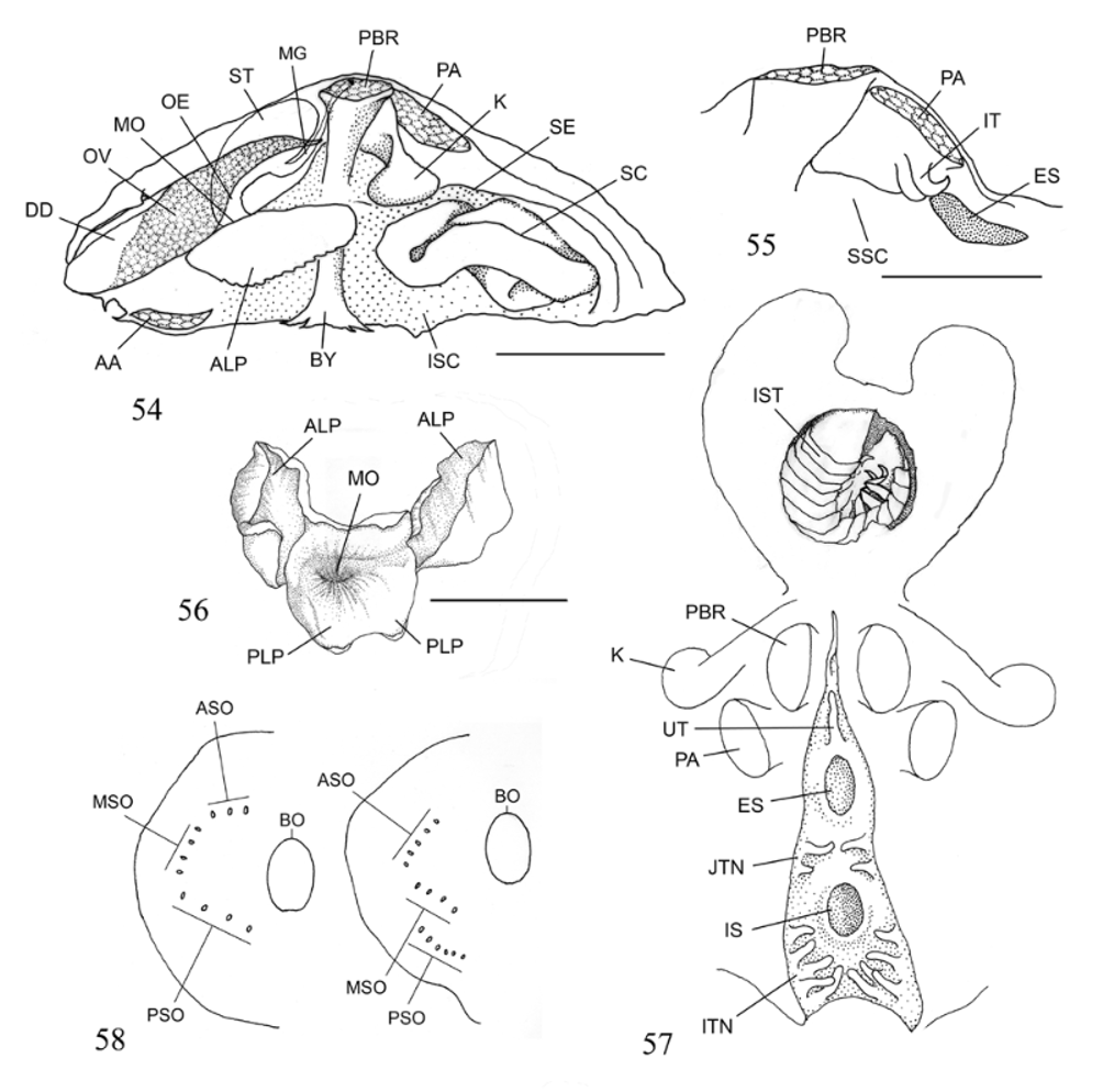
( Figures 1–33 View FIGURES 1 – 2 View FIGURES 3 – 24 View FIGURES 25 – 29 View FIGURES 30 – 33 , 58 View FIGURES 54 – 58 )
Diagnosis. Shell with sculpture of coarse growth lines; escutcheon not well separated from remainder of shell; foot filiform; byssus relatively thick, robust; siphonal cowl of moderate size; siphonal tentacles arranged around entire incurrent siphonal opening; septal ostia arranged to define three pairs of line segments deployed around byssal opening, roughly delineating a hexagon; siphonal ostia distributed as follows: three anterior, four median, and four posterior.
Description. SHELL ( Figures 1–29 View FIGURES 1 – 2 View FIGURES 3 – 24 View FIGURES 25 – 29 , 33 View FIGURES 30 – 33 ): Apparently nacreous internally, thin, equivalve (except for slight anteroposterior overlap at umbo), inequilateral, strongly compressed in anteroposterior direction, cardioid in lateral outline. Large byssal gap present in anteroventral position, on central portion of anterior shell surface.
Shell sculpture consisting of coarse growth lines, with well-developed carina separating anterior from posterior shell regions. Carina sometimes set by a constriction that resembles a “pinched” line parallel to main shell outline.
Umbones ( Figure 26 View FIGURES 25 – 29 , U) projecting dorsally, located in anterior position. Umbonal cavity, large, spacious.
Hinge with subtriangular, cardinal-like tooth ( Figure 26 View FIGURES 25 – 29 , CT) in each valve, tooth in left valve with cleft distal extremity and more developed than that in right. Corresponding sockets ( Figure 26 View FIGURES 25 – 29 , HS) present in each valve, that in right valve deeper than that in left valve. Posterior lateral tooth elongate, about 1/10 shell height, low, present on right valve only ( Figures 25, 26, 27 View FIGURES 25 – 29 , LAT) interlocking with notched depression on left valve ( Figure 27 View FIGURES 25 – 29 , LG).
Ligament ( Figures 25, 26, 27 View FIGURES 25 – 29 , LI) external, sunken; inner ligamental layer ( Figure 28 View FIGURES 25 – 29 , IL) in part white, hard, possibly calcified ( Figure 28 View FIGURES 25 – 29 , CIL); outer ligamental layer ( Figure 28 View FIGURES 25 – 29 , OL) brown, with dorsal periostracum adhering to outer layer.
Anterior adductor muscle scar ( Figures 25, 26, 29 View FIGURES 25 – 29 , AAS) located on projection close to margin of valve, dorsal to byssal gap. Posterior adductor muscle scar ( Figure 25 View FIGURES 25 – 29 , PAS) about same size as anterior adductor scar. Anterior byssal retractor scar ( Figure 26, 29 View FIGURES 25 – 29 , ABS) small, relatively deep, situated on internal entrance to umbonal cavity. Posterior byssal retractor scar ( Figure 25 View FIGURES 25 – 29 , PBS) larger than posterior adductor scar, elongate. Pallial line ( Figures 25, 29 View FIGURES 25 – 29 , PAL) continuous, strong.
Lunule absent. Escutcheon relatively poorly defined, not separated by a prominent groove, but distinguishable from remainder of shell by slightly smoother texture and commarginal band of lighter color.
In specimens examined, shell color ranging from dull light-brown to olive-brown, sometimes suffused with peach-orange, usually on posteroventral part of valves. Periostracum light-brown, laid in coarse commarginal lamellae, mostly flaking in live-collected specimens.
Macroanatomy. MANTLE MARGIN AND SIPHONS: Mantle margins fused ( Figure 31 View FIGURES 30 – 33 , FM) leaving two siphonal openings ( Figures 30, 32, 33 View FIGURES 30 – 33 , ES, IS) and byssal gape ( Figure 31 View FIGURES 30 – 33 , BGA). Byssal gape circular to elliptical. Siphonal apertures separate; siphonal area sometimes colorful in freshly preserved animals, siphons formed by fusion of inner mantle folds (“ Type A” of Yonge, 1982).
Incurrent siphonal opening ( Figures 30, 32 View FIGURES 30 – 33 , IS) located ventrally, much larger (at least twice as wide) than that of excurrent siphonal opening ( Figures 30, 32, 33 View FIGURES 30 – 33 , ES); siphonal cowl ( Figure 32 View FIGURES 30 – 33 , SC) inverted, resting inside infraseptal chamber, covering incurrent siphonal opening internally, probably eversible for prey capture, relatively short. Base of incurrent siphon surrounded by 15 tentacles ( Figures 30–32 View FIGURES 30 – 33 , TN); tentacles simple, tapered. Large unpaired tentacle ( Figures 30, 32 View FIGURES 30 – 33 , UT) present along fused mantle margin dorsal to excurrent siphonal opening; following six (3+3) tentacles deployed along membranous hem-like projection of middle mantle fold on each side of intersiphonal junction. Remaining eight tentacles arranged around incurrent siphonal opening.
Incurrent siphonal opening located almost ventrally, at least twice as wide as excurrent siphonal opening. Excurrent siphonal opening located posteriorly; angle formed by junction of margins of two valves in posterior direction subtending excurrent siphonal opening ( Figures 30, 33 View FIGURES 30 – 33 ).
MANTLE CAVITY: Septum ( Figure 32 View FIGURES 30 – 33 , SE) located dorso-ventrally within mantle cavity; septum roughly parallel to anterior shell surface. Septum thin, strong, dividing the mantle cavity into two chambers, the supraseptal (posterior) ( Figure 32 View FIGURES 30 – 33 , SSC) and infraseptal (anterior) ( Figure 32 View FIGURES 30 – 33 , ISC) chambers. Septum attaching to dorsal region of shell posterior to anterior adductor muscles but attaching to ventral region of shell anterior to posterior adductor muscle.
Septum perforated by byssal opening ( Figure 58 View FIGURES 54 – 58 [left], BO) and by groups of ostia arranged to define three pairs of line segments deployed around byssal opening, roughly delineating a hexagon. Ostia distributed as follows: three anterior ( Figure 58 View FIGURES 54 – 58 [left], ASO), four median ( Figure 58 View FIGURES 54 – 58 [left], MSO), and four posterior ( Figure 58 View FIGURES 54 – 58 [left], PSO).
MAJOR SHELL MUSCLES: Strong modification of shell shape associated with shape and positioning of adductor muscles: adductor muscles almost parallel to valve surfaces to which they are attached. Anterior adductor muscles ( Figures 31, 32 View FIGURES 30 – 33 , AA) short, flattened; posterior adductor muscles ( Figures 30, 32 View FIGURES 30 – 33 , PA) longer, also flattened. Posterior byssal retractor muscles ( Figure 30, 32 View FIGURES 30 – 33 , PBR) Y-shaped, very well developed, attaching to locations close to posterior margin of valves. Anterior byssal retractor muscles much thinner, attaching dorsally to internal surface of entrance to umbonal cavity. Septal attachment muscles difficult to observe given poor preservation of specimens.
BYSSUS AND FOOT: Byssus ( Figures 31, 32 View FIGURES 30 – 33 , BY) well-developed, prominent, circular in cross section, in preserved animals attached to grains of calcareous sediment, consisting of bundle of very fine, fused filaments, cylindrical in cross-section. Byssus originating from a thickened section of vestigial foot ( Figure 31 View FIGURES 30 – 33 , F), which is narrow and filiform. Position and size of vestigial foot indicates that it might never exit infraseptal chamber.
MOUTH AND LABIAL PALPS: Mouth ridged internally along entire circumference; posterior labial palps reduced to two vestigial projections flush with surrounding surface. Anterior labial palps very well developed, broad, folded over mouth, potentially expandable in posterodorsal direction.
ALIMENTARY SYSTEM: Esophagus with strong folds, very short, opening anterodorsally into stomach. Stomach ( Figure 30 View FIGURES 30 – 33 , ST) of Type II, compact, slightly elongate dorsoventrally, forming posteroventral projection, internally ridged, with ridges stronger in ventral surface. Crystalline style situated just ventral to esophagus opening, projecting slightly into stomach, surrounded by chitinous shield. Openings of digestive diverticula not observed because of poor fixation. Digestive diverticula located in dorsal “horns” ( Figures 30 View FIGURES 30 – 33 , DD). Midgut short, connecting to stomach anteroventral position, ventral to esophagus, curving to more posterodorsal position, anus opening in supra-septal chamber near opening of excurrent siphon between posterior byssal retractor muscles.
STOMACH CONTENTS: Examination of stomach contents of four paratypes revealed, in one specimen, partially digested remains of unidentifiable ostracod species.
KIDNEYS: Kidneys situated posterodorsally between the remainder of visceral mass and posterior byssal retractor muscle. Kidneys comprising two elongate structures, one on each side of the midline of posterior region ( Figure 30 View FIGURES 30 – 33 , K).
REPRODUCTIVE SYSTEM: Testes ( Figure 31 View FIGURES 30 – 33 , TE) elongated situated in anteroventral position in relation to ovaries, ovary overlying the testes. Ovaries ( Figures 30 View FIGURES 30 – 33 , OV) consisting of pair of elongated sacs symmetrically deployed on each side of midline. Ovaries and testis ventral to digestive diverticula.
Type material. Holotype, BMSM 15029, (length×height×width [in mm] = 7.41×20.37×16.55) Paratype 1, USNM 1112670 (6.61×16.76×14.87).
Paratype 2, UF 416419 (7.85×17.34×14.90), Paratype 3, collection of Steve Kern unnumbered (6.79×17.24×14.52), Paratype 4, collection of Frank Frumar (6×16×14; not measured with same accuracy as other types), unnumbered. All from type locality, Steve Kern coll. May 2006, dredged, lobster boat.
Type locality. Southwest of Key West, Monroe County, Florida, USA, 229 m depth.
Abbreviations: AAS = scar of anterior adductor muscle; ABS = scar of anterior byssal retractor muscle; ASC = scar of anterior septal muscle; CIL = calcified part of inner ligament layer; CT = cardinal-like tooth; HS = hinge socket; IL = inner ligament layer; LAT = lateral tooth; LG = lateral groove; LI = ligament; OL = outer ligament layer; PAL = pallial line; PAS = scar of posterior adductor muscle; PBS = scar of posterior byssal retractor muscle; PE = periostracum; OL = outer ligament layer; U = umbo.
Etymology. The new species is named simultaneously after Frank Frumar and Steve Kern, for kindly making the material available for study.
Remarks on habitat and type locality. The depth given above for the type locality is as related by the collector. The two other congeners (below) were collected much deeper, between 805 and 961 m depth. Remarks. See comparative remarks below, under Dilemma spectralis new species and Dilemma inexpectatum new combination.


No known copyright restrictions apply. See Agosti, D., Egloff, W., 2009. Taxonomic information exchange and copyright: the Plazi approach. BMC Research Notes 2009, 2:53 for further explanation.
|
Kingdom |
|
|
Phylum |
|
|
Class |
|
|
SuperOrder |
Anomalodesmata |
|
Order |
|
|
Family |
|
|
Genus |