
Lecithocera fascimaculata Oku, 2021
|
publication ID |
https://doi.org/10.11646/zootaxa.4996.3.7 |
|
publication LSID |
lsid:zoobank.org:pub:BA37E27E-087D-4451-BD43-43123645C301 |
|
persistent identifier |
https://treatment.plazi.org/id/083087FE-1976-FFD0-E8D0-ECF6375631DA |
|
treatment provided by |
Plazi |
|
scientific name |
Lecithocera fascimaculata Oku |
| status |
sp. nov. |
Lecithocera fascimaculata Oku View in CoL sp. nov.
[Japanese name: Kiiro-hosoba-higenaga-kibaga]
( Figs 1f View FIGURE 1 , 2d View FIGURE 2 , 3d View FIGURE 3 , 4d View FIGURE 4 , 5b View FIGURE 5 )
Lecithocera sp. 2 ; Oku, 2003: 42.
Diagnosis. The species is characterized by the following characteristics: forewing with a strigula at the end of the cell connected to the tornal stripe, and tegumen of the male genitalia with long hair-like scales situated dorsolaterally.
The new species is similar to L. aulias Meyrick, 1910 in genitalic features, but it separated by the following characteristics: the longer male cornuti of the aedeagus; the paired lobes of the sclerotized structure on the 7 th sternite of the male swollen and oblong on the cephalic side; the trapezoidal antrum of the female, and the ductus seminalis branched from the middle of ductus bursae. The new species is also very similar to L. bimaculata Park, 1999 from Taiwan on superficial and genitalic characteristics, but is distinguished by the following features: in male genitalia, valva with an obtuse apex, paired lobes of the sclerotized structure on the 7 th sternite widely swollen and oblong on the cephalic side, female genitalia with ductus bursae weakly sclerotized in near inception of ductus seminalis, and signum of corpus bursae transversely fiddle-shaped. It is also similar to L. quadtariella Park, 2018 from Taiwan in the genitalia, but is distinguished by the wingspan smaller than the latter, the basal plate of gnathos of the male genitalia slightly convex on caudal margin, and the costal bar angled in middle.
Description. Wingspan, 14.0–16.0 mm. Head: yellow; vertex concolorous. Frons light brown. Antenna yellow, 9/10 length of forewing; scape brown laterally; flagellum with light brown annulations on distal 1/5. Labial palpus reddish yellow on outer surface and light yellow on inner surface; 3rd palpomere slightly shorter than 2nd, blackish ventrally in male. Thorax: thorax and tegula orange yellow. Legs light yellow, ventral side randomly covered with brown scales. Mid-tibia brown at basal part dorsally, with spurs brown dorsally and light yellow ventrally. Hind tibia with numerous long and light yellow hairs and with two pairs of spurs light yellow dorsally and brown ventrally; hind tarsus covered with brown scales ventrally. Abdomen: brown dorsally, paler toward caudal side, reddish yellow ventrally.
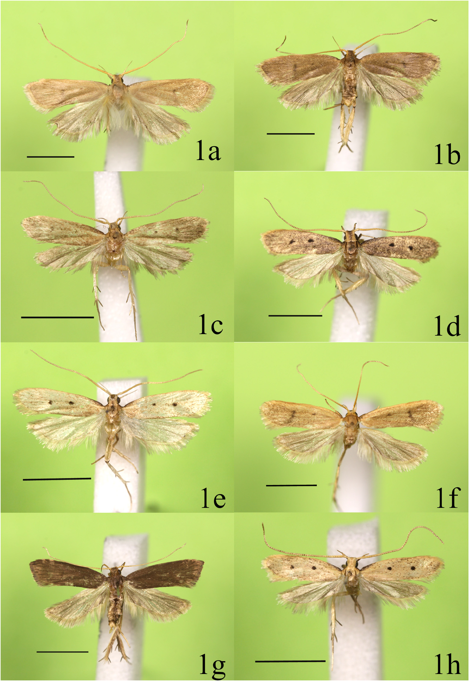
Forewing ( Fig. 1f View FIGURE 1 ) with oblique termen; ground color reddish yellow, slightly speckled with dark brown scales, becoming denser towards termen, with two discal spots present, small one at middle, and larger one at end of cell and conspicuously connected with tornal stripe; costa with dark brown suffusion on base 1/9; fringe reddish yellow. Hindwing color brown with oblique and slightly sinuate termen; fringe brown paler toward apex, pale yellow from basal part to middle of dorsum.
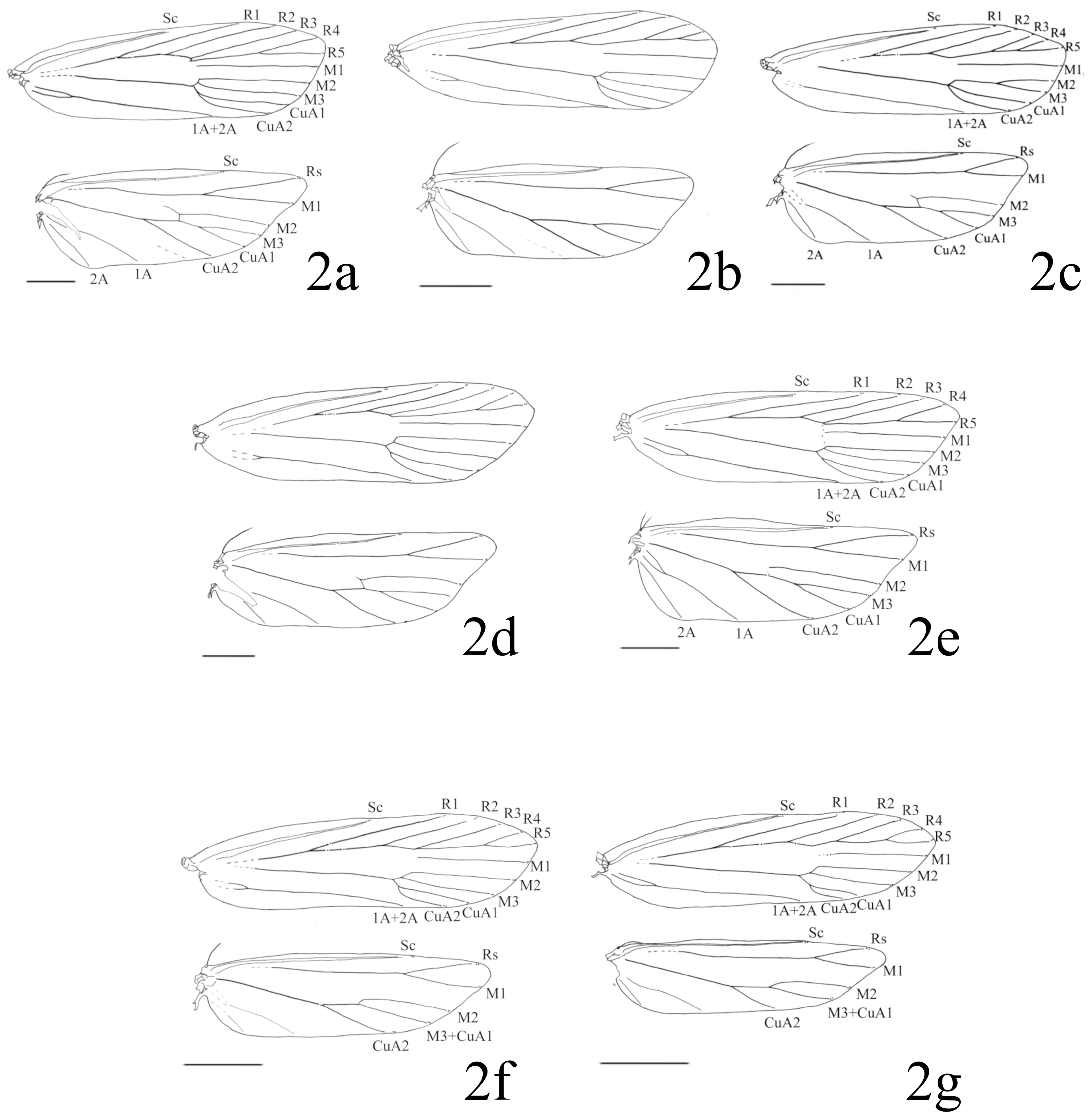
Wing venation ( Fig. 2d View FIGURE 2 ): Forewing: Sc ending middle of costa. R1 arising from basal 1/2 of cell; R2 from distal 1/8 of cell; R3 free, from upper angle of cell; R4 and R5 stalked for distal 1/3; R5 to costa. M1 and M2 parallel; M3 free; CuA1 and CuA2 short-stalked; 1A + 2A forked basally; cell opened. Hindwing: Sc ending at distal 1/3 of costa; Rs running to near apex; Rs and M1 stalked at distal 1/3; M2, parallel to M3 and CuA1; M3 and CuA1 short-stalked; CuA2 from distal 1/6 of cell; 1A to about 1/2 of dorsum; 2A to basal 1/3 of dorsum.
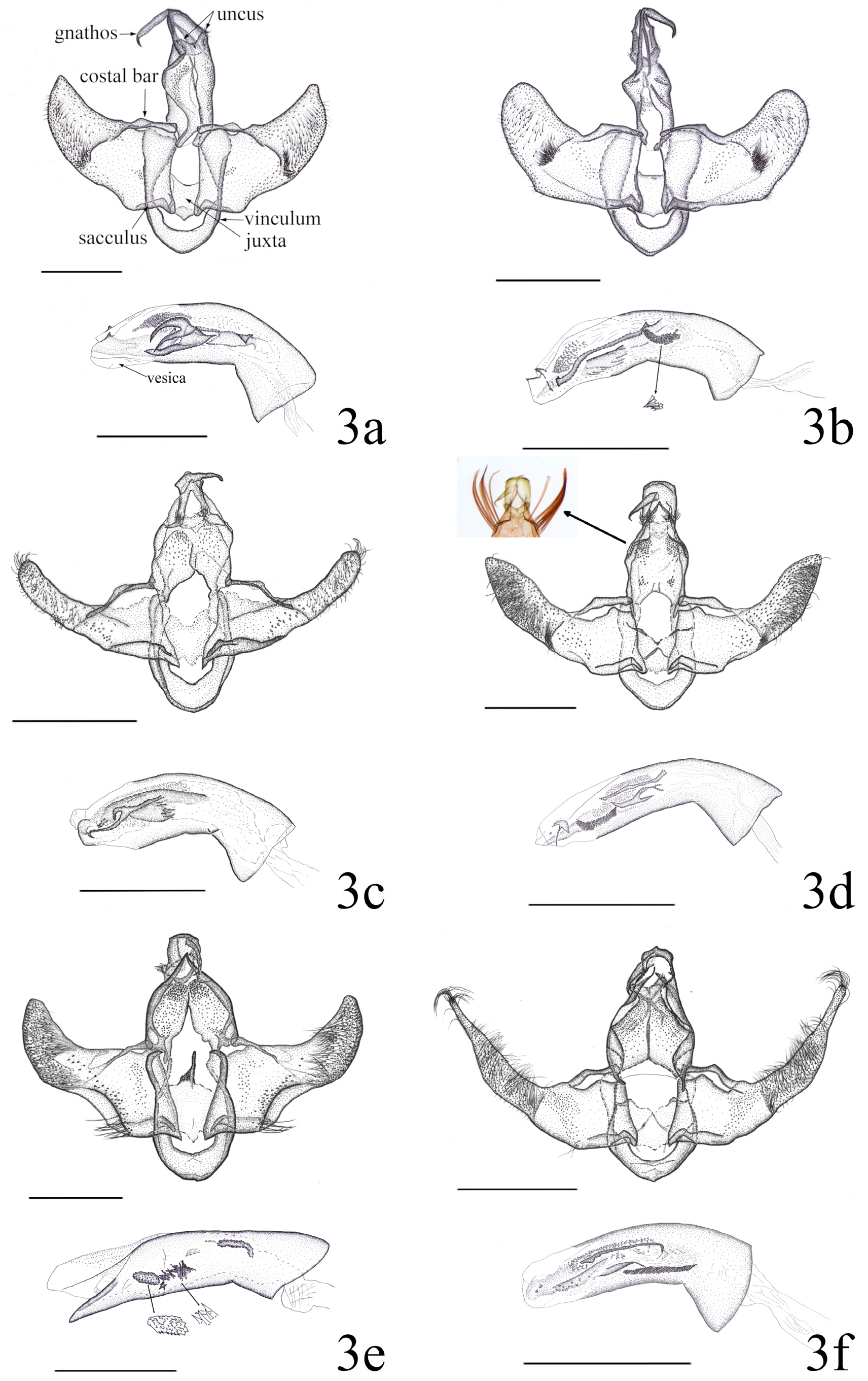
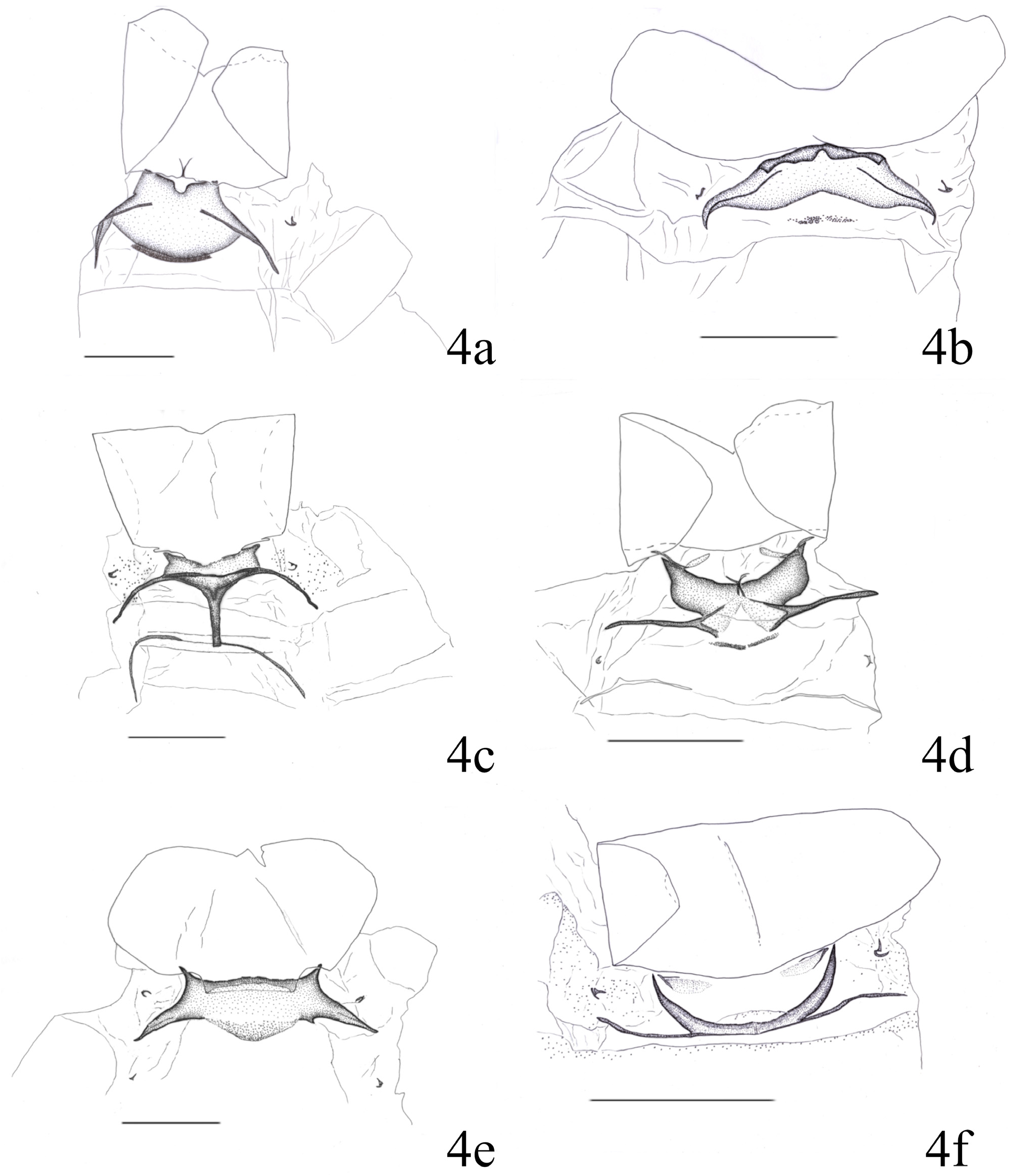
Male genitalia ( Figs 3d View FIGURE 3 , 4d View FIGURE 4 ). Uncus bilobed posteriorly, with some setae on surface. Median process of gnathos long, beak-shaped, five times as long as uncus, slender as wide as uncus, strongly bent subapically; basal plate of gnathos quadrate and slightly convex on caudal margin. Tegumen with long and removable hair-like scales dorsolaterally. Valva with obtuse apex, angled in basal 1/6 beneath costal bar, about three times as long as costal bar; cucullus about 3/5 times as wide as base, with numerous setae on ventral and inner surface; costal bar angled in middle; sacculus slender, cephalic side angled in basal 1/4, terminating near 1/4 of ventral margin. Juxta shieldshaped, caudal side slightly concave and with crescent process laterally, medial part of cephalic side moderately pointed. Aedeagus about 4/5 length as long as valva, about 1/5 times as wide as long; ventral margin strongly bent in proximal 1/4; apical part covered with membrane; proximal part about 1.4 times as wide as median part; vesica with cornuti; fan-shaped cornutus near apex; numerous oblong cornuti arranged in some rows bunched before apical part; long and liner-shaped, and bifurcated cornuti in middle. Seventh sternite with pair of lobes and long lateral rods plate, a pair of lobes projected on caudal side unilaterally and swollen and oblong on cephalic side, a pair of lateral rod-shaped plates bifurcated at basal 1/6.
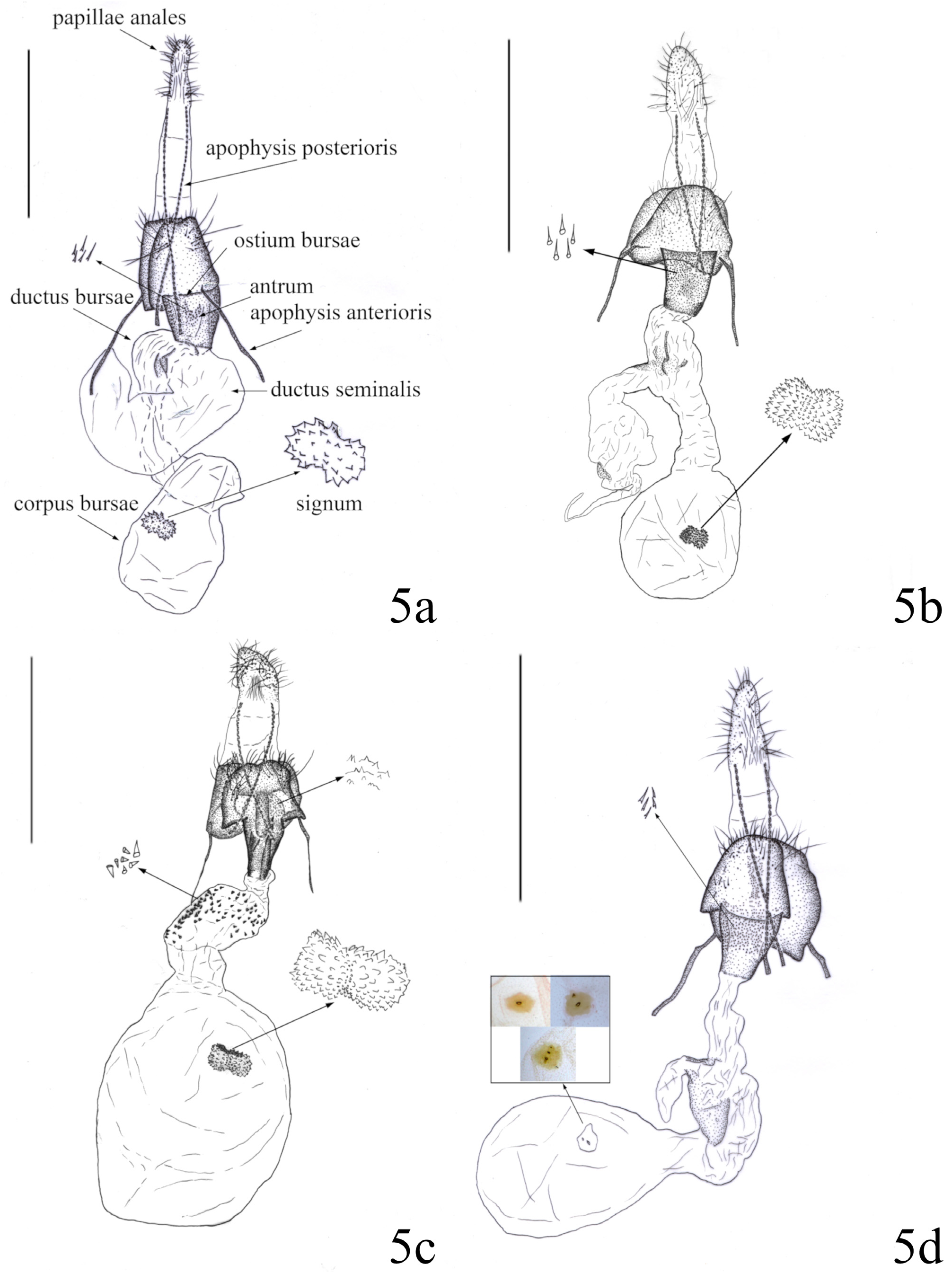
Female genitalia ( Fig. 5b View FIGURE 5 ). Papillae anales about 1/2 times as wide as long, with many short and long setae; joint membrane between papillae anales and eighth segment about equal length to papillae anales. Ostium bursae opening widely, occupying 5/8 width of eight segment. Antrum trapezoidal, cup-shaped, 5/6 times as long as papillae anales, with numerous minute and conical denticles on surface. Apophysis anterioris about 4/7 times as long as apophysis posterioris. Ductus bursae about 1.5 times as long as apophysis posterioris, weakly sclerotized in near inception of ductus seminalis; ductus seminalis relatively broad, branched from middle of ductus bursae. Corpus bursae ovate, about two times as long as papillae anales, about 5/6 times as wide as long. Signum transverse, fiddle-shaped, with many minute denticles on surface, placed on about middle of corpus bursae, 1/5 times as long as antrum, two times as wide as long.
Type materials. HOLOTYPE. JAPAN, [Shikoku]: ♂, Ishizuchi-skyline , Kumakogen Town, Ehime-Pref., 2.vii.2016, T. Ito leg. ( 1 ♂ Gen sl. no. Lecithocera No. 8), in ELKU.
PARATYPES. JAPAN, [ Shikoku]: 2 ♂, Okawa-mine, Uchiko Town, Ehime pref., 6.vii.2016, K. Kuroda leg. ( 1 ♂ Gen. sl. no. Lecithocera No. 9, wing sl. no. Lecithocera No. 9-2), in ELKU ; 1 ♂, Ishizuchi-skyline, Kumakogen Town , Ehime Pref., 20.vii.2016, J. Oku leg., in ELKU ; 1 ♀, Tengu-kogen, Kumakogen Town, Ehime Pref., 21.vii.2016, J. Oku leg. ( 1 ♀ Gen sl. no. Lecithocera No. 10, wing sl. no. Lecithocera No. 10-2), in ELKU ; [Kyushu]: 1 ♂, Terukuni shrine area, Kagoshima City, Kagoshima Pref., 11.v.2017, J. Oku leg. ( 1 ♂ Gen. sl. no. Lecithocera No. 18), in KGU ; 1 ♀, same locality, 25.v.2017, J. Oku leg. ( 1 ♀ Gen. sl. no. Lecithocera No. 25), in KGU .
Distribution. Japan: Honshu ( Oku 2003), Shikoku, Kyushu.
Biology. Adults fly from the mid-spring to the early autumn.
Etymology. The specific name is derived from the Latin “ fascia ” (=band) and “ maculatus ” (=spotted), because the forewings have large spots connected with the tornal stripe at the end of the cell.
Remarks. Oku (2003) treated this species as unidentified Lecithocera sp. 2 , and Sakamaki (2013) misidentified it as L. rotundata Gozmány, 1978 .
| KGU |
Geology and Mineralogy Museum |
No known copyright restrictions apply. See Agosti, D., Egloff, W., 2009. Taxonomic information exchange and copyright: the Plazi approach. BMC Research Notes 2009, 2:53 for further explanation.
|
Kingdom |
|
|
Phylum |
|
|
Class |
|
|
Order |
|
|
Family |
|
|
Genus |
Lecithocera fascimaculata Oku
| Oku, Johei, Sakamaki, Yositaka & Hirowatari, Toshiya 2021 |
Lecithocera sp. 2
| Oku, T. 2003: 42 |