
Microplana humicola Vejdovsky, 1890
|
publication ID |
https://doi.org/ 10.11646/zootaxa.4980.1.11 |
|
publication LSID |
lsid:zoobank.org:pub:984CA16F-FA9D-423D-B431-02740688E0B1 |
|
DOI |
https://doi.org/10.5281/zenodo.4945082 |
|
persistent identifier |
https://treatment.plazi.org/id/0873356D-DB61-FFA7-BCA9-DD05C1D8FB87 |
|
treatment provided by |
Plazi |
|
scientific name |
Microplana humicola Vejdovsky, 1890 |
| status |
|
Microplana humicola Vejdovsky, 1890 .
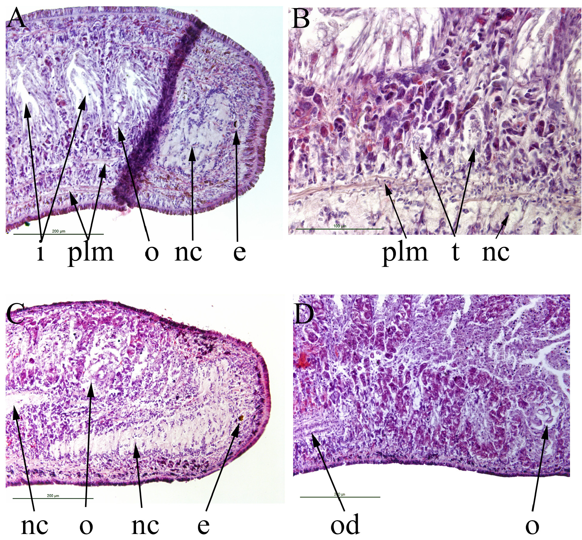
Vejdovsky (1890) described small white terrestrial planarians found in 1887 in a manure heap near Bechlin, Bohemia (now Czech Republic) as (translated from the original French): “shining body of snow-white”; “not flattened”; 1–2 mm long in July; 4–6 mm long in September; “and probably increased further after the genitals acquired their complete development”. Vejdovsky (1890) examined gently squashed whole live specimens by translucency and was unable to preserve or section any specimens.
A pair of eyes were present as very small black spots situated on the anterior lobe of the anterior expansion of the ventral nerve cord on each side, smaller than he has seen in any other planarian. There were two pairs of testes. Sperm ducts (his Plate II, Fig 15) are shown joining anterior to the penis to form a single seminal vesicle full of mature sperm, which opens into the base of the penis via a single opening. A blind sac (“glande accessoire”; his Plate II, Figs 12 & 15) opens near the junction of the ovovitelline ducts, but the description of the female ducts in this region is vague. There was no genito-intestinal connection.
Schneider (1935) described similar specimens found in Graz, Austria and in Cologne, Germany and considered them to be the same species as those of Vejdovsky (1890). These were 2.8–3.6 mm long and pure milk white (translated from the original German). The anterior end was transparent and the two eyes visible as fine dots. There were two pairs of testes. His Fig 2 View FIGURE 2 shows the sperm ducts as dashed lines approaching the base of the penis separately but that diagram has little detail. His Fig 3 View FIGURE 3 shows a “vesicula seminalis” at the base of the penis. It lacks stored sperm and unfortunately the diagram does not show the entry of the sperm ducts. The text states that “On my animals (sperm ducts) were not raised to false seminal vesicles”. He continues: “The well-formed, elongated cone-shaped penis protrudes from above into the atrium masculinium. The muscular penile bulb surrounds the elongated eggshaped seminal vesicle, which gradually narrows downwards and passes into the ejaculatory duct.” Thus, what he calls the seminal vesicle is inside the muscular bulb of the penis and appears to be the proximal, wider, part of the ejaculatory duct within the penis. The lack of stored sperm in any part of the male system could be because the specimens were either not fully mature or possibly spent. A genito-intestinal duct was present opening near the junction of the ovovitelline ducts and their opening into the common female duct. The opening of this duct into the intestine is shown (his Fig 5 View FIGURE 5 ) as an expanded, funnel-like opening.
None of Schneider’s material is known to survive.
Pantin (1953) concludes that there is a “fair probability” that Schneider’s (1935) specimens were the same as Vejdovsky’s (1890) and nominated M. humicola as the type species of the genus Microplana . This has been accepted by subsequent revisions (e.g. Ogren & Kawakatsu, 1988). However: there must be considerable doubt about the similarity of the specimens of Vejdovsky (1890) to those of Schneider (1935), despite Pantin’s (1953) conclusion. The approach of the sperm ducts to the base of the penis is different, and in Vejdovsky’s (1890) specimens there was a blind “glande accessoire” (equivalent to a copulatory bursa present in some species) joining at the posterior of the female duct, whereas Schneider’s (1935) specimens had a genito-intestinal connection. These two differences alone call into question the co-specificity of the specimens. There are apparently no known specimens of M. humicola in any collection. Thus any comparison has to rely solely on the descriptions of Vejdovsky (1890) and Schneider (1935).
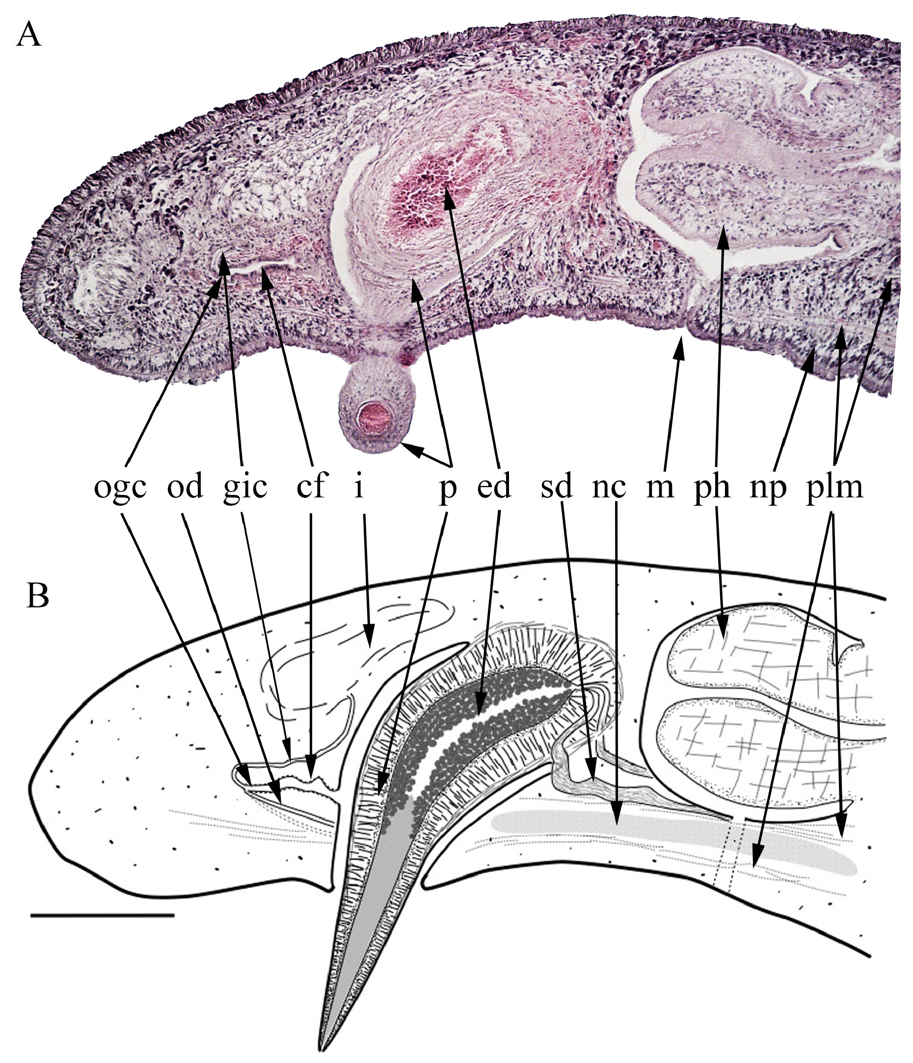
Our specimens are larger than those of either Vejdovsky (1890) or Schneider (1935). The eyes of our specimens are similarly small. Both Vejdovsky’s (1890) and Schneider’s (1935) specimens had two pairs of testes, stated to be dorsal in the latter. Our specimens have an uncertain number of testes but apparently more than two pairs. The sperm ducts in both our specimens are expanded and contain stored sperm (false seminal vesicles), whereas Vejdovsky’s (1890) specimens had a single seminal vesicle with stored sperm. Schneider (1935) did not note any stored sperm. The opening of the genito-intestinal duct into the intestine in our specimens is diffuse and indistinct, whereas Schneider (1935) shows the opening as a funnel-shaped opening. There was no genito-intestinal duct in Vejdovsky’s (1890) specimens, but a blind “glande accessoire” was present.
Thus our specimens have more in common with those of Schneider (1935) than with those of Vejdovsky (1890). But the number of testes and the form of the genito-intestinal opening are different. Thus we consider them not to be Microplana humicola .
Jones (2005) reported that M. humicola had been found in three localities in the U.K. This identification must be considered extremely doubtful and should be disregarded. The specimens were small and white, thus superficially similar to M. edwardsi . One of these specimens (in the collection of HDJ, collected 23 April 1983 from Noss Mayo, Devon, 50.311143, -4.045371) was sectioned. Sections are 2 mm long, the mouth and gonopore are respectively 1.25 mm and 1.6 mm from the anterior end (62.5% and 80% of body length). The pharynx is cylindrical, 180 μm long and 175 μm in diameter. The gonopore opens into an atrium but there is no development of the penis. Ovaries, ovovitelline ducts, testes and sperm ducts are not discernable. Sub-epidermal longitudinal muscle bundles are present but no parenchymal longitudinal muscle bundles, unlike M. edwardsi . The eyecups are 30 μm in diameter, larger than in M. edwardsi . Both the latter characteristics suggest that this specimen is not M. edwardsi , though what species it is remains unknown.

No known copyright restrictions apply. See Agosti, D., Egloff, W., 2009. Taxonomic information exchange and copyright: the Plazi approach. BMC Research Notes 2009, 2:53 for further explanation.