
Microplana edwardsi, Jones & Mcdonald, 2021
|
publication ID |
https://doi.org/ 10.11646/zootaxa.4980.1.11 |
|
publication LSID |
lsid:zoobank.org:pub:984CA16F-FA9D-423D-B431-02740688E0B1 |
|
DOI |
https://doi.org/10.5281/zenodo.4945080 |
|
persistent identifier |
https://treatment.plazi.org/id/0873356D-DB67-FFAC-BCA9-DDE9C5B6FE63 |
|
treatment provided by |
Plazi |
|
scientific name |
Microplana edwardsi |
| status |
sp. nov. |
Microplana edwardsi sp. nov.
Etymology: the specific epithet is after the father of one of us (J. McD) who encouraged her interest in natural history.
Diagnosis. Microplana edwardsi is distinguished from its congeners by its small size, live length 6–7 mm, <1 mm wide; approximately cylindrical body; creamy-white color with diffuse dark pigment anteriorly. Anatomically it has the characters of the genus, a single pair of eyes; parenchymal longitudinal muscle; a cylindrical pharynx; a pair of anterior ovaries; an unknown number of testes; conical penis papilla; genito-intestinal duct.
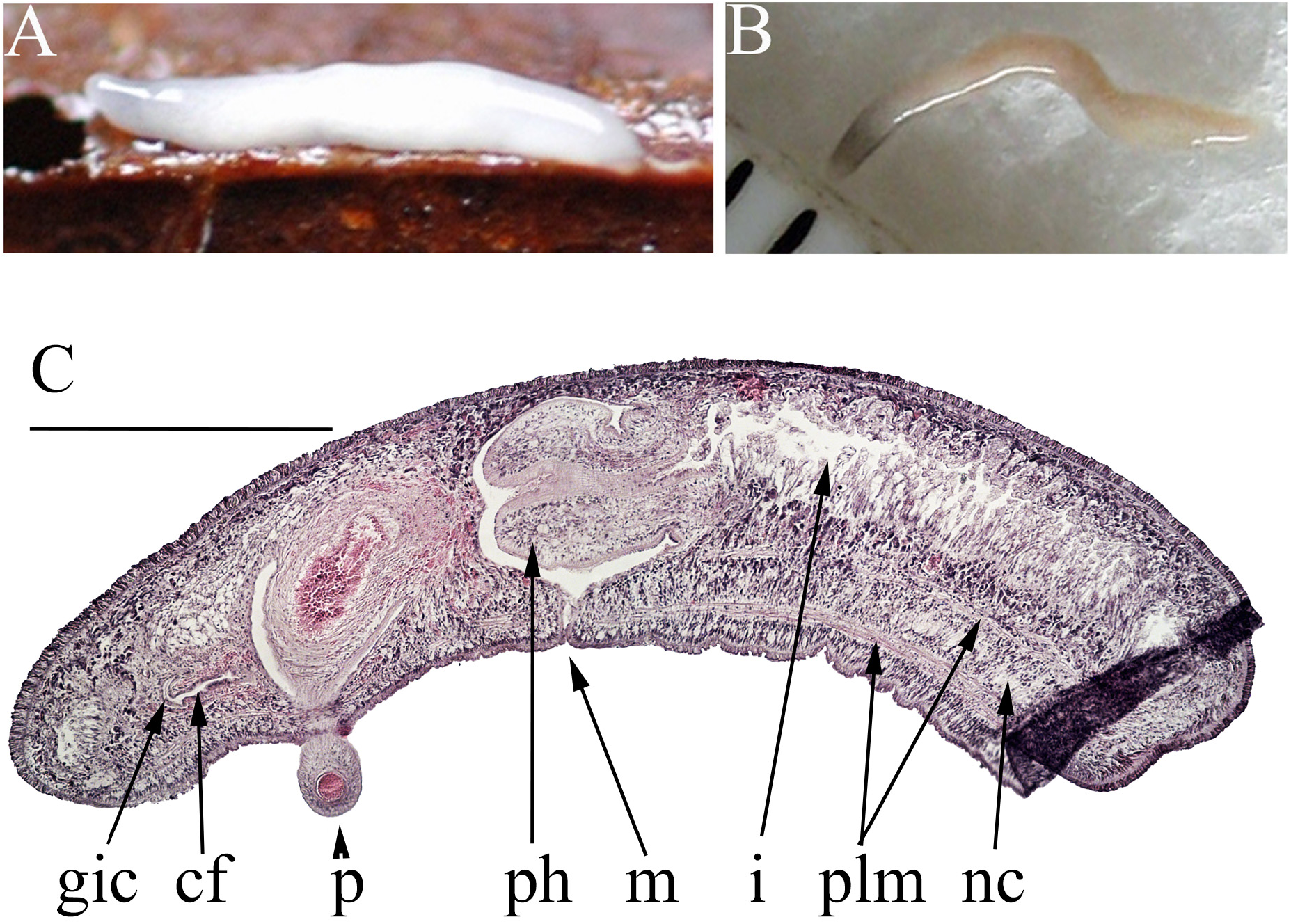
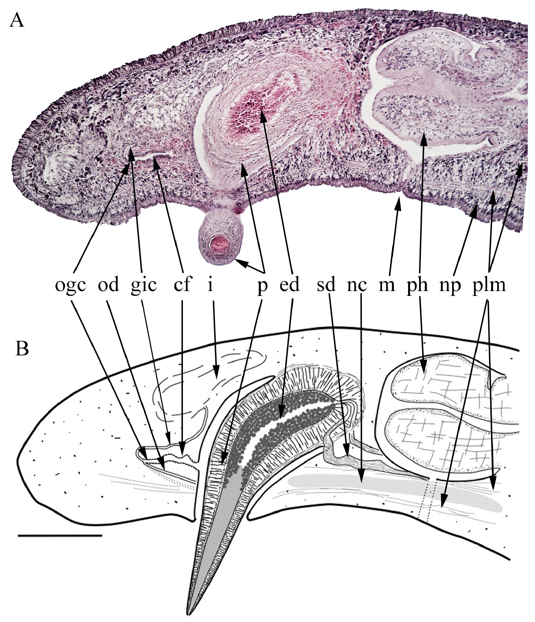
Description. Living worms ( Fig 1 A, B View FIGURE 1 ) are 6–7 mm long, <1 mm in width, cylindrical though slightly wider than high, and bluntly rounded anteriorly and posteriorly though the anterior end is narrower. They are creamywhite, with diffuse dark grey pigment particularly concentrated towards the anterior end. The ventral creeping sole is about half the width of the animal (based on the lateral extent of the ventral ciliated epidermis in sections of WF06, and an approximate transverse section of the posterior end of WF09). Sections of WF06 are about 2.3 mm long ( Fig 1 C View FIGURE 1 ), the mouth (pharyngeal aperture) and gonopore are respectively about 1.1 mm and 1.6 mm from the anterior end (55% and 77% of body length). WF09 was twisted making measurements difficult, though the mouth is 1.3 mm behind the anterior end.
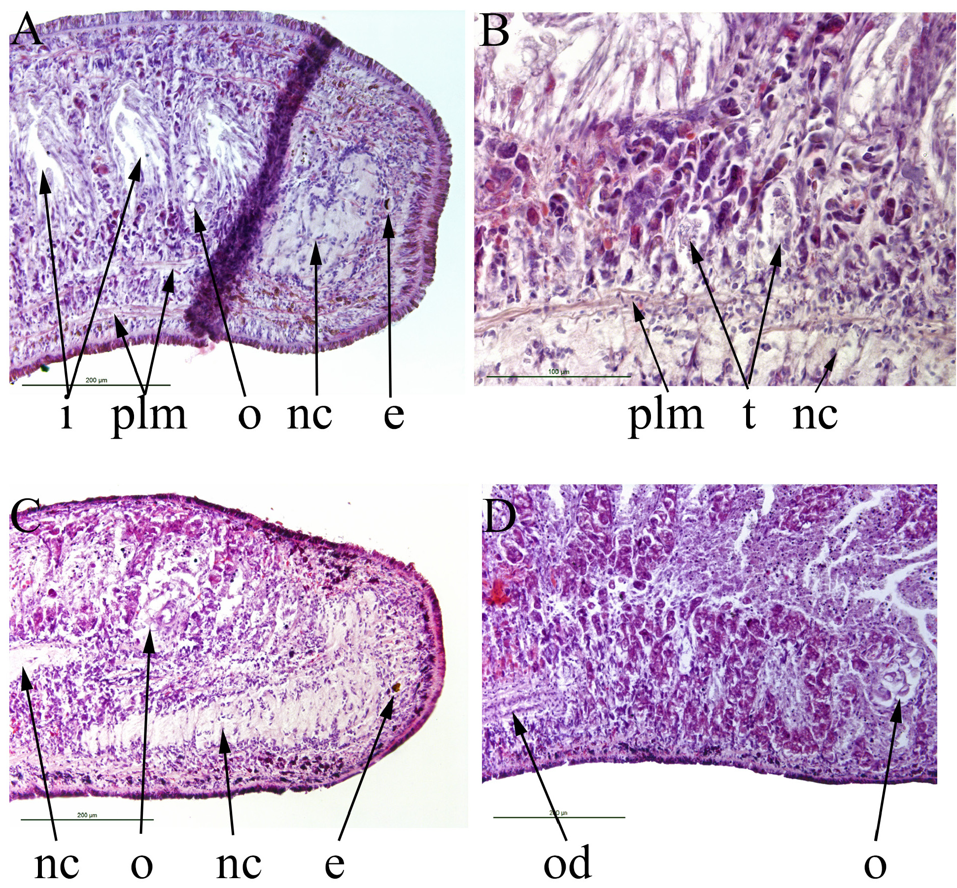
There are two eyes near the anterior end ( Fig 2 A, C View FIGURE 2 ), each a shallow pigmented cup 15–20 μm in diameter and about 10 μm deep. A pair of ventral nerve cords ( Figs 1 C View FIGURE 1 ; 2 A, B, C View FIGURE 2 ; 3 B View FIGURE 3 ; 4 E View FIGURE 4 ; 5 B, C, D, E, F View FIGURE 5 ), expanded anteriorly, run the length of the body, about 150 μm apart. There is a sub-epidermal nerve plexus above the creeping sole ( Fig 3 A View FIGURE 3 ).
Ventral epidermis is a ciliated monolayer about 0.25 μm thick, the cilia are 5 μm long. There is a layer of subepidermal circular muscle about 2 μm thick. Parenchymal longitudinal muscle fibres are present, one layer below and one above the ventral nerve cords, respectively 30–40 μm and 90–100 μm above the ventral epidermis ( Figs 1 C View FIGURE 1 ; 2 A, B View FIGURE 2 ; 3 A, B View FIGURE 3 ). Dorsally there is a weak single layer of parenchymal longitudinal muscle about 15 μm in from the dorsal epidermis, ental to the rhabdite-secreting layer. Dorsal epidermis is a non-ciliated monolayer, about 15 μm thick.
The retracted pharynx ( Figs 1 C View FIGURE 1 ; 3 A, B View FIGURE 3 ; 5 B View FIGURE 5 ) is cylindrical, about 300 μm long and 275 μm in diameter, the walls about 120 μm thick. Pharyngeal musculature consists of loose longitudinal and radial fibres. The pharyngeal aperture (mouth) is 57% along the pharyngeal pouch. The gut is triclad but the lateral diverticula are indistinct and the number is uncertain.
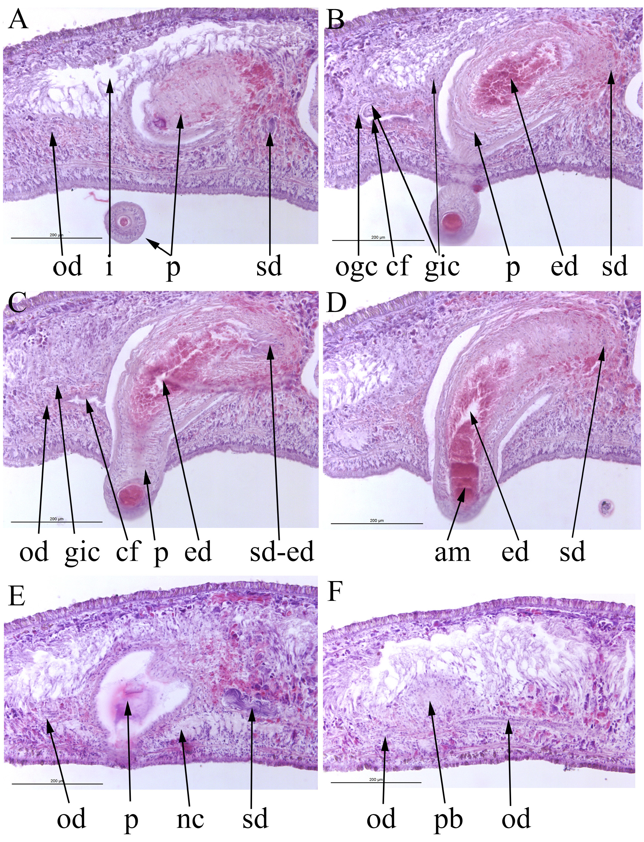
A pair of ovaries is near the anterior end ( Fig 2 A, C, D View FIGURE 2 ), in WF06 and WF09 they are respectively about 450 μm and 500 μm from the (contracted) anterior end. In WF06 each ovary is about 100 μm high by 80–100 μm antero-posteriorly. In WF09 the ovaries are about 150 μm high by 100 μm long antero-posteriorly (presumably distorted due to longitudinal contraction of the worm). Each ovary contains several presumed oocytes about 10 μm in diameter. An ovovitelline duct ( Figs 2 D View FIGURE 2 ; 3 B View FIGURE 3 ; 4 A, C, E, F View FIGURE 4 ; 5 B, C, D, E, F View FIGURE 5 ), lumen diameter about 10 μm, external diameter about 20 μm, opens onto the ventral surface of each ovary, runs posteriorly on the outer dorsal surface of the ventral nerve cord on each side to open together into the posterior of the common female duct. Vitellaria are not distinguishable with certainty. A ciliated genito-intestinal duct ( Figs 1 C View FIGURE 1 ; 3 A, B View FIGURE 3 ; 4 B, C View FIGURE 4 ; 5 D, E, F View FIGURE 5 ), internal diameter about 8 μm, joins at the junction of the ovovitelline ducts and common female duct, then runs anteriorly and dorsally, narrows to about 4 μm and opens through an indistinct opening into a digestive diverticulum on the right side. The common female duct ( Figs 1 C View FIGURE 1 ; 3 A, B View FIGURE 3 ; 4 B, C View FIGURE 4 ) is simple, about 150 μm long. In WF09 it is surrounded by eosinophylic cells ( Fig 5 F View FIGURE 5 ), presumed glandular and to secrete the shell of the egg cocoon. That of WF06 has no obvious glandular tissue surrounding it. In WF09, the posterior extremity of the ovovitelline ducts, and the proximal genito-intestinal duct are filled with an amorphous eosinophylic material.
In WF06, testes ( Fig 2 B View FIGURE 2 ) are present just above the nerve cord on each side between the ovaries and the pharynx. The number is uncertain because most appear spent and some are barely distinguishable. There are perhaps four discernable on one side and two on the other. Larger ones are ovate, about 50 μm high and 20 μm long (presumably distorted due to longitudinal contraction of the worm). Only one or two possible spent testes are visible in WF09. A narrow sperm duct, <10 μm wide, runs posteriorly on the inner dorsal surface of the ventral nerve cord on each side. In both specimens each sperm duct expands posterior to the pharynx to about 35 μm wide to form a false seminal vesicle containing stored sperm (cyanophylic) ( Figs 3 B View FIGURE 3 ; 4 A, B, D, E View FIGURE 4 ; 5 B, C View FIGURE 5 ). They then turn dorsally, narrow slightly to 15 μm wide, then to less than 10 μm and open separately but close together into the anterior end of the ejaculatory duct of the penis ( Figs 4 C View FIGURE 4 ; 5 A, C View FIGURE 5 ). The penis ( Figs 1 C View FIGURE 1 ; 3 A, B View FIGURE 3 ; 4 A, B, C, E View FIGURE 4 ; 5 B, C, D, E, F View FIGURE 5 ) is conical, outer diameter about 220 μm at its base and narrowing towards its distal end. In WF06 the penis is partly protruded through the gonopore and is about 400 μm long ( Figs 1 C View FIGURE 1 ; 3 A, B View FIGURE 3 ; 4 A, B, C, E View FIGURE 4 ). In WF09 the penis is inserted into the common female duct to the latter’s posterior extent ( Fig 5 View FIGURE 5 ). The musculature of the penis consists of outer longitudinal fibres, loose central tissue and a dense layer of circular muscle surrounding the ejaculatory duct. In the penis of WF06 the ejaculatory duct is centric throughout the length of the penis. In WF09 the ejaculatory duct is centric for most of the length of the penis, but is eccentric for a short distance in the mid part. The basal half of the ejaculatory duct in both specimens is lined with, and almost occluded by, eosinophylic cells, presumed to be glandular. The distal portion in both specimens contains amorphous eosinophylic material, almost plugging the duct.

No known copyright restrictions apply. See Agosti, D., Egloff, W., 2009. Taxonomic information exchange and copyright: the Plazi approach. BMC Research Notes 2009, 2:53 for further explanation.