
Pteronemobius (Pteronemobius) ruficeps Liu et Shi
|
publication ID |
https://doi.org/ 10.5281/zenodo.10084226 |
|
publication LSID |
lsid:zoobank.org:pub:D6C8CC8F-07D1-4AD2-B477-CF2E633A3BFC |
|
persistent identifier |
https://treatment.plazi.org/id/09180463-FFE1-FFB9-FF53-FA7FFE0AFB7C |
|
treatment provided by |
Carolina |
|
scientific name |
Pteronemobius (Pteronemobius) ruficeps Liu et Shi |
| status |
sp. nov. |
Pteronemobius (Pteronemobius) ruficeps Liu et Shi , sp. n.
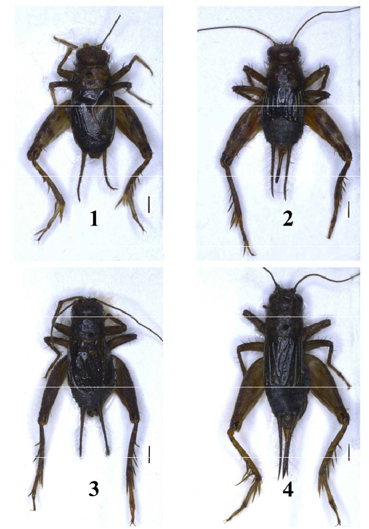
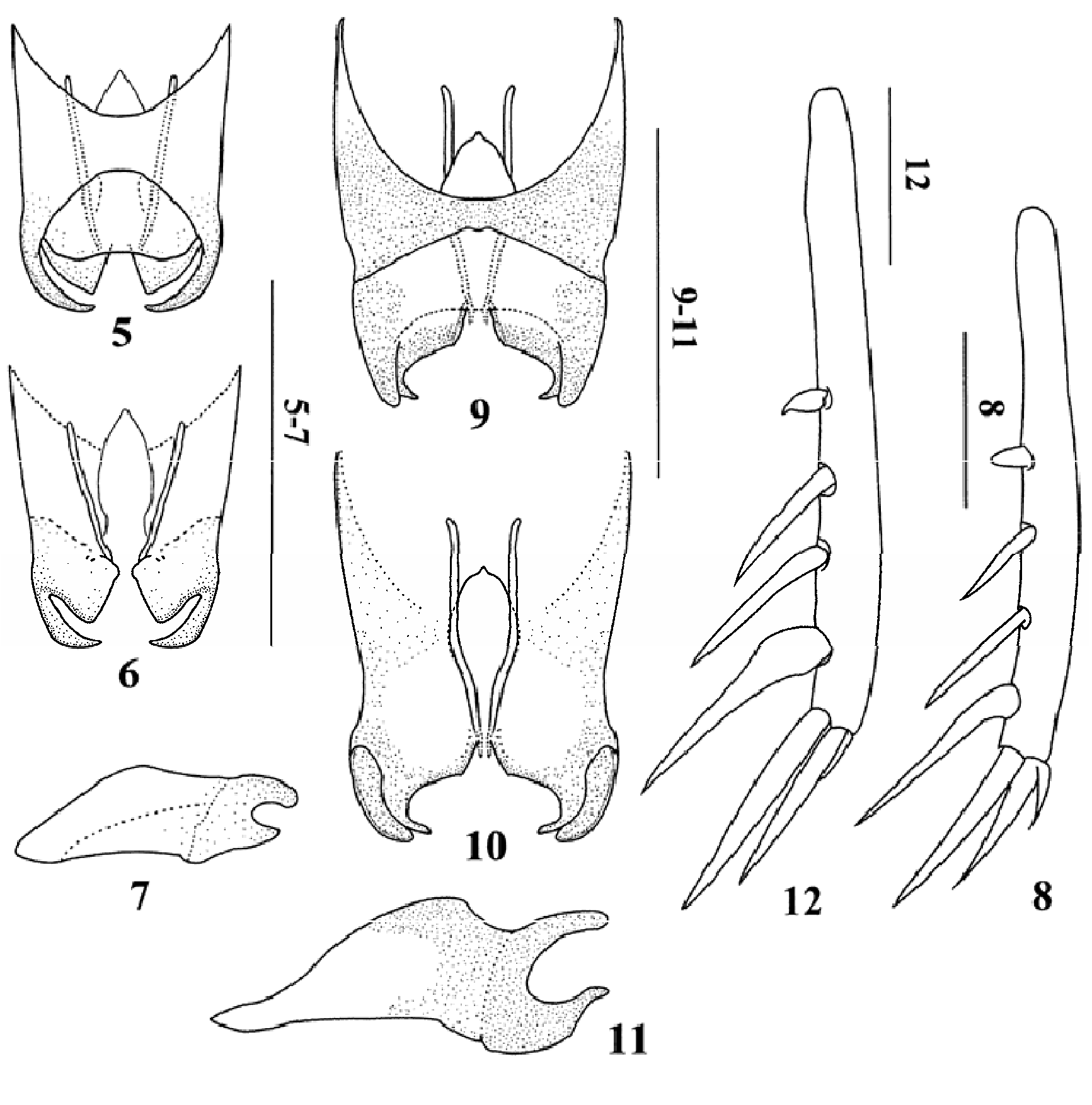
Figs 1–2 View Figs , 5–8 View Figs
MATERIAL. Holotype – ♂, China: Hainan province: Ledong, Jianfengling, 12-15.XI
2005, coll. Hao-Yu Liu ( MHBU). Paratypes: 19 ♂, 14♀, the same data as for holotype ( MHBU).
1 – male, 2 – female;
Scale bars= 1 mm.
– male, 4 – female. 3-4 – P. (Pteronemobius) kangdingensis sp. n.: 3 20
DESCRIPTION. MALE. Body small-sized and pubescent. Head as wide as width of anterior margin of pronotum, frontal rostrum very short and about 1.3 times as wide as scapus; median ocellus small and projecting forward; lateral ocelli large and slightly obliquely projecting outward; maxillary and labial palpi rather long, last joint of maxillary palpus wide and truncated at apex, and last joint of labial palpus claviform. Pronotum trapeziform, slightly widened posteriorly, about 0.6 times as long as width of posterior margin, anterior and posterior margins almost straight. Tegmina extending to apex of abdomen, present with one oblique vein, mirror small, distinctly wider than long, lateral field with 5 oblique subcostal veinlets; wings absent. Fore tibia with an oval tympanum on outer side. Hind tibia with 4 pairs of dorsal spines, the first inner spine very short and tuberculiform, the fourth inner spine obviously curved and slightly swollen at base ( Fig. 8 View Figs ). Supra anal plate slightly longer than wide, rounded at posterior margin, concave in centre of dorsum. Subgenital plate truncate at posterior margin and slightly emarginated in middle. Genitalia ( Figs. 5–7 View Figs ): apical part of epiphallus distinctly bent inwards, acute at apex; apical parts of ectoparamers distinctly widened, nearly triangular at apex, obviously shorter than epiphallus.
FEMALE. General appearance similar to that of male. Tegmina almost extending to middle of abdomen, present with 6 veins on dorsal field Hind tibia with the first and fourth internal spines normal. Subgenital plate widely rounded at posterior margin and deeply emarginated in middle. Ovipositor short, about 0.5 times as long as length of hind femur, with several small teeth on dorsal side.
Body yellow brown. Head and dorsal area of pronotum red when alive, lateral area of pronotum black. Tegmen light brown on dorsal field, lateral field black; Sc vein of male white, Cu 1 vein and transverse veins of female white. Hind femur with brown stripe on outer surface.
MEASUREMENTS. Length of body ♂ 5.9–6.2 mm, ♀ 6.2–6.5 mm; length of pronotum ♂ 1.2–1.3 mm, ♀ 1.3–1.5 mm; length of tegmen ♂ 3.4–3.5 mm, ♀ 2.0– 2.2 mm; length of hind femur ♂ 4.1–4.4 mm, ♀ 4.3–4.6 mm; length of ovipositor 2.1–2.3 mm.
DIAGNOSIS. The new species can easily be distinguished from other Pteronemobius species by the male genitalia: apical parts of ectoparamers distinctly widened, nearly triangular at apex, obviously shorter than epiphallus. By short apical parts of epiphallus and ectoparamers and by weakly widened near the base fourth inner spine of male hind tibia P. ruficeps sp. n. is similar to P. yezoensis (Shiraki, 1911) , but differs by the shape of apical part of epiphallus (in P. yezoensis apical part straight, with narrowly rounded apex).
ETYMOLOGY. The specific name is derived from Latin “ ruficeps ”, referring to its red head when alive.
No known copyright restrictions apply. See Agosti, D., Egloff, W., 2009. Taxonomic information exchange and copyright: the Plazi approach. BMC Research Notes 2009, 2:53 for further explanation.
|
Kingdom |
|
|
Phylum |
|
|
Class |
|
|
Order |
|
|
Family |
|
|
Genus |