
Compsodactylus, Fuhrmann, Juares, 2012
|
publication ID |
https://doi.org/10.11646/zootaxa.3577.1.2 |
|
publication LSID |
lsid:zoobank.org:pub:4BFAE75F-7BC0-4D5D-B1D1-DA2CC01F40C9 |
|
DOI |
https://doi.org/10.5281/zenodo.5874045 |
|
persistent identifier |
https://treatment.plazi.org/id/096F054C-FF98-547E-FF25-FE63FAB0EA50 |
|
treatment provided by |
Plazi |
|
scientific name |
Compsodactylus |
| status |
gen. nov. |
Compsodactylus new genus
( Figs. 1–76 View FIGURES 1 – 9. 1, 2, 4 View FIGURES 10 – 16. 10 View FIGURES 17 – 27. 17 – 26 View FIGURES 28 – 39. 28 – 35, 37 View FIGURES 40 – 45 View FIGURES 46 – 51 View FIGURES 52 – 62. 52 – 60 View FIGURES 63 – 75. 63 – 69 View FIGURE 76 )
Type species. Dicrania martinezi Frey, 1972 , here designated.
Etymology. Kompsos (Greek), elegant; daktylos (Greek), finger; in reference to distinct form of male metatibia; the name is masculine in gender.
Diagnosis. Mentum longer than wide; posterior area of frons and anterior area of pronotum with denticle-like setae; protibiae with 2 teeth, spur developed; external and internal margin of elytra bordered; matecoxae with short projection over trochanter basis; male metatibiae with internoapical spine, lacking spurs; abdominal spiracular area VII narrowed.
Description. Head ( Figs. 4 View FIGURES 1 – 9. 1, 2, 4 , 10–12 View FIGURES 10 – 16. 10 ). Epistomal suture poorly defined. Front with denticle-like setae. Clypeus trapezoid; anterior angles acute, upturned. Labrum thin, slightly emarginated. Epipharynx ( Fig. 13 View FIGURES 10 – 16. 10 ) setose; posterior area covered by seta-like processes ( Fig. 13 View FIGURES 10 – 16. 10 details); medial and posteromedial area with sensilla. Mandible ( Figs. 17–22 View FIGURES 17 – 27. 17 – 26 ) with prosthecae greatly developed; incisor rounded; molar ( Figs. 19, 22 View FIGURES 17 – 27. 17 – 26 ) striatoserrate, with posterior depression. Maxilla ( Figs. 23, 24 View FIGURES 17 – 27. 17 – 26 ) with lacinia toothed, galea ( Figs. 25–27 View FIGURES 17 – 27. 17 – 26 ) 7–8-toothed; palpi with 4 palpomeres, IV dorsally striate. Hypopharynx ( Fig. 15 View FIGURES 10 – 16. 10 ) laterally setose posteriorly covered with seta-like processes, medial area with sensilla. Labium ( Figs. 15, 16 View FIGURES 10 – 16. 10 ) with prementum prominent and fused with mentum; mentum longer than wide, with longitudinal groove, palpi with 3 palpomeres. Antenna with 9 antennomeres ( Fig. 14 View FIGURES 10 – 16. 10 ); club as long as or slightly shorter than pedicel-funicle.
Pronotum ( Figs. 28–30 View FIGURES 28 – 39. 28 – 35, 37 ) wider than long, maximum width at middle; all margins bordered; anterior margin with external membrane. Prosternal posterior process small, tubercle-like. Furca broad, truncate. Proleg ( Fig. 33 View FIGURES 28 – 39. 28 – 35, 37 ). Tibia with 2 teeth; spur present at middle of tibial length. Tarsus as long as or slightly shorter than tibia; male tarsomere I ( Fig. 32 View FIGURES 28 – 39. 28 – 35, 37 ) with internoapical area granulostriate. Pretarsus with 2 similar bifid claws; empodium short, with 2 setae.
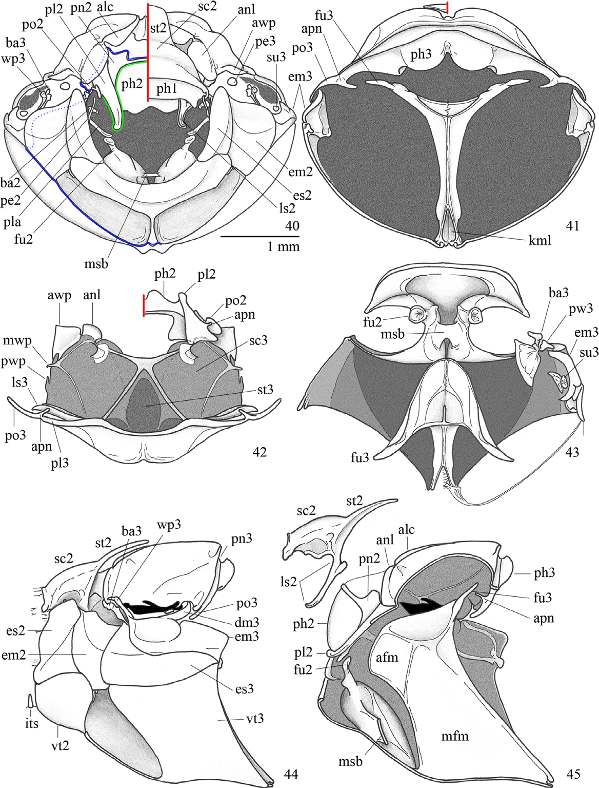
Mesothorax ( Figs. 40–48 View FIGURES 40 – 45 View FIGURES 46 – 51 ). Scutellum with anterior margin rounded, not bordered. Episternum as wide as epimeron or near so, not reaching the mesocoxal cavity. Elytra ( Fig. 58 View FIGURES 52 – 62. 52 – 60 ) about 1.2 times longer than wide (combined), striatopunctate; anterior margin abruptly rounded, not bordered; external and internal margins bordered; lateral margin without external membrane. Mesoleg ( Fig. 34 View FIGURES 28 – 39. 28 – 35, 37 ). Femur with internal area longitudinally carinate. Tibia conical, transversal carina with denticle-like setae; 2 continuous spurs present.
Metathorax ( Figs. 40–48 View FIGURES 40 – 45 View FIGURES 46 – 51 ). Episternum not reaching the mesocoxal cavity, extended between mesepimeron and metepimeron. Metendosternite with anterior flange rounded. Wing ( Figs. 52–61 View FIGURES 52 – 62. 52 – 60 ) with anterodistal margin setose, RA3 and RA4+ RP 1 not reaching the apex, MP3 indistinct. Metaleg ( Figs. 35–39 View FIGURES 28 – 39. 28 – 35, 37 ). Coxa medially contiguous, area of ventral articulation with trochanter enlarged. Tibia with reduced transversal carina more developed in female; male tibial apex ( Figs. 37, 39 View FIGURES 28 – 39. 28 – 35, 37 ) with internal tooth, lacking spurs; female tibial ( Fig. 38 View FIGURES 28 – 39. 28 – 35, 37 ) apex with two contiguous spurs.
Abdomen ( Figs. 49–51 View FIGURES 46 – 51 ). Spiracular area VII ( Fig. 49 View FIGURES 46 – 51 ) narrowed. Male pygidium longer than wide, downturned; female pygidium as long as wide or near so, backwardly projected. Ventrite V longer than ventrite IV. Intersegmentar membrane VII–VIII (between ventrite V and VI) exposed. Spiculum gastrale ( Fig. 63 View FIGURES 63 – 75. 63 – 69 ) with cranial piece longer than wide and 0.66 times shorter than caudal arms. Sternite IX ( Fig. 63 View FIGURES 63 – 75. 63 – 69 ) triangular, longer than wide, with apical row of short setae. Aedeagus with phallobasic apodeme longer than phallobase. Parameres longer than phallobase (with apodeme), symmetrical (except by the ventral side), glabrous; pieces free, longer than wide; apex acute and ventrally deflected; laterolongitudinal sulcus present. Intestine with 6 posterior caeca ( Fig. 72 View FIGURES 63 – 75. 63 – 69 ).
Discussion. Even with some recently published works the differentiation between Macrodactylini and Tanyproctini is difficult. Tanyproctini were recognized by their strong sexual dimorphism, male antennal club with 3–7 antennomeres, reduced mouthparts, epistomal suture well defined or replaced by angular border, intersegmental membrane VII–VIII well developed ventrally. Macrodactylini were recognized by their poorly defined epistomal suture, contiguous metatibial spurs not separated by tarsal insertion, propygidium and ventrite V not separated by a suture, and ventrite V evidently longer than preceding ventrites ( Sanmartín & Martín-Piera 2000, Lacroix 2007, Katovich 2008). Neotropical Tanyproctini occur predominantly in temperate regions ( Bolivia, Peru, Uruguay, Argentina, Chile) and Macrodactylini species occur predominantly in tropical areas, especially in Brazil ( Sanmartín & Martín-Piera 2000, Katovich 2008). Compsodactylus share some features with Tanyproctini , like the presence of intersegmental membrane VII–VIII, the structure of aedeagus with well-developed phallobasic apodeme and long and symmetrical parameres, but these characteristics are also found in some Macrodactylini . The poorly-developed epistomal suture and enlarged ventrite V are features more commonly present in Macrodactylini . Cladistic studies on Macrodactylini and Tanyproctini are necessary and certainly will change the present scenario and composition of both tribes.
The transfer of C. martinezi and C. parvulus from Dicrania to Compsodactylus is based on the following characters (in opposition to Dicrania ): frons with denticle-like setae (thin or scale-like setae); pronotum posteriorly bordered (not bordered); protibia with a spur (without spur); scutellar anteriorly rounded (angled); elytron anteriorly rounded (angled); elytron posteriorly bordered (without defined border); external elytral margin without membrane (with membrane); female elytra covering pygidial base (exposing the pygidium); mesofemur internal side longitudinally carinate (without carina); metacoxae weakly enlarged over trochanter basis (without enlargement over trochanter); abdominal spiracular area VII ( Fig. 53 View FIGURES 52 – 62. 52 – 60 ) narrowed (unmodified); ventrite IX large and shield-like (reduced a marginal sclerite); lateral area of parameres with a longitudinal line (with depression).


No known copyright restrictions apply. See Agosti, D., Egloff, W., 2009. Taxonomic information exchange and copyright: the Plazi approach. BMC Research Notes 2009, 2:53 for further explanation.
|
Kingdom |
|
|
Phylum |
|
|
Class |
|
|
Order |
|
|
Family |
|
|
SubFamily |
Melolonthinae |