
Caligus nuenonnae, Andrews, Melanie, Bott, Nathan, Battaglene, Stephen & Nowak, Barbara, 2009
|
publication ID |
https://doi.org/ 10.5281/zenodo.185016 |
|
DOI |
https://doi.org/10.5281/zenodo.6217714 |
|
persistent identifier |
https://treatment.plazi.org/id/0A2E0A3A-FF81-FFEE-7BF7-FAD1FA29FE39 |
|
treatment provided by |
Plazi |
|
scientific name |
Caligus nuenonnae |
| status |
sp. nov. |
Caligus nuenonnae n. sp.
( Figs 1–3 View FIGURE 1 View FIGURE 2 View FIGURE 3 )
Type material. Holotype Ψ (QM W28171), allotype ɗ (QM W28172) and 4 paratypes (2ɗ and 2Ψ) (QM W28173), ex body surface of Latris lineata (Forster) reared at the TAFI, Marine Research Laboratories, Tasmania, Australia (43°35’S, 147°35’E), 10 October, 2006, leg. M. Andrews.
Other material examined. 15 ɗ and 15 Ψ, ex body surface of L. lineata reared at the TAFI, Marine Research Laboratories, Tasmania, Australia, 10 October, 2006, leg. M. Andrews.
Description of female. Body as in Fig. 1 View FIGURE 1 A. Total length (excluding setae on caudal rami) 4.27–4.82 (4.56), based on 9 specimens. Cephalothoracic shield ovate, marginally wider than long, 2.58–2.88 (2.75) × 2.62–2.94 (2.79) (excluding marginal membrane). Posterior margin of thoracic zone extends past lateral zone. Fourth pedigerous somite wider than long, 0.38–0.58 (0.47) × 0.40–0.64 (0.54). Genital complex indented laterally, almost equal in length and width, 1.29–1.58 (1.43) × 1.29–1.60 (1.46). Dorsal and ventral surfaces with lateral setules; posterior margin concave. Abdomen ( Fig. 1 View FIGURE 1 B) 1-segmented, almost equal in length and width, 0.44–0.46 (0.45) × 0.45–0.47 (0.46); with 10 setules on dorsal surface. Caudal ramus ( Fig. 1 View FIGURE 1 B) slightly longer than wide, 0.06–0.08 (0.07) × 0.05–0.06 (0.06); with row of setules along medial margin, 1 dorsal setule, and 3 long and 3 short pinnate setae.
Antennule ( Fig. 1 View FIGURE 1 C) 2-segmented; proximal segment with 27 setae (18 plumose, 9 naked); distal segment slightly shorter than proximal segment, with 1 subterminal seta on posterior margin, 11 apical setae and 2 apical aesthetascs. Antenna ( Fig. 1 View FIGURE 1 D) 3-segmented; proximal segment with large spatulate process; middle segment with adhesion pad on dorsal surface; distal segment uncinate, longer than middle segment, with 1 short proximal seta and 1 long seta at mid-length of claw. Postantennal process ( Fig. 1 View FIGURE 1 E) slender, distally curved, pointed at tip, with proximal pair of bisetose papillae and 1 bisetose papilla alongside process. Mandible ( Fig. 1 View FIGURE 1 F) modified into stylet, with 12 teeth on medial margin of apex. Maxillule ( Fig. 1 View FIGURE 1 G) reduced, with 3 unequal setae anteriorly and slender, bluntly pointed dentiform process posteriorly. Maxilla ( Fig. 1 View FIGURE 1 H) 2-segmented, brachiform; lacertus unarmed, broader than brachium; brachium elongate, with triangular flabellum at midlength and 2 unequal apical elements. Short canna with serrate marginal membranes; longer calamus with smooth marginal membranes. Corpus of maxilliped ( Fig. 1 View FIGURE 1 I) large, with proximal ridged protrusion on posterior surface. Subchela with relatively long, straight shaft and sharply pointed claw; shaft with distal hyaline element; claw with basal setiform barb. Sternal furca ( Fig. 1 View FIGURE 1 J) with broad box; tines slender, widely separated, apically truncate.
Armature on rami of legs 1–4 ( Figs 2 View FIGURE 2 A–D) as follows (Roman numerals indicating spines and Arabic numerals setae):
Leg 1 ( Fig. 2 View FIGURE 2 A) protopod with 1 outer setule, 1 anterolateral seta, and 1 distal seta. Endopod vestigial, represented by unarmed lobe near joint of sympod and first exopodal segment. First exopodal segment with row of setules along posterior margin and small anterodistal spine. Second exopodal segment with 3 large apical spines (each with lateral flange; inner 2 with secondary process), 1 long apical seta, and 3 long inner setae (each seta with denser row of setules along outer margin).
Leg 2 ( Fig. 2 View FIGURE 2 B) coxa with striated membrane on posterior margin, 1 inner setule and 1 large inner plumose seta. Basis with striated membrane on posterior margin, outer naked seta, and small inner setule. First and second exopodal segments with striated membrane on lateral margin. Spine on first exopodal segment with flange on both margins and extends beyond medial margin of second exopodal segment. Spine on second exopodal segment with flange on lateral margin and extends to medial margin of third exopodal segment. Distal exopodal segment bears 1 small and 1 large lateral spine (latter with flange on medial margin) and 6 inner plumose setae. All exopodal segments with row of setules along medial margin; all endopodal segments with row of setules along lateral margin; second endopodal segment with row of setules along medial margin.
Leg 3 ( Fig. 2 View FIGURE 2 C) protopod with 2 small adhesion pads on dorsolateral surface (indicated by arrows), striated membrane on lateral and medial margins, 1 short pinnate seta on posterior surface, 1 small setule, and 1 plumose inner seta. First exopodal segment with striated membrane along lateral margin, 1 ventral setule, and 1 large naked spine. Second exopodal segment with row of setules along both margins. Terminal exopodal segment with row of setules along lateral margin. First endopodal segment expanded laterally into velum, with one inner plumose seta. Second endopodal segment with row of setules along lateral margin.
Leg 4 ( Fig. 2 View FIGURE 2 D) protopod with plumose outer seta. First exopodal segment with 1 naked distolateral spine. Second exopodal segment with 1 outer, laterally flanged spine situated 2/3 along length of segment and 3 apical, flanged spines; small pectinate membrane present at base of outer spine and apical spines 1 and 3; innermost apical spine about twice as long as middle and outermost apical spines.
Leg 5 ( Fig. 2 View FIGURE 2 E) highly reduced, represented by 2 small lobes on genital complex; anterior lobe with 1 pinnate seta; posterior lobe with 2 unequal pinnate setae.
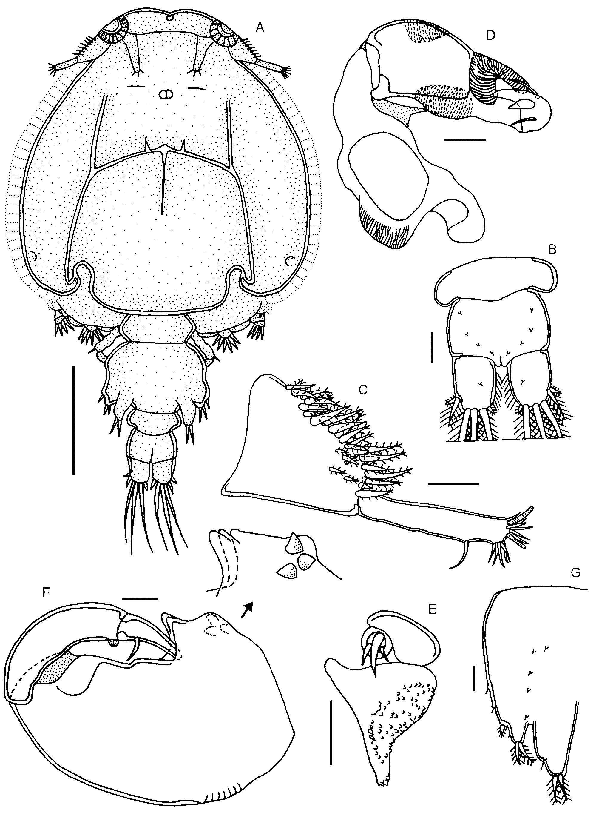
Male. Body as in Fig. 3 View FIGURE 3 A. Total length (excluding seta on caudal rami) 3.99–5.26 (4.58), based on 9 specimens. Cephalothoracic shield ovate, marginally longer than wide, 2.56–3.20 (2.82) × 2.38–3.24 (2.70) (excluding marginal membrane). Thoracic zone extends past posterior margins of lateral zones. Fourth pedigerous somite slightly wider than long, 0.33–0.52 (0.42) × 0.43–0.58 (0.51). Genital complex laterally indented, longer than wide, 0.91–1.38 (1.17) × 0.84–1.17 (0.98); with 10 setules on ventral surface. Abdomen ( Fig. 3 View FIGURE 3 B) 2-segmented, both segments wider than long [proximal segment 0.17–0.21 (0.19) × 0.40–0.56 (0.48); distal segment 0.25–0.36 (0.31) × 0.38–0.47 (0.43)]; with 8 setules on dorsal surface. Caudal ramus ( Fig. 3 View FIGURE 3 B) longer than wide, 0.075–0.087 (0.080) × 0.055–0.063 (0.059); with row of setules along medial margin, 1 dorsal setule, and 3 long and 3 short pinnate setae.
Antennule ( Fig. 3 View FIGURE 3 C) 2-segmented; proximal segment with 29 setae (22 plumose, 7 naked); distal segment longer than that of female, with 1 subterminal seta on posterior margin, 11 apical setae, and 2 apical aesthetascs. Antenna ( Fig. 3 View FIGURE 3 D) 3-segmented; proximal segment with large adhesion pad; second segment with 3 corrugated adhesion pads (2 dorsal, 1 ventral); distal segment uncinate, with 1 large accessory process and 2 setae near apex. Postantennal process slender, without papillae. Dentiform process of maxillule ( Fig. 3 View FIGURE 3 E) blunt, with numerous small points embossed on medial and apical surfaces. Corpus of maxilliped ( Fig. 3 View FIGURE 3 F) broad, with corrugated area on proximolateral margin, 3 large medial processes on myxal region, and 3 small proximal processes on anteromedial surface; subchela with relatively long, curved shaft. Legs 5 and 6 ( Fig. 3 View FIGURE 3 G) highly reduced, each represented by lobiform projection on genital complex; leg 5 with 1 small lateral seta and 2 apical pinnate setae; leg 6 larger than leg 5, with 2 apical pinnate setae.
Etymology. The specific name refers to the south-eastern region of Tasmania, known as ‘nuenonne’ by indigenous Australians, where the new caligid species occurs.
Remarks. Caligus nuenonnae n. sp. shares an accessory process on apical spines two and three of the distal exopodal segment of leg 1, three well developed inner setae on the distal exopodal segment of leg 1, a 2- segmented leg 4 exopod with I-0; I, III armature and a 1-segmented abdomen in the female that is about a third the length of the genital complex in common with C. acanthopagri Lin, Ho & Chen, 1994 , C. asymmetricus Kabata, 1965 , C. dieuzeidei Brian, 1933 , C. latigenitalis Shiino, 1954 , C. pomacentrus Cressey, 1991 , C. serratus Shiino, 1965 , C. willungae Kabata, 1965 and C. zei Norman & T. Scott, 1906 .
The following comparisons refer to female characteristics unless stated otherwise. Caligus nuenonnae n. sp. differs from C. acanthopagri and C. latigenitalis by having a spatulate rather than pointed proximolateral process on the first segment of the antenna, a proximal ridged protrusion on the maxilliped corpus, widely separated furcal tines, an outer flange on each apical spine of the terminal exopodal segment of leg 1, a relatively shorter accessory process on the middle and inner apical spines of the terminal exopodal segment of leg 1, a 1-segmented instead of 2-segmented leg 1 endopod, a relatively longer distolateral spine on the terminal exopodal segment of leg 2 and two small adhesion pads on the leg 3 protopod (see Ho & Lin 2004; Izawa & Choi 2000).
Caligus nuenonnae n. sp. can be distinguished from C. asymmetricus by the presence of a large rather than small proximolateral process on the first segment of the antenna, widely separated furcal tines, a relatively shorter proximolateral spine on the terminal exopodal segment of leg 2 and a lateral row of setules rather than large coarse teeth on the middle endopodal segment of leg 2. Further differences include the absence of a medial tooth-like protrusion and basal irregular outgrowth on the maxilliped corpus and spinules on the protopod of legs 1 and 3 in the new species (see Ho & Lin 2004).
Caligus dieuzeidei differs from the new taxon by having a slender rather than broad proximolateral process on the first segment of the antenna, an accessory basal process on the postantennal process, a secondary process on the maxillulary dentiform process and a relatively longer sternal box.
Caligus nuenonnae n. sp. can be distinguished from C. pomacentrus by having a nearly subequal instead of distinctly wider abdominal somite, a spatulate rather than pointed proximolateral process on the first segment of the antenna, relatively slimmer furcal tines, a flange rather than fine teeth on the outer margin of the middle and inner apical spines on the terminal exopodal segment of leg 1, a flange rather than denticles on the outer margin of the lateral spine on the middle exopodal segment of leg 2 and a considerably longer distolateral spine on the second exopodal segment of leg 4.
Caligus serratus differs from the new species by having furcal tines that are spaced closer together, relatively slimmer apical spines on the terminal exopodal segment of leg 1, a relatively slimmer lateral spine on the proximal exopodal segment of leg 2 and a relatively shorter distolateral spine on the terminal exopodal segment of legs 2 and 4. The former taxon also lacks a proximolateral process on the first segment of the antenna, a proximal ridged protrusion on the maxilliped corpus and an outer flange on each apical spine of the terminal exopodal segment of leg 1.
Caligus nuenonnae n. sp. can be distinguished from C. willungae by possessing a subcircular rather than pyriform cephalothoracic shield, a recurved postantennal process, a pointed maxillulary dentiform process, a proximal ridged protrusion on the maxilliped corpus, widely separated furcal tines and a relatively shorter middle apical spine on the terminal exopodal segment of leg 4. The new species also lacks an accessory basal process on the postantennal process and an apical flange on the postantennal process, maxillulary dentiform process and each furcal tine.
Caligus zei differs from C. nuenonnae n. sp. by having a genital complex that is nearly as large as the cephalothoracic shield, a relatively shorter outer apical spine on the terminal exopodal segment of leg 1 and a relatively shorter inner apical spine on the distal exopodal segment of leg 4 (see Kabata 1979).
Caligus nuenonnae n. sp. can be further distinguished from those eight congeners by having a mid-lateral indentation and strongly concave distal margin on the female genital complex, a distinctly broader first abdominal somite relative to the second abdominal somite in the male, numerous small points embossed on the surface of the male maxillulary dentiform process and male legs 5 and 6 represented by distinct lobiform projections on the genital complex.
In summary, C. nuenonnae n. sp. is characterised by the following combination of features: 1) female genital complex with a mid-lateral indentation and highly concave posterior margin; 2) 1-segmented abdomen in the female that is about one-third the length of the genital complex; 3) distinctly broader first abdominal somite relative to the second abdominal somite in the male; 4) antenna with a spatulate process on the first segment; 5) recurved postantennal process lacking a basal process; 6) female maxilliped with a ridged protrusion on the posterior surface of the corpus; 7) sternal furca with widely separated, apically truncate tines; 8) distal exopodal segment of leg 1 with a lateral flange on each apical spine and an accessory process on apical spines two and three; 9) leg 3 with two adhesion pads on the protopod; 10) 2-segmented leg 4 exopod with I- 0; I, III armature; 11) terminal exopodal segment of leg 4 with the outer apical spine being slightly shorter than the middle apical spine; 12) male maxillulary dentiform process with numerous small points embossed on the surface; and 13) relatively well developed, lobate male legs 5 and 6.
The presence of C. nuenonnae n. sp. in high numbers (40 parasites per fish) on cultured striped trumpeter has been associated with the development of small lesions on the host’s body surface, which heal within several weeks once the parasites have been removed. It is uncertain whether C. nuenonnae n. sp. will have a negative effect on striped trumpeter health in a commercial setting. Further, it is unknown at this juncture whether C. nuenonnae n. sp. occurs on wild populations of striped trumpeter (and other marine fish) in Australia or elsewhere. To date, only four parasite species, i.e. two monogenean species: Allomegalocotyla johnstoni ( Robinson, 1961) and Pseudomegalocotyla latridis ( Robinson, 1961) and two unidentified nematode species each belonging to the genus Anisakis Dujardin, 1845 and Cucullanellus Törnquist, 1931 , have been reported from wild striped trumpeter collected off the coast of New Zealand ( Brunsden 1956; Robinson 1961). Clearly, a detailed parasite survey of wild striped trumpeter would be advantageous before intensive sea-cage farming of striped trumpeter commences in Australia.
Molecular analysis of the Caligidae is of particular interest, especially for the highly speciose genus Caligus , to delineate morphologically similar taxa and further understand the phylogenetic relationships amongst this diverse parasite group. For example, Øines & Heuch (2005) and Øines & Schram (2008) recently demonstrated the presence of sibling species for Caligus elongatus Nordmann, 1832 based on differences in morphology and mitochondrial gene (CO1 and 16S) sequences. We have sequenced the mtCO1 gene of C. nuenonnae n. sp. and made it available on Genbank (accession number EF452642 View Materials ) for future investigators.


No known copyright restrictions apply. See Agosti, D., Egloff, W., 2009. Taxonomic information exchange and copyright: the Plazi approach. BMC Research Notes 2009, 2:53 for further explanation.