
Pseudosinella meganporteri, Soto-Adames, Felipe N., 2010
|
publication ID |
https://doi.org/ 10.5281/zenodo.275457 |
|
DOI |
https://doi.org/10.5281/zenodo.6208546 |
|
persistent identifier |
https://treatment.plazi.org/id/0B21878A-636E-FD03-F7EF-CE62FCAF8C66 |
|
treatment provided by |
Plazi |
|
scientific name |
Pseudosinella meganporteri |
| status |
sp. nov. |
Pseudosinella meganporteri sp. nov.
Figs 21–33 View FIGURES 21 – 29 View FIGURES 30 – 34 , Table 4 View TABLE 4
Material Examined. Holotype: RANDOLPH Co., Simmon’s-Mingo Cave, 6–25 July 2005, D. Culver, D. Fong & H. Hobbs, col., 1 on slide. Paratypes: same collection information as the holotype, 2 individuals in alcohol. POCAHONTAS Co., Tub Cave, 2 June 2004, D. Culver, D. Cowan & H. Hobbs, col., 1 on slide; Dreen Cave, 6–25 July 2005, D. Culver, D. Fong & H. Hobbs, col., 1 on slide, 45 in alcohol, mostly in poor condition; High Hopes Cave, 6–25 July 2005, D. Culver, D. Fong & H. Hobbs, col., 1 on slide.
Etymology. This species is dedicated to Dr. Megan Porter a speleobiologist currently at University of Maryland-Baltimore County and one of the primary collectors for the West Virginia cave fauna survey.
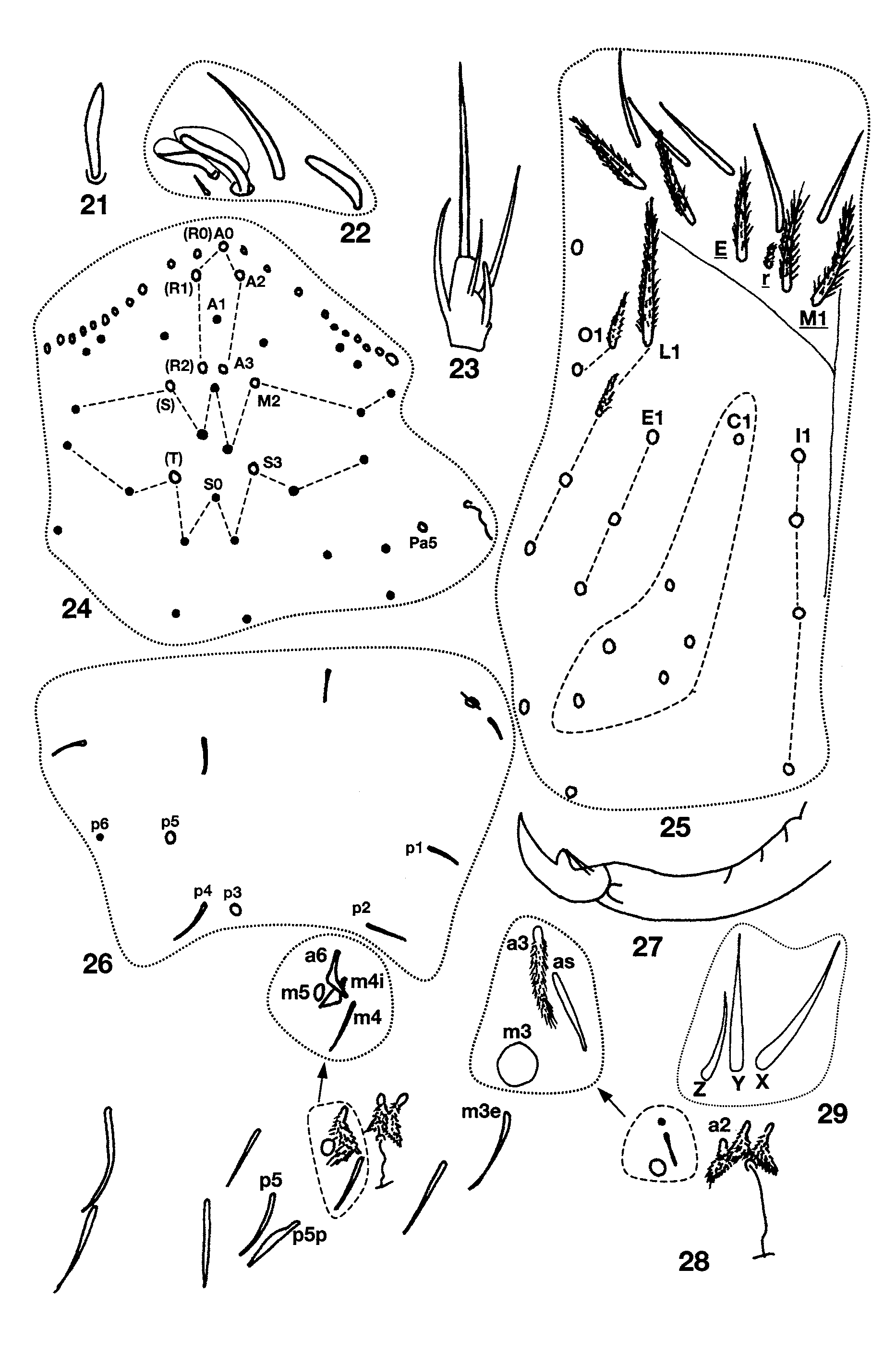
Description. Length to 2.2mm; color in alcohol white, without trace of pigment. Ant. 4 subapical sensilla weakly clubbed ( Fig. 21 View FIGURES 21 – 29 ). Ant. 3 sense organ ( Fig. 22 View FIGURES 21 – 29 ) inserted in individual shallow pits and formed by two paddle-shaped sensilla, with dense raquis and thin, translucent lateral extensions; additional basally-swollen, thin walled setae present near usual sense organ. Eyes absent. Dorsal head chaetotaxy ( Fig. 24 View FIGURES 21 – 29 ) includes 10– 11 macrosetae along antennal base and macrosetae A0, A2, A3, M2, S3 and Pa5; macrosetae M2 displaced forward, almost forming a row with A3; microseta A1 ciliate; M0, S0, S1 and S2 smooth microsetae; S0 inserted closer to S3 than S2, M1/S1 closer to S3 than M2. Prelabral setae ciliate, all labral setae smooth. Outer labral papillae larger than inner pair, both sets of papillae smooth. Pleural fold setae ciliate. Peristomal seta pss0 ciliate normal, slightly shorter than pss1; pss1–2 slender, ciliate, bothriotricha-like. Maxillary ungulum with 3 teeth. Lateral process of labial papilla E blunt ( Fig. 23 View FIGURES 21 – 29 ), slightly curved forward, barely reaching papillar tip. Labial palp proximal seta Z clearly shorter than seta Y ( Fig. 29 View FIGURES 21 – 29 ). Labial chaetotaxy M1M2rEL1L2A1–5. All post-labial setae strongly ciliate ( Fig. 25 View FIGURES 21 – 29 ); 4+4 setae along ventral groove; column C with 3–6 setae posterior to C1; column E with 2–5 setae; L with 3–5 setae, L2 reduced, similar to labial r; O with 2 setae, O1 modified as L2. Body formula for inner macrochaetotaxy as 22/0100+2. Mesothorax macrosetae p3 and p5 present ( Fig. 26 View FIGURES 21 – 29 ). Metathorax with macrosetae p2 and p3. Chaetotaxy of Abd. 1 linear, seta a6 absent. Chaetotaxy of Abd. 2 as in Fig. 28 View FIGURES 21 – 29 ; m3 and m5 normal macrosetae; a2, a6 and all supplementary setae fan-shaped; a2p absent; a3 ciliate, external to, and reaching as; as reaching socket of macroseta m3; m3e smooth or weakly ciliate, not reaching socket of m3; p5p short and ciliate. Abd. 3 ( Fig. 30 View FIGURES 30 – 34 ) with a2, a6, am6 and all supplementary setae fan-shaped; a3 ciliate, anterior to and far from as; as about half the length and reaching m3; m3 reaching as socket; d2 reaching p5; im fan-shaped (triangle in Fig. 30 View FIGURES 30 – 34 ) and subequal to em; a7, m7 and p7 normal microsetae, a7 ciliate, inserted close to, and reaching base of am6, m7a a macroseta. Chaetotaxy of Abd. 4 bothriotrichal complex as in Fig. 31 View FIGURES 30 – 34 : setae a, m, s, and D1 fanshaped; s posterior to a; C1p, T3 and D1p ciliate; T3 anterior to, and surpassing base of D1p; D1p reaching base of Pe; Pe and Pi fan-shaped. General chaetotaxy of Abd. 4 ( Fig. 32 View FIGURES 30 – 34 ) with macrosetae B5, B6, T6, T7, D2, D3, E1, E2, E3, F1 and F3; macroseta B5 anterior to line drawn between A5 and C2; A5 displaced externally with respect to other setae in row A and inserted near the position usually occupied by B5; microseta F2 closer to E3 than E2. Microseta posterior to E3 present. Abd. 4 without posterior setae.
Male genital plate multisetaceous. Trochanteral organ with 21–27 setae. Metathoracic femora with three acuminate macrosetae on basal half. Foot complex as in Figures 33–34 View FIGURES 30 – 34 . Tenet hair acuminate, shorter than unguiculus and metathoracic posterior smooth setae. Unguiculus lanceolate with basal swelling; inner lamella weakly truncate on prothoracic legs; posterior lamella of all legs smooth. Unguis with 3 small, but distinct teeth near base: basal pair unequal in size, smaller member of pair shorter than unpaired tooth; outer teeth short and inconspicuous, not attaining base of inner teeth, visible only on anterior views. Collophore not seen clearly: anterior face apparently with at least 9+9 setae, lateral setae 9–10 (3–5 ciliate, others smooth), posterior face with 4 paired and 1 unpaired setae along distal margin, and 4+4 setae forming two columns. Manubrial plate with 5–6 outer and 2–5 inner setae separated by 2 pseudopores. Apical mucronal tooth longer and narrower than basal tooth ( Fig. 27 View FIGURES 21 – 29 ).
Variation. Second abdominal segment seta m 4i is present only in the holotype. Second abdominal segment seta Li is present only in one individual, although detached from bothriotricha a5.
Remarks. Among North American Pseudosinella only P. v i t a Christiansen & Bellinger, 1980 and P. espanita Christiansen & Bellinger, 1996 are blind and have four paired dorsal head macrosetae. Pseudosinella meganporteri sp. nov. is most similar to P. espanita , but it keys out to P. vita in Christiansen and Bellinger (1998) due to the absence of an outer tooth on the unguiculus. The three forms can be easily separated by the number of body macrosetae and foot complex characters as indicated in Table 4 View TABLE 4 . The single individual from Tube Cave was found together with 26 individuals of P. gisini .

No known copyright restrictions apply. See Agosti, D., Egloff, W., 2009. Taxonomic information exchange and copyright: the Plazi approach. BMC Research Notes 2009, 2:53 for further explanation.
|
Kingdom |
|
|
Phylum |
|
|
Class |
|
|
Order |
|
|
Family |
|
|
Genus |