
Pseudosinella collina Wray, 1952
|
publication ID |
https://doi.org/ 10.5281/zenodo.275457 |
|
DOI |
https://doi.org/10.5281/zenodo.6208550 |
|
persistent identifier |
https://treatment.plazi.org/id/0B21878A-6377-FD1C-F7EF-CE62FD3F8C66 |
|
treatment provided by |
Plazi |
|
scientific name |
Pseudosinella collina Wray, 1952 |
| status |
|
Pseudosinella collina Wray, 1952
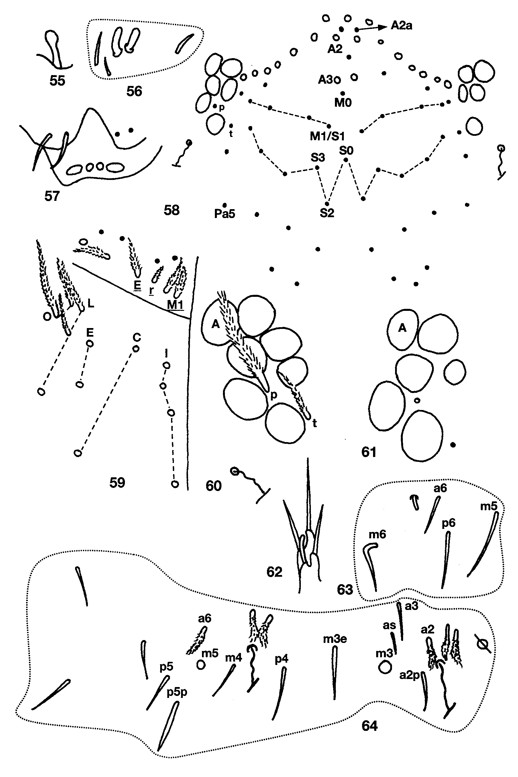
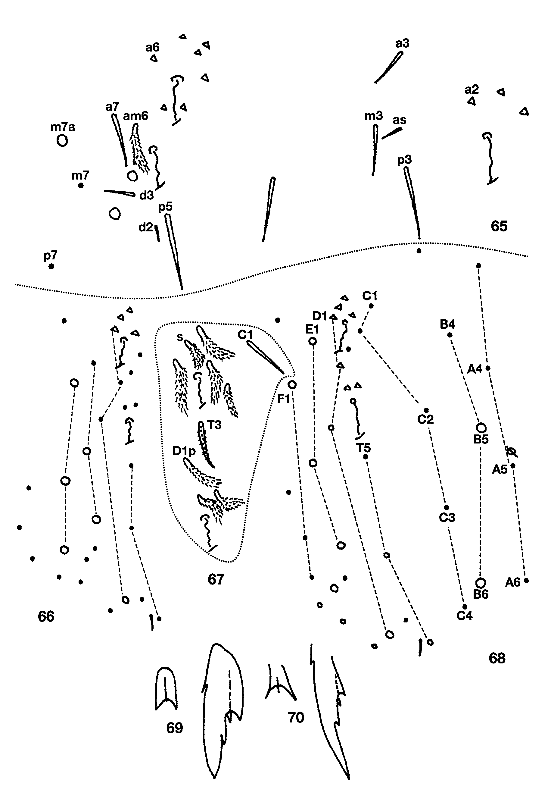
Figs 55–70 View FIGURES 55 – 64 View FIGURES 65 – 70
Material Examined. GREENBRIER Co., Trillium Cave, 28 May 2004, 1 on slide; Spencer Trap, 2 on one slide; Dyer’s Cave, 24 May 2004, 2 on slides, 3 in alcohol; Klevi’s Gap Cave, 20 May 2006, 1 mounted, 1 in alcohol; Stream Cave, Cassel-Windy System, 1 mounted.
Descriptive notes. Length up to 1.7mm. Color in alcohol variable among caves: individual from Trillium Cave, white with purple antennae, coxae and margin of Th. 2–Abd. 1; from Spencer Trap Cave white with a uniform blue wash over head and body; from Dyer’s and Stream caves deep blue or purple over antennae, head, body and all leg segments; specimens from Klevi’s Gap Cave were in very poor condition and the actual pigment distribution was not evident. Ant. 4 with subapical sense organ capitate ( Fig. 55 View FIGURES 55 – 64 ). Ant. 3 sense organ two thin walled rods ( Fig. 56 View FIGURES 55 – 64 ). Dorsal head chaetotaxy ( Fig. 58 View FIGURES 55 – 64 ) includes 6–7 macrosetae along antennal base, and macrosetae A0, A2, and A3, setae A2a enlarged but not typical macrosetae, Pa5 a microseta; setae A1 and M0 ciliate, all other dorsal microsetae smooth; seta S0 anterior to S3; M1/S1 near, but posterior to M2. Eyes 6+6, subequal; one individual from Dyer’s Cave has a distinct, although small, eye H only on one side ( Fig. 60 View FIGURES 55 – 64 ) whereas one individual from Stream Cave has eye C clearly smaller than the others ( Fig. 61 View FIGURES 55 – 64 ). Eye patch with setae p and t ( Figs. 58, 60 View FIGURES 55 – 64 ). Prelabral setae ciliate, all labral setae smooth. Inner labral papillae smaller than outer papillae, all papillae smooth ( Fig. 57 View FIGURES 55 – 64 ). Peristomal setae pss 0–2 normal, ciliate; setae on pleural fold ciliate. Ungulum of maxilla with 3 teeth. Labial papilla E with lateral process slightly curved dorsally and not reaching tip of papilla ( Fig. 62 View FIGURES 55 – 64 ). Proximal labial palp seta Y and Z subequal. Labial chaetotaxy M1M2rEL1L2A1–5, in one individual from Dyer’s cave setae E is smooth; the individual measuring less than 1 mm has only one seta m. All postlabial setae ciliate ( Fig. 59 View FIGURES 55 – 64 ), with 4+4 setae along ventral groove; columns C, E, L and O with 2, 2, 3 and 2 setae respectively, L1–2 and O1 shorter than other postlabial setae. Body formula for inner macrochaetotaxy as 00/0100+2. Abd. 1 setae organization linear; seta a6 present ( Fig. 63 View FIGURES 55 – 64 ). Chaetotaxy of Abd. 2 as in Fig. 64 View FIGURES 55 – 64 : a2, a6 and all supplementary setae fan-shaped (two individuals measuring ≤ 1mm with most supplementary setae ciliate instead of fan-shaped); a2p present, and from slightly to clearly longer than a2; a3 inserted between as and a2, and reaching as; as not reaching socket of m3; m3e reaching socket of m3; m5 a normal macroseta; p5 smooth, p5p ciliate. Abd. 3 ( Fig. 65 View FIGURES 65 – 70 ) with a2, a6, am6 and all supplementary setae fan-shaped; a3 not reaching as; as shorter than, but reaching m3; d2 and d3 present; a7, m7, and p7 normal microsetae, m7a a macroseta; a7 weakly ciliate and inserted very close to am6, m7 anterior to p6. Chaetotaxy of Abd. 4 bothriotrichal complex ( Fig. 67 View FIGURES 65 – 70 ) with all supplementary setae anterior to T2 fan-shaped; s smaller than surrounding supplementary setae and posterior to a; c1p and T3 ciliate; tip of T3 reaching base of D1p; D1p fan-shaped, posterior to T3 and reaching Pe; Pe and Pi fanshaped. General chaetotaxy of Abd. 4 ( Fig. 68 View FIGURES 65 – 70 ) with, macrosetae B5, B6, D2, T6, T7, D3, E1, E2, E3, F1; D3 and E1 are slender and acuminate but have unusually large sockets, and in the absence of the hair would be interpreted as stout macrosetae; in the two individuals measuring ≤ 1mm, D2 and E1 are microsetae whereas F2 and F3 are macrosetae ( Fig. 66 View FIGURES 65 – 70 ); macroseta B5 crossed by, or just anterior to a line drawn between A5 and C2; F2 closer to E3 than E2; microseta posterior to E3 present. Abd. 4 with 5+5 posterior setae. Metathoracic femora with posterior blunt macrosetae, but not well differentiated; lateral and anterior macrosetae acuminate. Trochanteral organ with up to 20 setae. Tenet hair clavate, as long or longer than unguiculus; unguiculus lanceolate with posterior lamella appearing serrate in some individuals. Unguis with 3 inner and 3 outer teeth ( Figs. 69–70 View FIGURES 65 – 70 ): basal inner pair unequal in size, but difference not well marked, unpaired tooth distinct; outer teeth varying in size from short, not reaching base of inner teeth in specimens from Stream Cave ( Fig. 70 View FIGURES 65 – 70 ), to long and reaching base of basal inner pair in individuals from Dryer’s Cave ( Fig. 69 View FIGURES 65 – 70 ). Ventral tube with 7+7 anterior setae, posterior and disto-lateral setae obscured in all specimens studied. Chaetotaxy of manubrial plate with 5–6 outer and 1–2 inner setae separated by 2 pseudopores (one individual has 3 pseudopores). Apical mucronal tooth longer than basal tooth; mucronal spine smooth and reaching basal tooth.
Remarks. The identification of these specimens as P. collina is based on the description provided by Christiansen and Bellinger (1998) and the characters available in the online Pseudosinella database (www.unav.es/unzyec/collembola). The specimens from West Virginia differ from the material described by Christiansen & Bellinger in having outer ungual teeth, and from the information in the Pseudosinella database online in having 5–6 outer setae on the manubrial plate (2 setae in the database). Pseudosinella collina appears most similar to P. georgia Christiansen & Bellinger, 1998 , but the two species are easily distinguished by the sculpturing of prelabral and some labial setae, number of setae on anterior face of ventral tube, and general structure of tenent hair and claw complex.
No known copyright restrictions apply. See Agosti, D., Egloff, W., 2009. Taxonomic information exchange and copyright: the Plazi approach. BMC Research Notes 2009, 2:53 for further explanation.