
Massisteria marina Larsen and Patterson 1990
|
publication ID |
https://doi.org/10.4467/16890027AP.15.005.2192 |
|
DOI |
https://doi.org/10.5281/zenodo.12522295 |
|
persistent identifier |
https://treatment.plazi.org/id/0B708784-E66A-8926-FCBA-FB7D206CD419 |
|
treatment provided by |
Felipe (2024-04-18 19:01:46, last updated by Guilherme 2025-02-03 16:49:55) |
|
scientific name |
Massisteria marina Larsen and Patterson 1990 |
| status |
|
Massisteria marina Larsen and Patterson 1990 ( Fig. 3c View Fig )
Observation: Cells are 3 to 6.5 µm and dorso-ventrally flattened irregular body with delicate pseudopodia bearing extrusomes. The pseudopodia extend radially from the cell and normally adhere to the substrate. Two short curved flagella arise from the dorsal side of the cell and are relatively inactive. Rarely observed.
Remarks: Generally, the observations are in good agreement with those of Larsen and Patterson (1990) and Lee and Patterson (2000). Previously reported size ranges are 2 to 12 µm and this species was found at marine sites in Agean Sea ( Turkey), subtropical and tropical Australia, Brazil, Denmark, Gulf of Finland, equatorial Pacific, Korea and Panama (Larsen and Patterson 1990; Patterson and Fenchel 1990; Vørs 1992a, b; Vørs et al. 1995; Ekebom et al. 1996; Tong 1997a; Tong et al. 1998; Lee and Patterson 2000; Al-Qassab et al. 2002; Lee 2002b, 2006b; Aydin and Lee 2012).
Al-Qassab S., Lee W. J., Murray S., Simpson A. G. B., Patterson D. J. (2002) Flagellates from stromatolites and surrounding sediments in Shark Bay, Western Australia. Acta Protozool. 41: 91 - 144
Aydin E. E., Lee W. J. (2012) Free-living heterotrophic flagellates from intertidal sediments of Saros Bay, Aegean Sea (Turkey). Acta Protozool. 51: 119 - 137
Ekebom J., Patterson D. J., Vors N. (1996) Heterotrophic flagellates from coral reef sediments (Great Barrier Reef, Australia). Arch. Protistenkd. 146: 251 - 272
Larsen J., Patterson D. J. (1990) Some flagellates (Protista) from tropical marine sediments. J. Nat. Hist. 24: 801 - 937
Lee W. J. (2002 b) Some free-living heterotrophic flagellates from marine sediments of Inchon and Ganghwa Island, Korea. Korean J. Biol. Sci. 6: 125 - 143
Lee W. J. (2006 b) Some free-living heterotrophic flagellates from marine sediments of tropical Australia. Oce. Sci. J. 41: 75 - 95
Patterson D. J., Fenchel T. (1990) Massisteria marina Larsen & Patterson 1990, a widespread and abundant bacterivorous protist associated with marine detritus. Mar. Ecol. Prog. Ser. 62: 11 - 19
Patterson D. J., Lee W. J. (2000) Geographic distribution and diver- sity of free-living heterotrophic flagellates. In: The flagellates: unity, diversity and evolution, (Eds. Leadbeater B. S. C., Green J. C.). Taylor & Francis, London and New York. 49: 267 - 287
Tong S. M. (1997 a) Heterotrophic flagellates from the water column in Shark Bay, Western Australia. Mar. Biol. 128: 517 - 536
Tong S. M., Nygaard K., Bernard C., Vors N., Patterson D. J. (1998) Heterotrophic flagellates from the water column in Port Jack- son, Sydney, Australia. Europ. J. Protistol. 34: 162 - 194
Vors N. (1992 a). Heterotrophic amoebae, flagellates and heliozoa from the Tvarminne area, Gulf of Finland in 1988 - 1990. Ophelia 36: 1 - 109
Vors N., Buck K. R., Chavez F. P., Eikrem W., Hansen L. E., Oster- gaard J. B., Thomsen H. A. (1995) Nanoplankton of the equatorial Pacific with emphasis on the heterotrophic protists. Deep-Sea Res. 42: 585 - 602
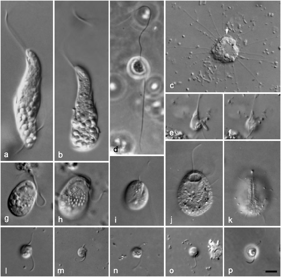
Fig. 3. a–b –Rhynchobodo formica; c –Massisteria marina, general appearance of cell showing pseudopodia and flagella (arrow); d–f – Cercomonas sp.; d – note two acronematic flagella; e – general appearance; f – note flagellar orientation; g–h – Roombia truncata, note cell attached to the substrate by the tip of the posterior flagellum and note extrusomes; i – Protaspa obliqua, note anterior protrusion;j–k – Thaumatomastix setifera, note spines around the body; j – general appearance of cell; k – ventral face showing a deep groove and pseudopodia; l–m – Ancyromonas sigmoides of different cells; l – note slightly thick anterior flagellum with acronematic tip and broad rostrum; m – note thick anterior flagellum and acute rostrum; n–p – Ancyromonas impluvium nov. spec., showing general appearance of cell and note flagellar insertion.All micrographs are DIC images with the exceptions of (d) and (p) which are phase contrast images. Scale bar: 5 µm for all figures.
No known copyright restrictions apply. See Agosti, D., Egloff, W., 2009. Taxonomic information exchange and copyright: the Plazi approach. BMC Research Notes 2009, 2:53 for further explanation.
|
Kingdom |
|
|
Phylum |
|
|
Class |
|
|
Order |
|
|
Family |
|
|
Genus |