
Hughjonestrongylus arfakiensis, Smales, 2020
|
publication ID |
https://doi.org/ 10.11646/zootaxa.4861.4.4 |
|
publication LSID |
lsid:zoobank.org:pub:2297FB97-3C02-42B0-B811-019646E33C0C |
|
DOI |
https://doi.org/10.5281/zenodo.4416879 |
|
persistent identifier |
https://treatment.plazi.org/id/08207AC1-C37B-46AF-906C-889EEF5D7C53 |
|
taxon LSID |
lsid:zoobank.org:act:08207AC1-C37B-46AF-906C-889EEF5D7C53 |
|
treatment provided by |
Plazi |
|
scientific name |
Hughjonestrongylus arfakiensis |
| status |
sp. nov. |
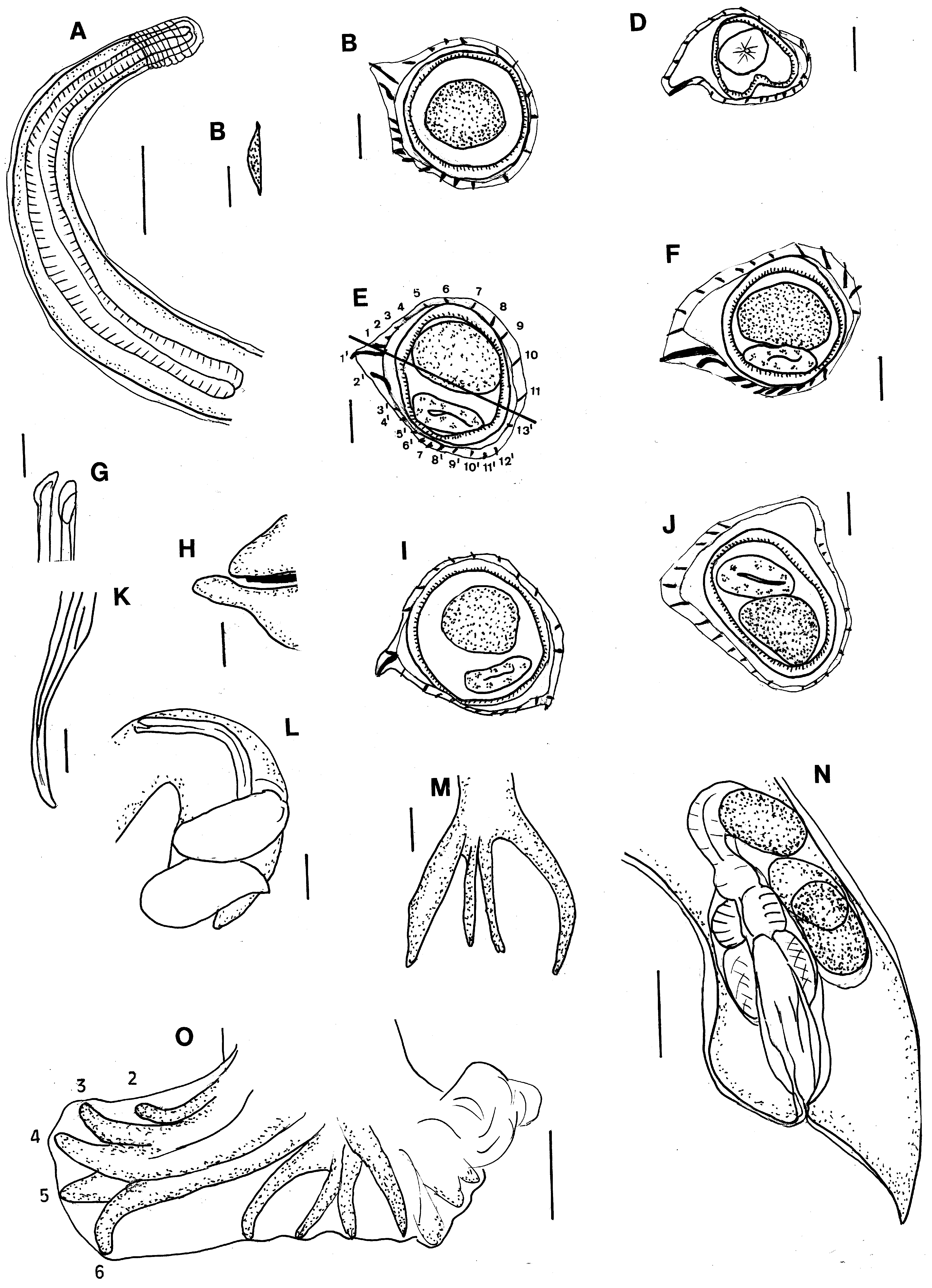
Hughjonestrongylus arfakiensis n. sp.
( Figs 5 View FIGURE 5 A–O)
Type host. Paramelomys mollis (Thomas)
Type specimens. Holotype male AM W. 53158, allotype female AM W. 53159, paratypes 5 males, 8 females AM W. 53152 from Paramelomys mollis Bichate, Arfak Mountains , Papua, Indonesia (1° 06´S 133° 56´E); coll. T. Flannery, A. Szalay 10. x. 1992. GoogleMaps
Other material examined. From P. mollis From Indonesia, Papua; 5 males, 2 females, Bichate, Arfak Mountains , AM W. 53151.
Site in host. Small intestine.
Etymology. The species name is taken from the type locality.
Description. General: Small worms, slightly or tightly coiled. Cephalic vesicle prominent with 6–8 transverse annulations. Buccal capsule vestigial, mouth opening triangular with rudimentary lips; cephalic and labial papillae not seen, 2 lateral amphids. Oesophagus claviform, nerve ring not seen, excretory pore and deirids at same level in mid region of oesophagus.
Synlophe: (based on sections from 15 worms). Longitudinal pointed ridges extend from posterior margin of cephalic vesicle to anterior to bursa or vulva; 21–26 ridges; 21 in males, 17–21 in females in anterior body; 24–25 in males, 22–24 in females in mid body; 26 in males, 23–25 in females in posterior body. Ridges markedly different in size. Axis of orientation of ridges oblique from right ventral to left dorsal; 9–12 ridges dorsal side, 10–13 ventral side. Left ridge distinct from ridge 1´; lateral left and lateral right ridges largest, ridge 2´smaller than ridge 1´ventral ridges 3´–13´about same size, more robust than dorsal ridges, right ventral quadrant without ridges in female mid body; dorsal ridges 1–7 decreasing in size, ridges 8/9– 12 /13 increasing in size. Ridges losing size gradient, reducing in size posteriorly.
Male: (measurements of 10 specimens) Length 2.7–4.0 (3.7) mm, maximum width 87–114 (87). Cephalic vesicle 36–43 (40) long. Oesophagus 320–440 (378) long; nerve ring, deirids and excretory pore not seen. Bursa relatively large, tightly curled, shape constrained by cuticular folds in most specimens, dissymmetrical, left lobe larger; pattern of rays 2–3. Dorsal lobe shorter than laterals; dorsal trunk symmetrical, divided at about ½ its length, terminal divisions rays 9, 10 symmetrical, rays 8 arising from dorsal trunk at about same level, slightly dissymmetrical, left ray larger, reaching margins of bursa; lateral rays 6, 5, 4, all about same size, reaching margin of bursa, rays 3, 2 arise together from common trunk, reaching margin of bursa. Genital cone prominent, ventral lip elongated. Spicules equal, filiform, posterior part of spicule curved, tips straight, 250–410 (399) long; spicule to body length ratio 11.7 %. Gubernaculum 20–43 (32) long.
Female: (measurements of 10 specimens) Length 2.6–3.8 (3.3) mm, maximum width 67–107 (78). Cephalic vesicle 33–43 (39) long. Oesophagus 350–620 (451) long; nerve ring not seen, deirids, excretory pore at about same level, 150, 220 (2 measurements) from anterior end. Vulva opens 65–120 (96) from tail tip; region of ovejector bulbous, tail more or less straight. Ovejector monodelphic, infundibulum looped posteriorly 60–80 (70) long, sphincter shortest element 30 long, vestibule 50–60 (56) long, vagina thick walled 60 long; posterior end of uterus lying parallel with ovejector. Tail conical with blunt tip, 45–95 (73) long. Eggs thin shelled, ellipsoidal, up to 18 eggs in utero, 51.0–66.0 (60.3) long, 33.0–39.0 (35.8) wide.
Remarks. This new species belongs to the genus Hughjonestrongylus because it has a synlophe showing the same arrangement of pointed ridges as described in the diagnosis of Durette-Desset & Digiani (2015) and a dissymmetrical bursa with a type 2–3 pattern of bursal rays (Digiani & Durette-Deset, 2014). In having a synlophe of 24 – 25 ridges at the mid body Hughjonestrongylus arfakiensis n. sp. is placed between species with more than 25 ridges and species with fewer than 25 ridges at the mid body in the key to the species of Hughjonestrongylus of Smales (2019) and comes closest to H. mirzai ( Smales, 2009) with 21–24 ridges and H. amplicauda ( Smales & Heinrich, 2010) with 20 –24 ridges at the midbody. Having spicules 215–410 long, 11.7% of body length males of H. arfakiensis can be distinguished from both H. mirzai (spicules 250–320, 9.8 % of body length) and H. amplicauda (spicules 280–365, 14.5 % of body length) ( Smales 2009; Smales & Heinrich 2010). Females of H. arfakiensis can readily be distinguished from all other species of Hughjonestrongylus by the shape of the posterior end and the relationship of the uterus to the ovejector.
| AM |
Australian Museum |
| T |
Tavera, Department of Geology and Geophysics |
No known copyright restrictions apply. See Agosti, D., Egloff, W., 2009. Taxonomic information exchange and copyright: the Plazi approach. BMC Research Notes 2009, 2:53 for further explanation.
|
Kingdom |
|
|
Phylum |
|
|
Class |
|
|
Order |
|
|
Family |
|
|
Genus |