
Desmodora striatocephala, Tchesunov, Alexei V., 2008
|
publication ID |
https://doi.org/ 10.5281/zenodo.183775 |
|
DOI |
https://doi.org/10.5281/zenodo.6231746 |
|
persistent identifier |
https://treatment.plazi.org/id/0C0087CA-5A4F-FFFF-FF51-83DBFD5BF99A |
|
treatment provided by |
Plazi |
|
scientific name |
Desmodora striatocephala |
| status |
sp. nov. |
Desmodora striatocephala sp. nov.
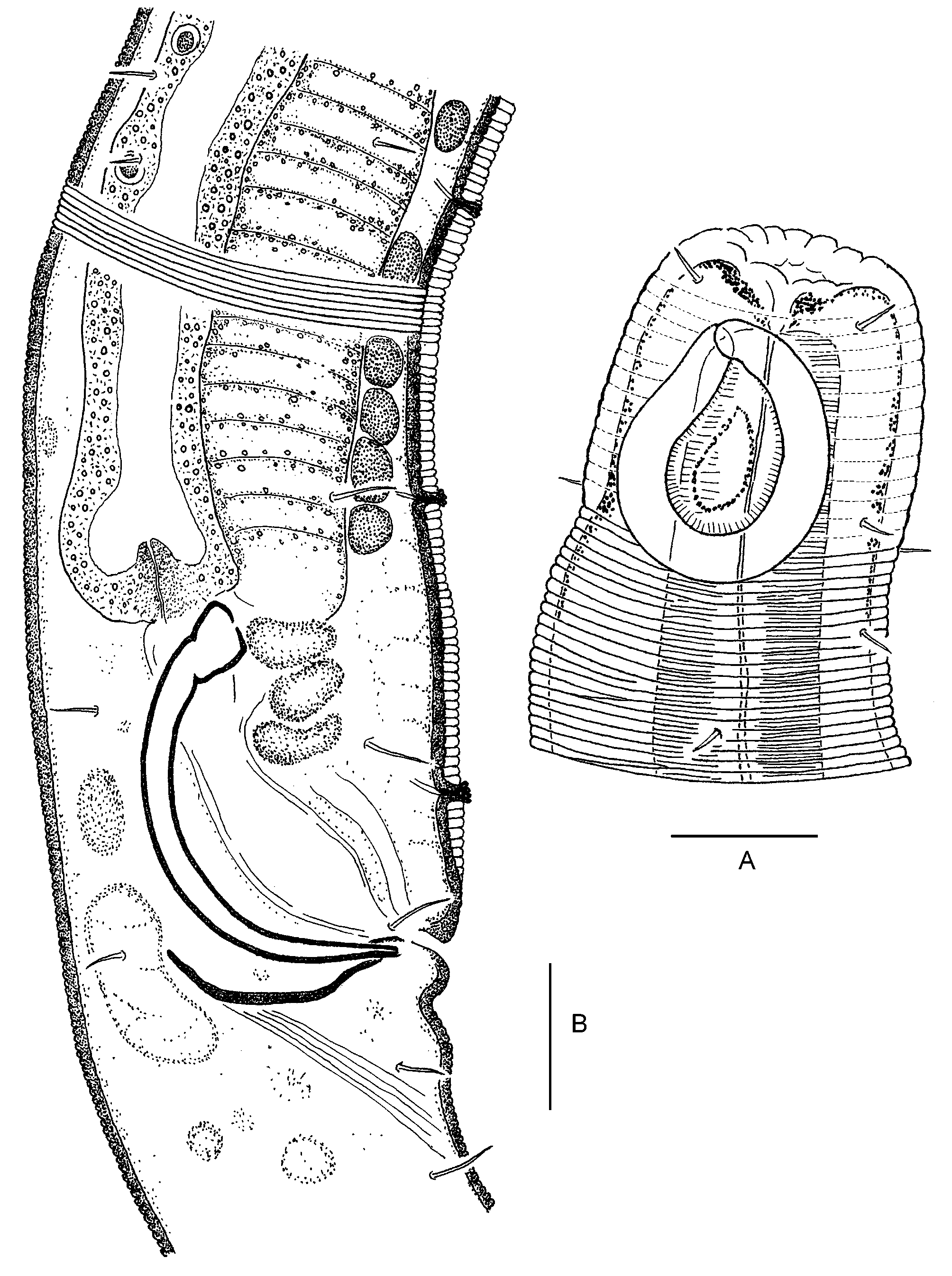
Figs 10–11 View FIGURE 10 View FIGURE 11
Type material: Holotype male. Type specimen deposited in the DIVA nematode collection.
Type locality: DIVA I, Meteor 48/1, Station 325/4, Multicorer 8: Southeast Atlantic Ocean, 19°58.3'S & 002°59.8'E, depth 5450 m. 14 July 2000.
Etymology: The species name reflects fine cross striation of the head.
Description. Body cylindrical, of brownishyellow colouration. Anterior end truncate. Somatic cuticle finely but distinctly annulated (just posterior to the cephalic capsule, nine annules for 10 µ m on convex body side and twelve annules on the opposite concave side; in the midbody, 17–20 annules for 10 µ m), with no lateral differentiation.
Body length 1009 µ m, a 34.8, b 7.6, c 7.4. Body diameter at the level of: cephalic setae 16 µ m, amphidial fovea 21 µ m, nerve ring 29 µ m, cardia 29 µ m, midbody 29 µ m, cloaca 24 µ m.
Labial region slightly drawn inward and therefore inner and outer labial sensilla not discernible. There are four short cephalic setae 3 µ m long at anterior cephalic capsule. Cephalic capsule 18 µ m long, shaped by thick cuticle with very fine traces of fused annules; the annules of the cephalic capsule by a factor of 1.5–2 wider and much less distinct than postcapsular annules.
Amphidial fovea very large and occupies nearly the whole lateral surface of the cephalic capsule. Amphidial fovea loopshaped, slightly longitudinally oval and narrowed to its anterior end. Exit site of the amphidial nerve is thus unusually situated at the anterior end of the fovea where descending and ascending branches contact with one another. Amphidial fovea width 16 µ m or 76% c.b.d; distance from the cephalic apex to the amphidial fovea 3–4 µ m.
Somatic setae arranged in longitudinal rows distinct in the pharyngeal region and becoming irregular in the intestinal region.
Cheilostoma indistinctly discernible because of the dense pigmentation. The subsequent part of the buccal cavity narrow, equal to the cephalic capsule. Walls of the buccal cavity and small but distinct dorsal tooth very weakly sclerotized.
Pharynx slender, evenly muscular, with sinuous cuticular lining of the internal lumen. Posteriorly, the pharynx widened into a clear roundish bulb. Pharynx width at the level of the nerve ring 13 µ m; posterior bulb 24 µ m wide and 25 µ m long. Nerve ring hardly visible. No indication of a renette cell.
Cardia short, trapeziumshaped. Intestine with yellowbrownish inclusion droplets.
Testis singular, outstretched, situated ventrally to the intestine. Spermatozoa relatively large, of uncertain shape, with granular content. Vas deferens divided into alternate regions made up of cells with transparent and coarsely granulated content.
Spicules arched, distally pointed and proximally widened into shovellike knobs, Gubernaculum paired, as two curved plates nearly parallel to the distal half of the spicules. Spicules length 29 µ m (chord) and 40 µ m (arch); gubernaculum plate 15 µ m long.
Precloacal ventral cuticle thickened and transparent. About five midventral supplementary organs inserted in the precloacal cuticle. Supplementary organ presents an intracuticular canal with a small papilla on the surface of the cuticle. The supplementary organs decrease in size from the cloaca anteriad.
Tail elongate conical, relatively slender, with terminal spinnerete tube. Tail length 5.32 anal diameters long. There a few pre and postanal subventral setae on the posterior body; their length up to 4.5 µ m, greater than those of the intestinal region.
Diagnosis. Body length 1009 µ m, a 34.8, c 7.38. There are four short cephalic setae 3 µ m long at anterior cephalic capsule. Cephalic capsule 18 µ m long, shaped by thick cuticle with traces of fused annules; the annules of the cephalic capsule by a factor of 1.5–2 wider and much less distinct than postcapsular annules. Amphidial fovea very large and occupies nearly the whole lateral surface of the cephalic capsule. Amphidial fovea loopshaped, slightly longitudinally oval and narrowed to its anterior end. Amphidial fovea width 16 µ m or 76% c.b.d. Precloacal ventral cuticle thickened and transparent. About five midventral supplementary organs inserted in the precloacal cuticle. Supplementary organ present as an intracuticular canal with a small papilla on the surface of the cuticle. Tail elongate conical, relatively slender. Tail length 5.32 anal diameters long.
Differential diagnosis: Desmodora striatocephala sp. nov. is characterized by very fine but distinct cross striation of the cephalic capsule. Some related desmodorid genera such as Paradesmodora Stekhoven, 1950 , Echinodesmodora Blome, 1982 and Stygodesmodora Blome, 1982 are also characterized by a crossstriated periamphidial cuticle hardly distinguishable from that of the neck region. However, the cephalic region of D. striatocephalata is clearly different in having a thickened cuticle with very fine transversal grooves thus forming a cephalic capsule set off the neck region with thinner cuticle and sharp grooves between annules.
Desmodora striatocephala sp. nov. is well characterized by a combination of very large amphidial fovea occupying nearly the entire lateral surface of the cephalic capsule and thickened midventral preanal cuticle with a few supplementary papillae inserted therein in males. The new species shares this set of features with D. cuddlesae Inglis, 1963 and to some lesser degree with D. inflexa Wieser, 1954 . D. striatocephala sp. nov. differs from D. cuddlesae by the shorter body (1009 µ m versus 1760–1980 µ m), relatively longer tail (c 7.38 versus 13.5–14.1, c’ 5.32 versus 2.65–2.75), absolutely smaller but relatively bigger amphidial fovea (16 µ m wide and 76% c.b.d. versus 26–37 m and 59% c.b.d.), about five versus twentynine preanal supplementary papillae in the ventral thickened transparent cuticle. D. striatocephala differs from D. inflexa with shorter body (1009 µ m versus 2450 µ m), relatively thicker body (a 34.8 versus 53.7), about five versus fourteen preanal supplementary papillae in the ventral thickened transparent cuticle and shorter tail (c 7.38 versus 25.8, c’ 5.32 versus 2.00). Other Desmodora species with ventrally thickened preanal cuticles are much more distinguished from D. striatocephala in other characters.

No known copyright restrictions apply. See Agosti, D., Egloff, W., 2009. Taxonomic information exchange and copyright: the Plazi approach. BMC Research Notes 2009, 2:53 for further explanation.