
Hersilia orvakalensis, Javed, Maqsood, Foord, Stefan H. & Tampal, Farida, 2010
|
publication ID |
https://doi.org/ 10.5281/zenodo.197848 |
|
DOI |
https://doi.org/10.5281/zenodo.6201705 |
|
persistent identifier |
https://treatment.plazi.org/id/0D3D6E57-9558-FFAD-FF7F-FA3EFDFE0302 |
|
treatment provided by |
Plazi |
|
scientific name |
Hersilia orvakalensis |
| status |
sp. nov. |
Hersilia orvakalensis View in CoL sp. nov.
Figs 3–27 View FIGURES 3 – 4 View FIGURES 5 – 8 View FIGURES 9 – 14 View FIGURES 15 – 18 View FIGURES 19 – 24 View FIGURES 25 – 27
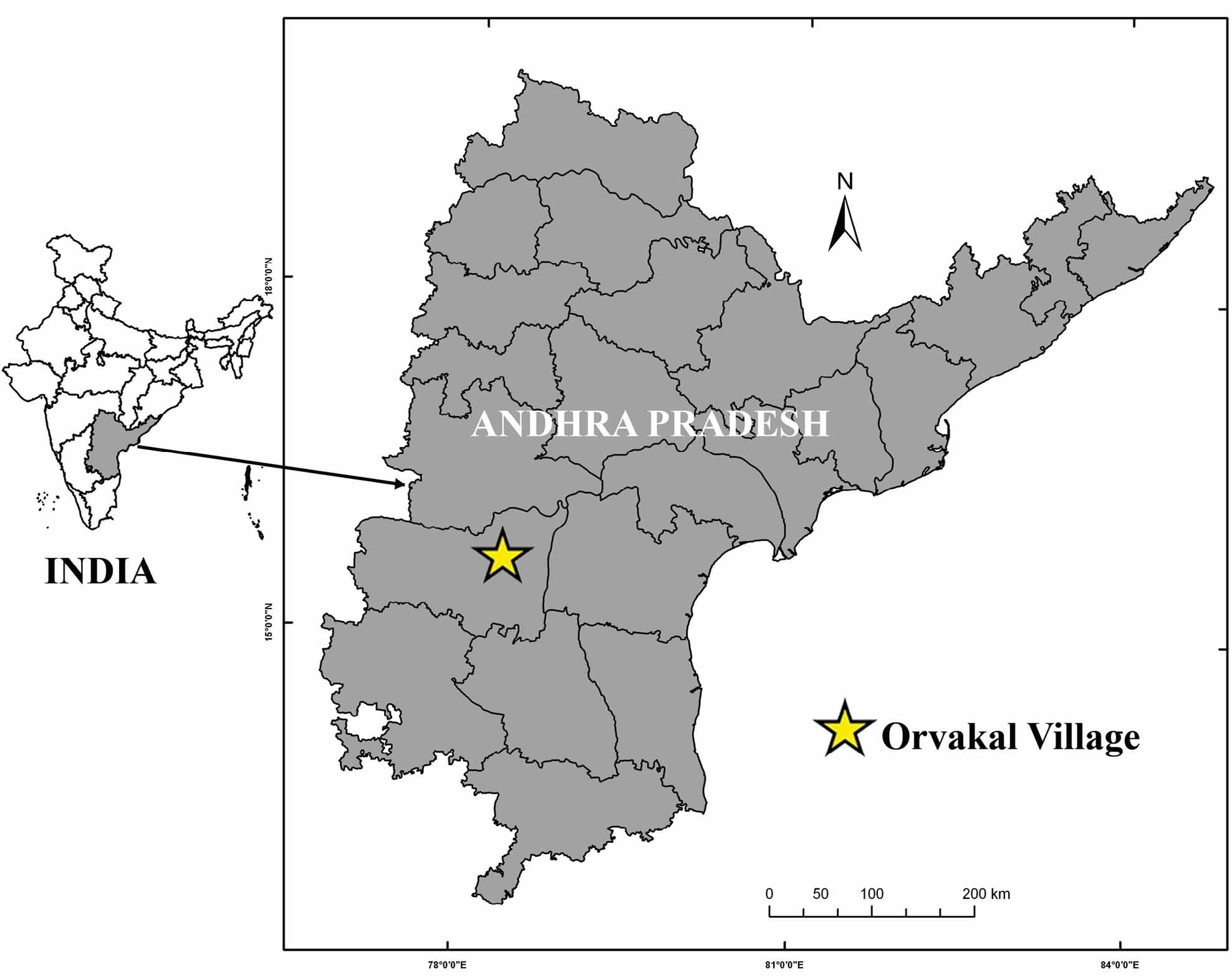
Type material. INDIA: Andhra Pradesh: holotype male, Orvakal, Kurnool District, 15º41’N, 78º10’E, 20 May 2010, S. M. Maqsood Javed (ZSI/ FBRC /A-27). Paratypes: allotype female, same data as holotype except 14 February 2010 (ZSI/ FBRC /A-28); 1 male, 1 female, same data as holotype (ZSI/ FBRC /A-29-30).
Etymology. The specific epithet is a noun in apposition, taken from the type locality at Orvakal Village, Kurnool District.
Affinities. In the classification of Baehr and Baehr (1993) Hersilia orvakalensis sp. nov. would be placed in the H. pectinata species group. This group includes three other species from the Oriental Region and four species from the Afrotropical Region. Males are characterized by a pronounced angular dorsal projection on the pedipalpal tibia with four-seven strong spines, by lamellar modifications on the bulb of the male pedipalp, by a complex median apophysis and by a long, ridged embolus. Females are characterized by a median epigynal plate with basal rippled pads. Hersilia orvakalensis seems otherwise closely related to H. tibialis from India and Sri Lanka in possessing a relatively short male pedipalp, with a short and wide tibia bearing a conspicuous ridge and five strong spines. Like H. tibialis , the cymbium is also stout and markedly pilose, rounded off at the apex, with numerous short spines; the embolus is curved and free, with an enlarged apex; the retrolateral projection of the median apophysis is flat and broad; and the epigynum is large, consisting of a median plate with distinct lateral borders and an extended base.
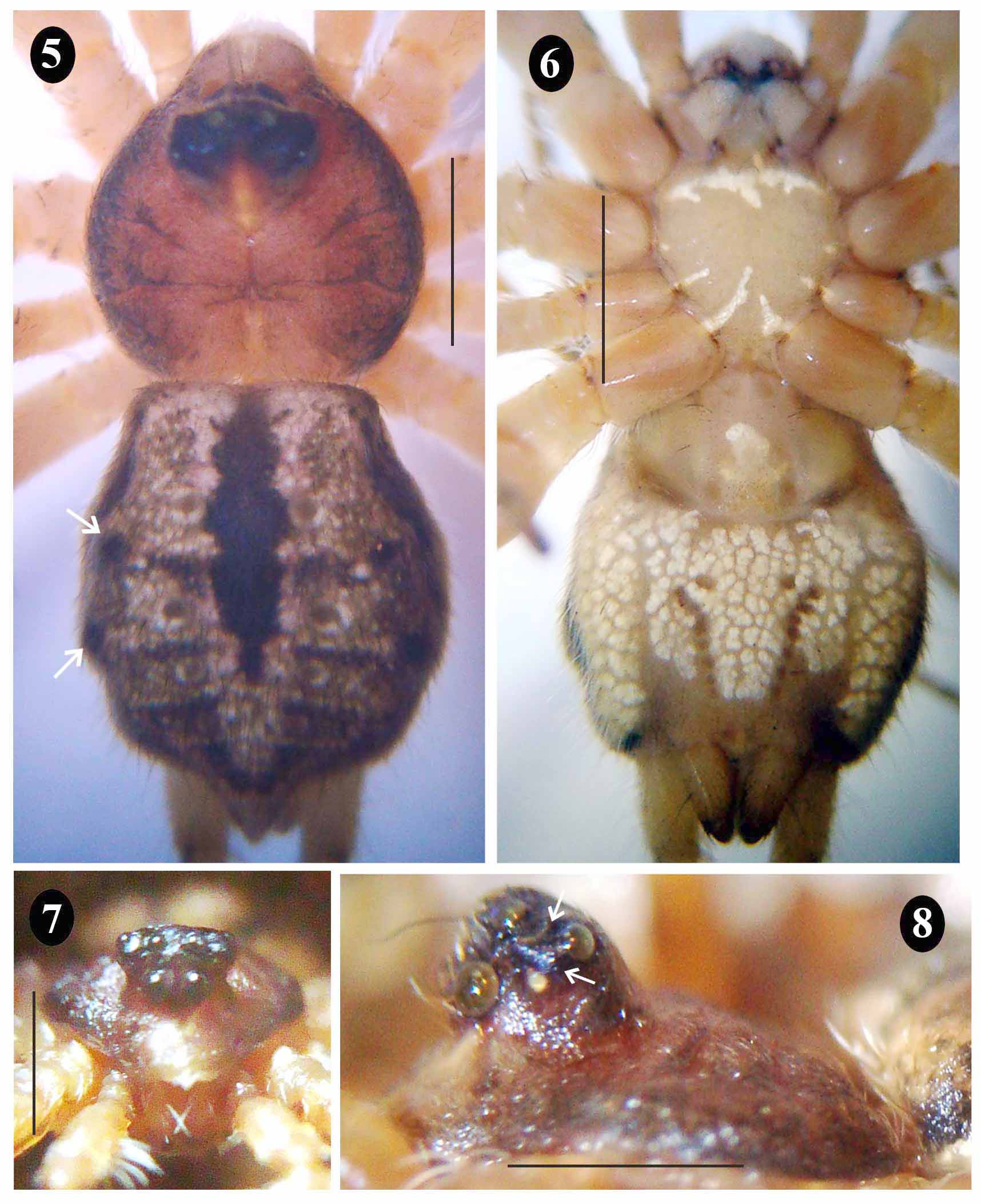
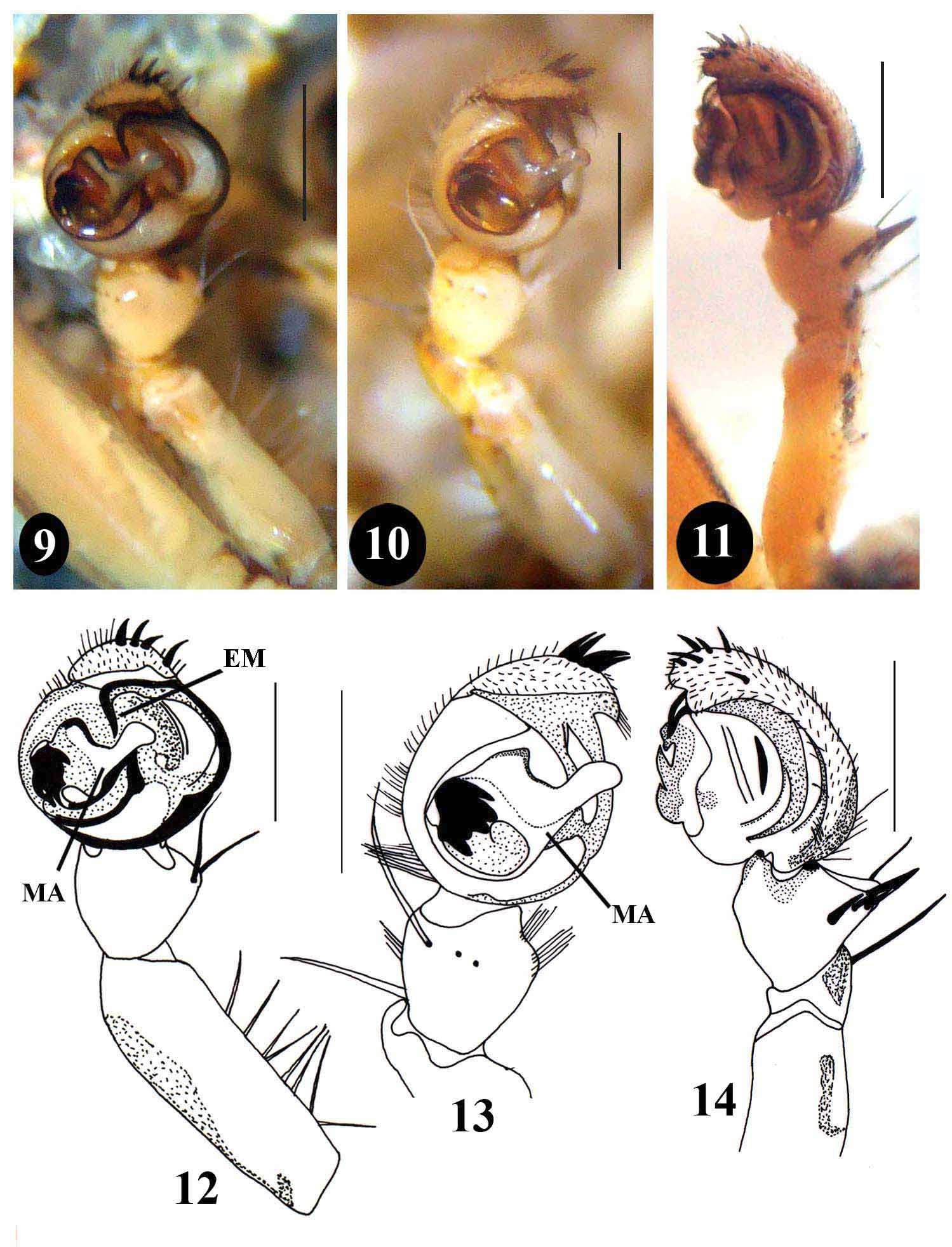
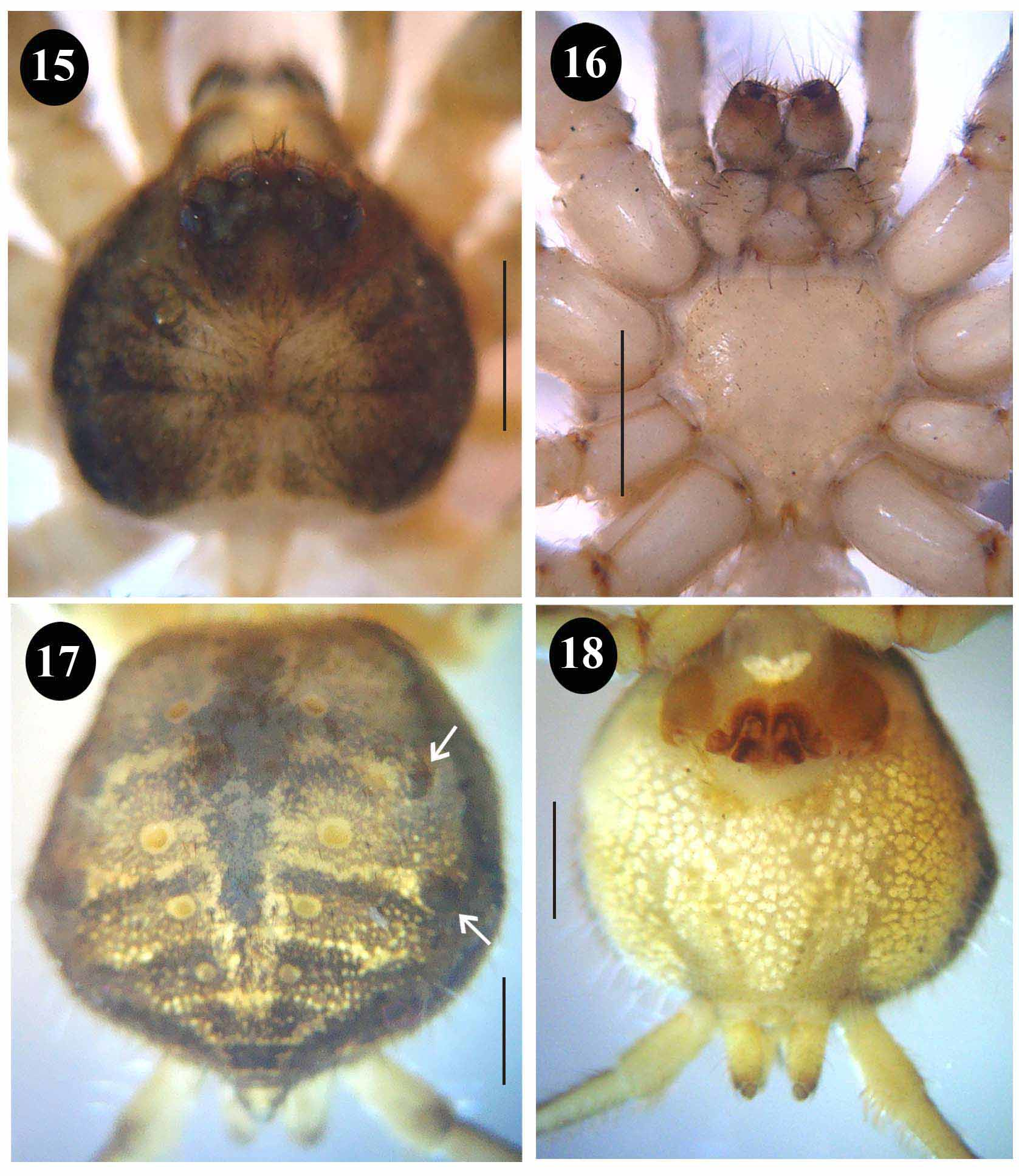
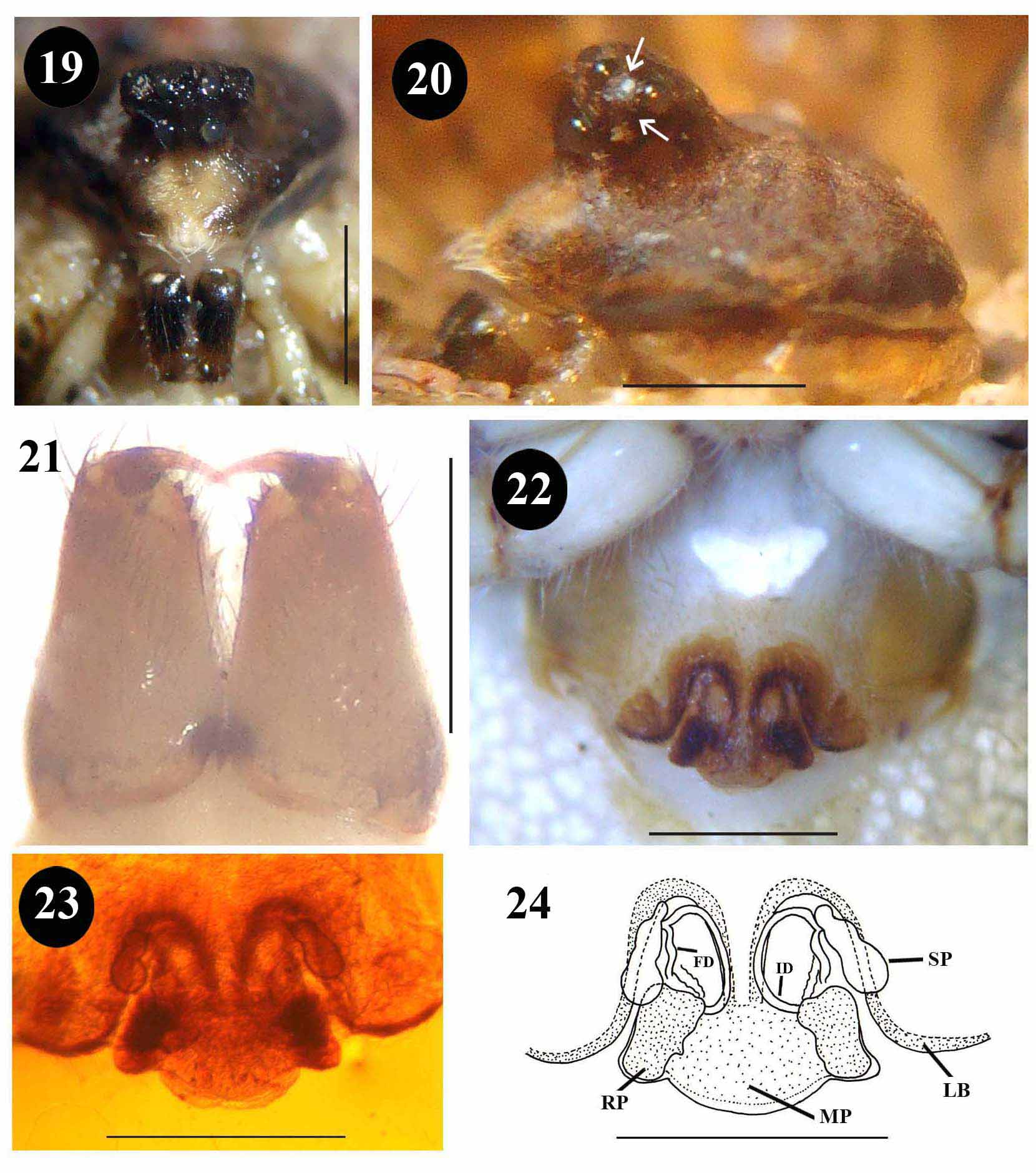
Diagnosis. Males of Hersilia orvakalensis sp. nov. can be distinguished from all other described congeners (including H. tibialis ) by the absence of crenulae on the retrolateral border of the median apophysis and by the short truncated distal area of the cymbium ( Figs 9 – 14 View FIGURES 9 – 14 ). Females can be distinguished by the shape of the spermathecae and accessory glands, which form a single elongate tri-lobed and pod-shaped structure, and by the shape of the median epigynal plate, which is extended posteriorly with a broad base ( Figs 23–24 View FIGURES 19 – 24 ). Both sexes are also characterized by the presence of two humps laterally on the abdomen ( Figs 5 View FIGURES 5 – 8 , 17 View FIGURES 15 – 18 ).
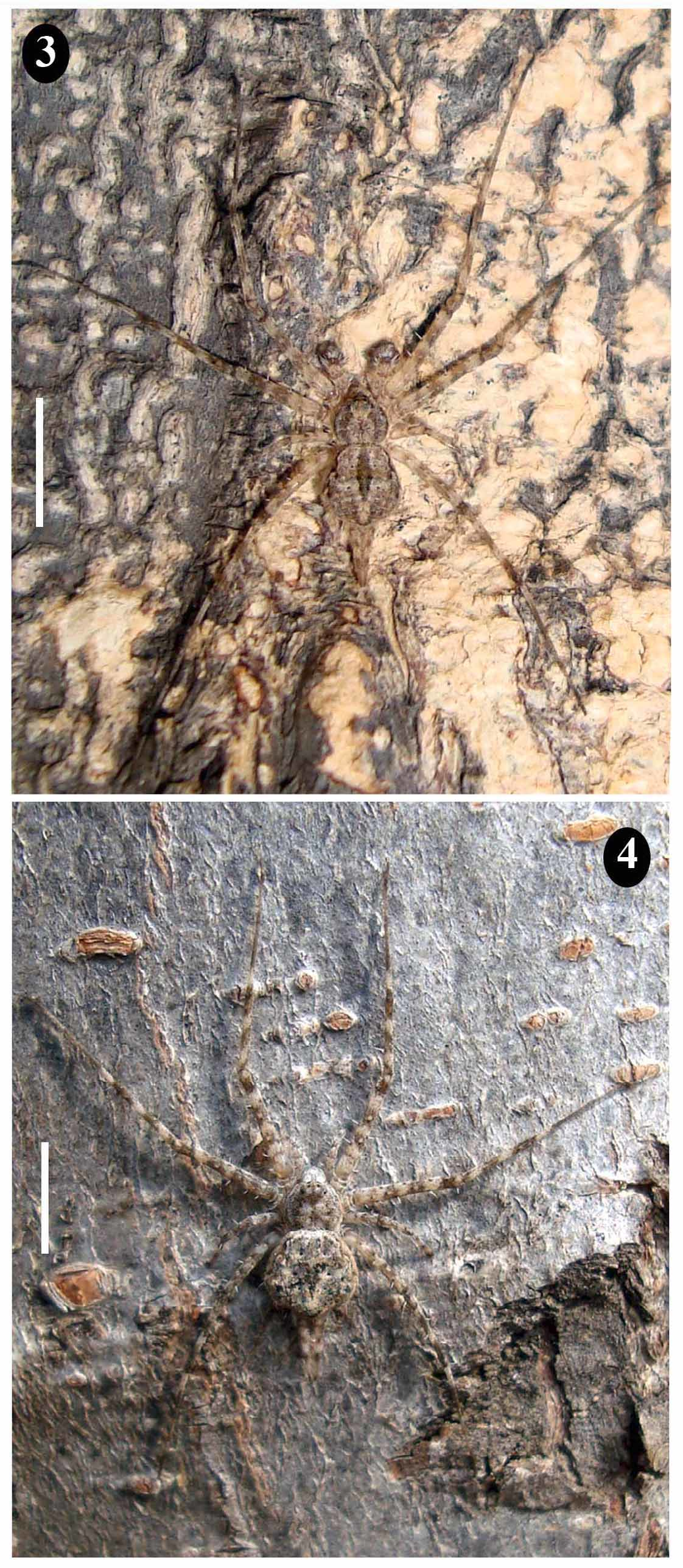
Description. Male (holotype): Cephalothorax ( Figs 3 View FIGURES 3 – 4 , 5 – 8 View FIGURES 5 – 8 ): Carapace slightly longer than wide (2.02 long, 1.84 wide), covered with short white pilose setae; ocular region strongly raised, slightly convex between PME, concave behind PER; AER and PER recurved; clypeus broad, 0.49 long. Cheliceral paturon 0.42 long; promargin with three robust, triangular teeth and retromargin with nine minute teeth. Maxillae 0.47 long, 0.27 wide; labium 0.29 long, 0.19 wide; sternum 0.98 long, 0.92 wide. Eyes small, with lateral eyes situated on a tubercle; AME, PME and PLE black, ALE pearly white; MOQ square, 0.38 long, 0.38 wide. Eye sizes and inter-distances: AME>PLE>PME>ALE (0.16, 0.12, 0.12, 0.06); AME–AME 0.04, AME–ALE 0.11, PME– PME 0.14, PME–PLE 0.14; AER 0.81 wide, PER 0.91 wide.
Legs: Leg formula 2143, lengths of legs [total length (femur + patella + tibia + metatarsus + tarsus)]: I = 13.22 (3.42 + 1.01 + 3.43 + 4.79 + 0.57); II = 13.75 (3.48 + 1.08 + 3.60 + 4.97 + 0.62); III = 4.79 (1.33 + 0.38 + 1.30 + 1.32 + 0.46); IV = 11.43 (3.14 + 0.65 + 2.76 + 4.18 + 0.70). Leg spination: legs I, II, III and IV have similar numbers and arrangement of spines (femur dorsal 1–1–1, prolateral 1–1–1–1, retrolateral 1–1–1–1; patella dorsal 1–1, prolateral 1–1, retrolateral 1–1; tibia dorsal 1–1, prolateral 1–1–1, retrolateral 1–1–1; metatarsus dorsal 1–1–1, prolateral 1–1–1, retrolateral 1–1–1).
Abdomen ( Figs 5 – 6 View FIGURES 5 – 8 ): Longer than wide, 3.06 long, 2.28 wide, widest posteriorly, subquadrate, with two slight humps on lateral edge. Dorsally with four pairs of spherical muscular sigillae, all dissimilar in size, with fourth pair smallest. Ventral muscular sigillae arranged in a wide V-shape. Posterior lateral spinnerets slightly shorter than abdomen, 3.03 long (basal segment 0.72, terminal segment 2.31); spigots on median border dense and elongate.
Pedipalp ( Figs 9 – 14 View FIGURES 9 – 14 ): Total length (femur + patella + tibia + tarsus): 2.40 (0.91+ 0.40 + 0.39 + 0.70). Tibia short, as long as patella, with angulated projection dorsally and five strong dorsal spines arranged in continuous row. Cymbium very short and stout, markedly pilose on anterior dorsal region; apex widely rounded, with five short and stout spines distally and separate spine present on lateral side. Embolus widely curved, free, apex slightly excised. Retrolateral projection of median apophysis flat and broad, without any crenulae on retrolateral border.
Colouration and markings ( Figs 3 View FIGURES 3 – 4 , 5 – 8 View FIGURES 5 – 8 ): In life dorsum of carapace, abdomen and legs grey with black streaks and spots along with several brown and cream markings. In ethanol carapace reddish brown with darker radiating grooves, dark ocular region and broad black band around margin; clypeus with broad white longitudinal streak below MOQ; chelicerae uniform brown and ventrally pale. Sternum, maxillae and labium pale grey; anterior edge of maxillae bordered with thin black streak; sternum marked with irregular mottled white radiating markings towards the centre. Dorsum of abdomen grayish intermingled with mottled brown, white and black patches; lateral borders narrowly dark; medially with broad, dark lancet-shaped stripe extending to third pair of dorsal muscular sigillae. Dorsal muscular sigillae black with horizontal black streaks present between each pair. Ventrum of abdomen (posterior to epigastric furrow) with uniform mottled white patches. Legs, pedipalps and spinnerets with grey and black annulate markings, on retrolateral femora and patellae of legs.
Female (allotype) ( Figs 4 View FIGURES 3 – 4 , 15 – 21 View FIGURES 15 – 18 View FIGURES 19 – 24 ): As for male except as follows. Cephalothorax: Carapace 2.32 long, 2.01 wide; clypeus 0.66 long; chelicerae 0.52 long with three large prominent teeth on promargin ( Fig. 21 View FIGURES 19 – 24 ); maxilla 0.39 long, 0.21 wide; labium 0.24 long, 0.16 wide; sternum 1.35 long, 1.31 wide; MOQ 0.40 long, 0.38 wide. Eye sizes and inter distances: AME>PLE>PME>ALE (0.18, 0.14, 0.14, 0.06); AME–AME 0.04, AME–ALE 0.12, PME–PME 0.14, PME–PLE 0.14); AER 0.92 wide, PER 0.98 wide.
Genitalia ( Figs 22 – 24 View FIGURES 19 – 24 ): Epigynum large, consisting of median plate with distinct lateral borders and broad, rounded base extending posteriorly; basal rippled pads behind epigynal plate visible in dorsal view; spermathecae elongate, tri-lobed, pod-shaped with small basal and large distal lobe; insemination ducts curved and elongate; fertilization ducts short.
Distribution. This species is known only from the type locality at Orvakal Village, Andhrah Pradesh, India ( Fig. 1 View FIGURE 1 ).
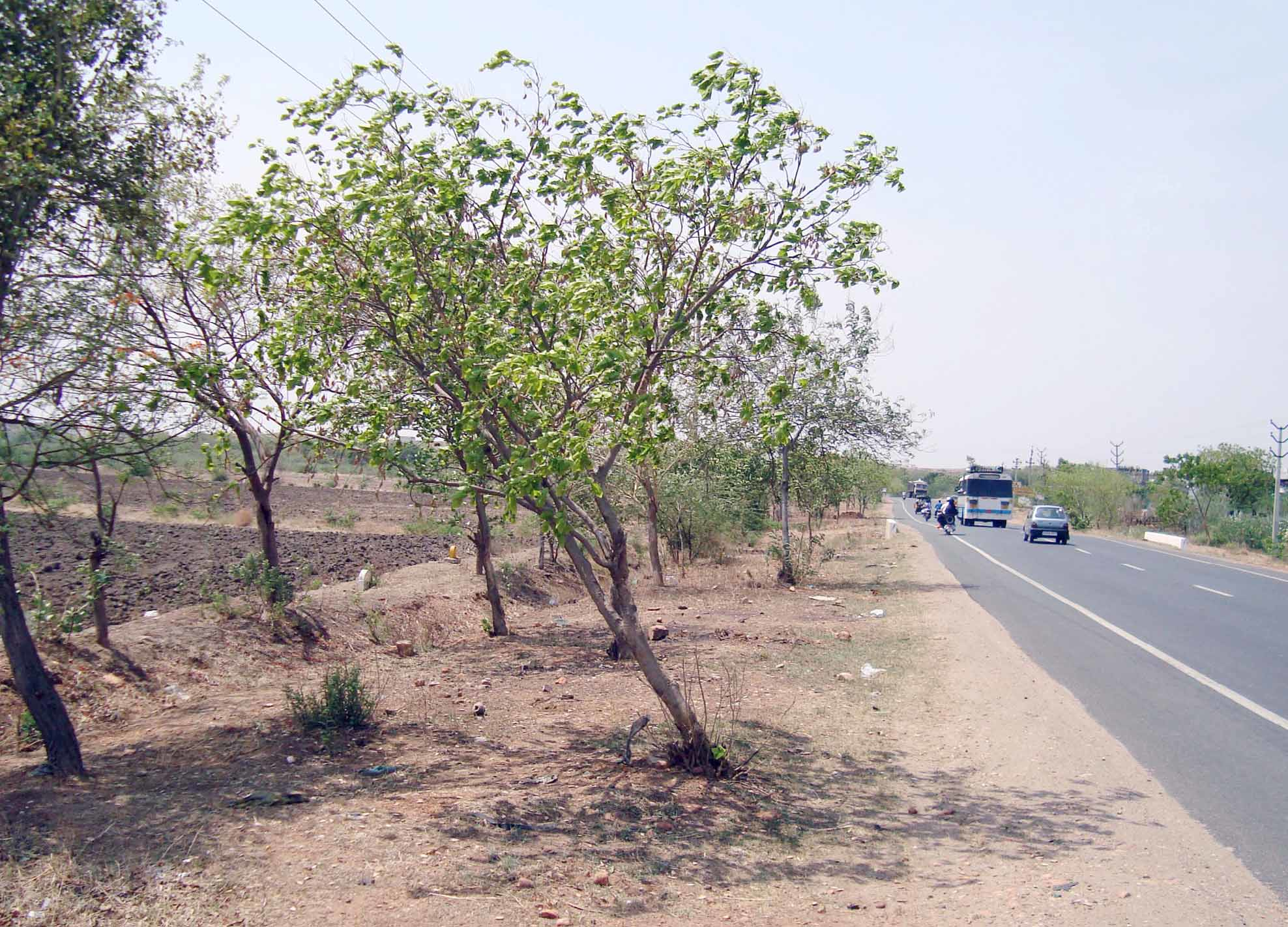
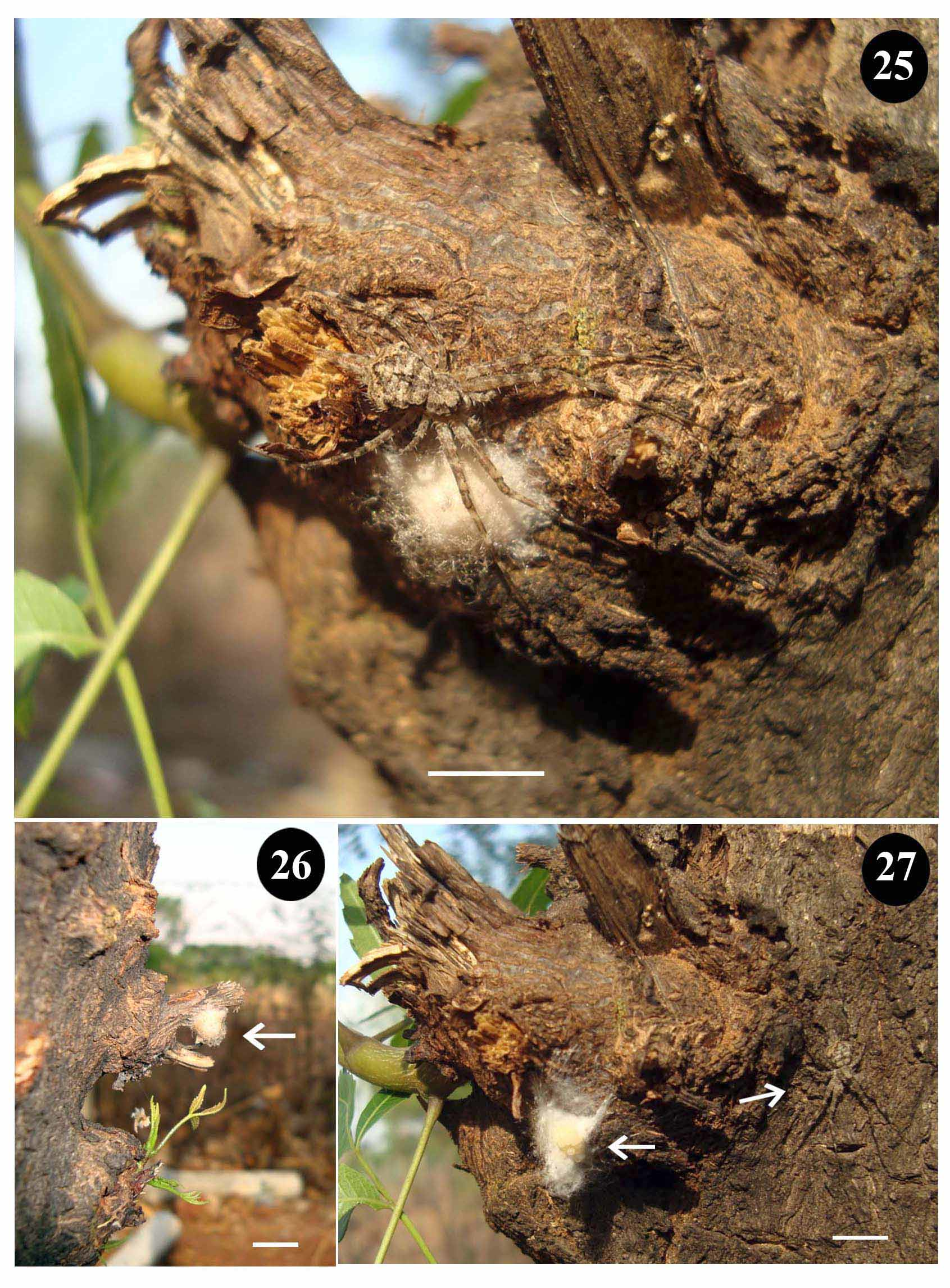
Natural history. Hersilia orvakalensis is an arboreal species, found on Pongamia pinnata (Fabaceae) and Azadirachta indica (Meliaceae) tree trunks close to the Kurnool-Nandyal State Highway near Orvakal Village ( Fig. 2 View FIGURE 2 ). Three individuals (two males and a female) were located on P. pinnata , while one female, guarding an egg sac, was located on A. indica ( Figs 25 – 27 View FIGURES 25 – 27 ). Egg sacs were pale cream to white, while the older sacs were pale and dry in appearance. The egg sac forms a spherical knob on tiny, broken twigs near the base of a broken branch. We located two egg sacs on the same branch, 10 cm apart, and on opposite sides of the branch. One fresh egg sac was opened and a clutch of 42 small spherical eggs were observed. All adult specimens were recorded in February and May.
No known copyright restrictions apply. See Agosti, D., Egloff, W., 2009. Taxonomic information exchange and copyright: the Plazi approach. BMC Research Notes 2009, 2:53 for further explanation.