
Rizalthus anconis, Mendoza & Ng, 2008
|
publication ID |
https://doi.org/ 10.5281/zenodo.5340846 |
|
persistent identifier |
https://treatment.plazi.org/id/0E16272E-FF82-FF80-FC3A-FB52FD13FD40 |
|
treatment provided by |
Diego |
|
scientific name |
Rizalthus anconis |
| status |
sp. nov. |
Rizalthus anconis View in CoL , new species
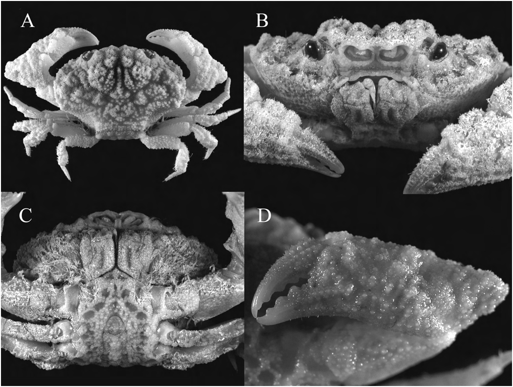
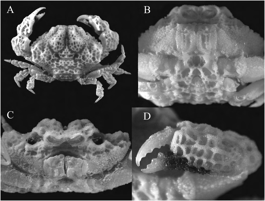
( Figs. 1 View Fig , 2 View Fig , 9A View Fig )
Material examined. – Holotype: 1 male (12.5 × 7.9 mm) (NMCR- 27507), Station R 30, reef slope with black coral, 15–37 m, 9°37.1'N 123°46.1'E, Napaling , Panglao Island , coll. by Panglao 2004 Marine Biodiversity Project, 8 Jun.2004. GoogleMaps
Paratypes: Two females (7.8 × 4.5 mm to 11.2 × 7.3 mm) (MNHN- B30700), 1 ovigerous female (12.7 × 8.1 mm) ( ZRC 2008.0215 View Materials ), Station B 39, reef wall with small caves, 17–25 m, 9°32.8'N 123°42.1'E, Pontod Lagoon 1, Panglao Island , coll. by Panglao 2004 Marine Biodiversity Project, 2 Jul.2004; 1 male (4.2 × 2.7 mm) ( ZRC 2008.0216 View Materials ) GoogleMaps , Station B 10, reef wall with small caves, 3–14 m, 9°36.5'N 123°45.6'E, Momo Beach , Panglao Island , coll. by Panglao 2004 Marine Biodiversity Project, 10 Jun.2004; 1 female (7.0 by 4.3 mm) ( ZRC 2008.0217 View Materials ) GoogleMaps , Station B 17, reef wall with small caves, 3–21 m, 9°37.5'N 123°46.9'E Bingag , Panglao Island , coll. by Panglao 2004 Marine Biodiversity Project, 19 Jun.2004 GoogleMaps .
Description. – Carapace ( Fig. 1A View Fig ) much broader than long, regions well-defined; epigastric, protogastric, mesogastric and metagastric regions raised, together forming a dorsally convex anterior portion on carapace; 2M partially divided longitudinally, lateral lobe larger, 4M almost indistinct, with 5 widely-spaced granules; 1L, 2L and 3L modified into large, granular, dorsally directed tubercles/teeth, 3L most prominent; all dorsal regions of carapace with large granules, surrounded by short setae basally, suborbital, subhepatic and pterygostomial regions with smaller, lower granules. Front about 0.4 times carapace width, bilobed; lobes separated by broad V-shaped cleft, continuous with a deep median fissure on frontal region; each lobe broadly triangular, bluntly tipped, continuing laterally and separated from rounded inner orbital angle by a short groove. Supraorbital margin granulose with 2 small notches laterally, relatively short, no obvious external orbital tooth, not clearly meeting anterolateral margin. Orbits relatively small, width about 0.1 times carapace width. Anterolateral margin lined with tooth-like granules, appearing unevenly serrated, feebly cristate, anterior-most part not meeting orbital margin but becoming lower and less distinct and eventually indiscernible as it curves ventrally to meet pterygostomial region; anterior two-thirds strongly arcuate; posterior third slightly convex, curving posteriad to posterolateral margin, junction of margins marked by prominent, dorsally directed, granular tooth. Posterolateral margin granulose, deeply concave, anterior portion almost parallel to frontal margin, then curving sharply to converge posteriad with posterior carapace margin. Last ambulatory leg coapted against concavity in posterolateral margin. Posterior carapace margin granulose, straight, with rows of granules anterior to it.
Eye with short eyestalks, distal edge with cornea lined with small, tooth-like granules; corneas well developed ( Fig. 2A View Fig ). Antennules ( Fig. 2A View Fig ) folding transversely. Basal antennal segment large, granulose, subrectangular, occupying entire space between antennular fossa and internal orbital angle, filling orbital hiatus; long flagellum arising from distal margin, reaching outer edge of orbit. Epistome ( Fig. 1B View Fig ) with small granules, distal edge slightly sweeping outward, central region slightly undulate, concave. Endostome without oblique or longitudinal ridges.
Outer surface of third maxilliped ( Figs. 1B, C View Fig , 2B View Fig ) granulose, appearing eroded. Merus subquadrate, median length about half that of ischium, with 2 shallow depressions on either side of a submedian, granular ridge; margins with small granules, anterior and external margins slightly concave, internal margin straight; anteroexternal angle auriculiform. Ischium subrectangular, inner margin with large granules and short, stiff setae; with deep, longitudinal median sulcus bordered by granules; separated from basis by distinct suture. Exopod granulose, tapering toward distal end which almost reaches anterior edge of merus, flagellum long.
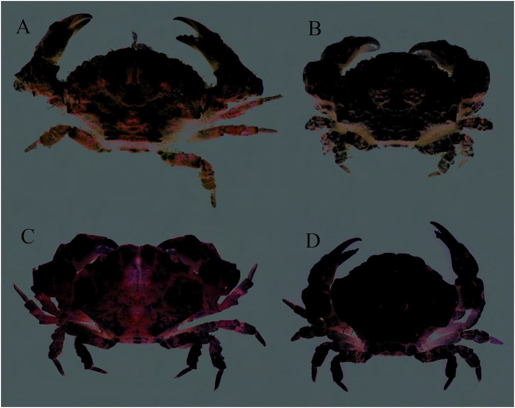
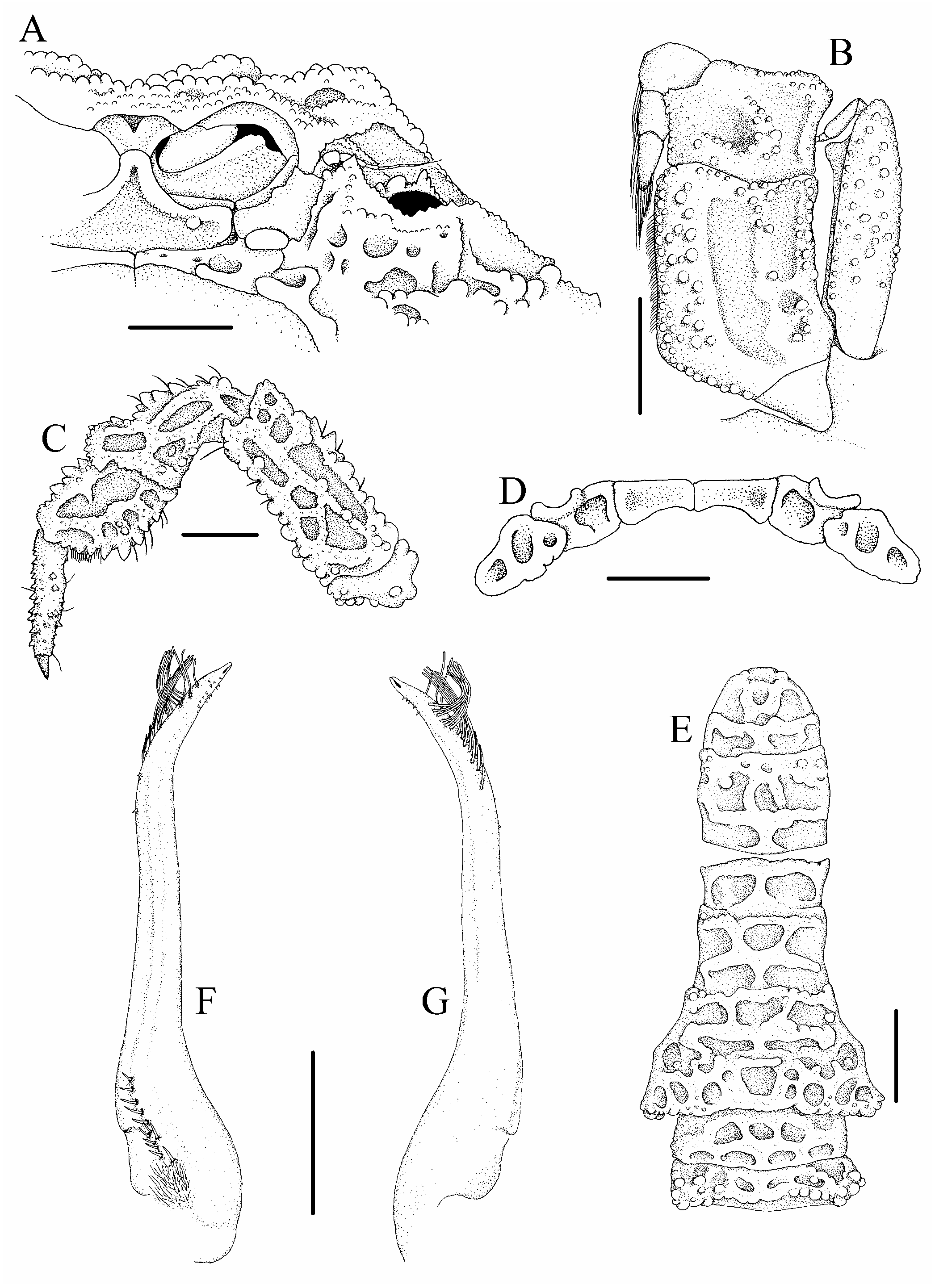
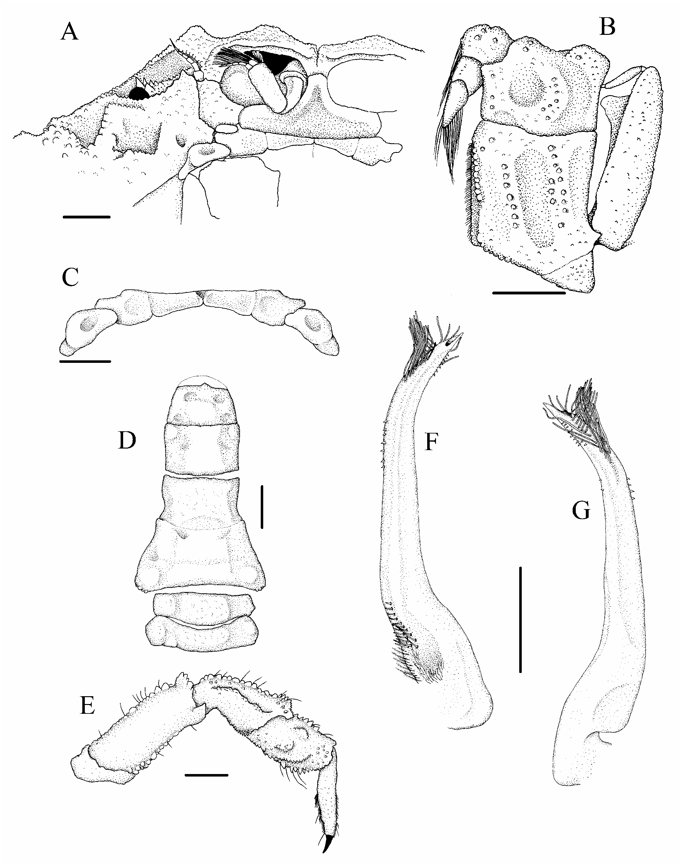
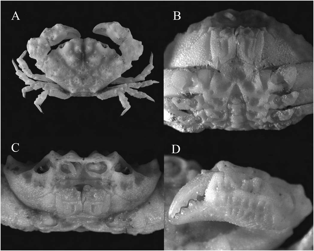
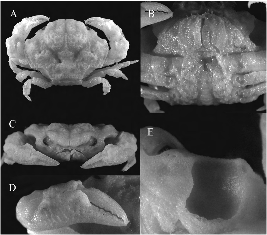
Surface of thoracic sternum ( Fig. 1C View Fig ) eroded, with granules and several distinct depressions on anterior region and on ( Fig. 2I View Fig ) short, as figured. either side of abdomen, anterior region elongate. Sternites 1 and 2 completely fused into triangular plate, separated from Live colouration. – The carapace and pereiopods (Fig. sternite 3 by deep transverse suture. Sternites 3 and 4 partially 9A) have a light pinkish or white base; certain regions of fused, with partial suture seen near anterior edge of cheliped the carapace (frontal, epigastric, protogastric, branchial, coxae; median suture present within 1 median depression intestinal) and ambulatory legs light orange; individual anterior to telson, 3 depressions obliquely anterior and lateral granules on carapace (mesogastric, metagastric, cardiac) and to it, arranged in an oblique line. Abdominal cavity deep, ambulatory legs dark orange to reddish-pink. Cheliped fingers tubercle for locking mechanism (sternal condyle) on sternite are light-brown at the base and white at the tips. This colour 5 slightly nearer to suture with sternite 4, abdomen reaching pattern probably helps it blend into the reef environment. to imaginary line joining posterior edge of cheliped coxae. Etymology. – The specific epithet anconis is derived from the Chelipeds ( Fig. 1A, D View Fig ) similar, subequal. Fingers shorter than Greek word for elbow, alluding to the pronounced elbow-like palm, cutting edges with 3 or 4 teeth, not pigmented, tips projection on the carpus of this animal’s chelipeds. Used as pointed. Dactylus slightly curved, with 2 rows of granules a noun in apposition. and deep submarginal groove along length, stiff short setae on upper margin. Fixed finger slightly deflexed with 2 rows Remarks. – See Remarks for the genus. Rizalthus anconis , of granules and broad submarginal groove continuing from new species, is known thus far only from the type locality, palm. Palm outer surface rugose, upper margin irregular, Panglao island in the Bohol Sea, central Philippines, and is wedge-like and granulose, with 2 irregular rows of granules associated with coral reef walls, occurring at depths between near convex proximal-lower margin; inner surface relatively 3 and 37 m. It has also been collected from reef walls in smoother, with fewer and smaller granules when compared at least one station (B17) together with the two species of with outer surface. Carpus short, dorsal and ventral surface Visayax , new genus, described in this paper. granulose, inner distal angle with low triangular tooth, outer surface with prominent truncatiform wedge-like expansion. Inner surfaces of fingers, palm and carpus coapted against Visayax , new genus anterolateral margin of carapace. Merus granulose, slightly longer than carpus, with rectangular, ventro-distal tooth Diagnosis. – Carapace broader than long; regions wellapposed against carpus. defined; 1M, 2M and 2F raised, with prominent granulose tubercle on 2M; branchial region with 2 dorsally-projecting Ambulatory legs ( Figs. 1A View Fig , 2D, E View Fig ) relatively short, granulose, tubercles; 3M and 1P distinct; suborbital region eroded with edges with setae; second leg longest, coxa-to-dactylus length small depressions; subhepatic and pterygostomial regions about 0.7 times carapace width. Merus subrectangular, with low, discrete granules; carapace surface with distinct subtriangular in cross-section; anterior and posterior edges, or faint reticulate pattern of fused granules; front bilobed, especially in third and fourth ambulatory legs, lined with lobes triangular at the tips; anterolateral margin arcuate, large, pointed granules; dorsal surface in fourth ambulatory descending into subhepatic region, meeting with anterolateral leg more granulose than rest. Dorsal surface of carpus with corner of buccal frame, communicating with raised orbital 1 median row of pointed granules, another row of pointed margin through 2 or 3 thin ridges radiating from external granules on serrated anterior edge. Propodus subrectangular, orbital angle and suborbital margin; posterolateral margin anterior edge serrated. Dactylus straight, dorsal surface and concave, with dorsolaterally directed tubercle at junction edges covered with fine granules, short setae; terminates with anterolateral margin; posterior margin straight, with distally in curved chitinous claw. dorsally projecting tubercle at junction with posterolateral margin. Antennules folding almost transversely. Basal Male abdomen and telson ( Fig. 2C View Fig ) very granulose, appearing antennal segment large, trapezoidal, proximal end with medial eroded. Telson subtriangular with rounded tip; lateral margins extension toward the antennulary fossa, granulose, orbital raised due to presence of granules, slightly convex. Segment hiatus completely filled; long flagellum almost reaching 6 rectangular, about 1.5 times length of telson, lateral margins outer edge of orbit. Epistome broadly concave. Endostome concave. Segments 3–5 completely fused, without any trace without ridges. Outer surface of third maxilliped granulose, of sutures; lateral margins markedly concave; median region eroded; merus subquadrate, median length about half that raised. Segment 2 subtrapezoidal, with 2 shallow longitudinal of ischium; ischium subrectangular, inner margin with grooves on either side of a central raised granular region. granules, short, stiff setae; with deep, longitudinal median Segment 1 trapezoidal with large granules, no transverse sulcus; exopod granulose, tapering toward distal end which ridge. almost reaches anterior edge of merus, flagellum long. Surface of thoracic sternum eroded, large distinct depression Gonopod 1 ( Fig. 2F–H View Fig ) long and slender, mostly straight, just anterior to sternal cavity, no discernible suture within curving outward and tapering to a fine point distally; this anterior depression, smaller depressions seen on either terminally located opening unadorned by flaps or folds; side of sternal cavity; sternites 1 and 2 completely fused, several long, thick and stiff plumose setae located subdistally; separated from sternite 3 by deep transverse suture; sternites distal third sparsely covered with tiny spines, mostly found 3 and 4 almost fused except for short sutures near base of on the inside of the subterminal bend, some spines found in cheliped. Chelipeds similar, subequal; fingers shorter than a row on the inner margin; basal region setose. Gonopod 2
palm, cutting edges with 4 or 5 teeth, tips pointed, curving inward; palm outer surface eroded in reticulate pattern, inner surface relatively smoother; fingers, palm and carpus coapted against anterolateral margin of carapace. Ambulatory legs relatively short, coapted against carapace. G1 long and slender, mostly straight, curving outward and tapering distally; with terminally located opening; several long, thick and stiff simple setae located subdistally on medial edge and internal (dorsal) surface; basal external (ventral) region with row of stiff simple setae, surrounded by finer setae.
Comparative material. – Glyptoxanthus erosus ( Stimpson, 1859) , 1 male (53.1 × 38.1 mm) ( ZRC 1998.9 View Materials ; USNM 168865 View Materials ex.), Sapelo Island , 4.75 miles (7.6 km) off Whistle Buoy, Georgia, U.S.A., 60–65 feet (18.2–19.7 m), coll. by M. Gray, 7 Apr.1966 ; Cranaothus deforgesi Ng, 1993 , 1 male (7.5 × 5.3 mm) (NMCR-1521), Station D-1, Maluso Bay, Basilan Island, Philippines, 27 fathoms (49.38 m), coll. by Pele-Sulu Sea Expedition, 15 Feb.1964 .
Type Species. – Visayax osteodictyon View in CoL , new species, by present designation.
Etymology. – This genus is an arbitrary name derived from the Visayas islands, which comprise the central region of the Philippines. Gender masculine.
Remarks. – Visayax , new genus, is superficially similar to a monotypic genus Lipaesthesius Rathbun, 1898 , in the general outline of the carapace, wherein the anterolateral margins are arcuate and the posterolateral margins are concave, and the eroded/reticulate surface of the carapace, sternum and chelae (cf. Rathbun, 1930: 272, pl. 112). However, it can be distinguished from Lipaesthesius primarily by having the distal end of the basal segment of the antenna free, with the attachment of the antennal flagellum visible ( Figs. 4A View Fig , 6A View Fig ) (vs. distal end fixed against front, with attachment of flagellum not visible in Lipaesthesius ). Other distinguishing features are the presence of the dorsally projecting tubercles on the gastric and branchial regions ( Figs. 3A View Fig , 5A View Fig ) (vs. absent in Lipaesthesius ); the presence of thin ridges linking the anterolateral margin of the carapace to the orbits ( Figs. 3C View Fig , 5C View Fig ) (vs. absent); and the presence of a tubercle each at the junctions between the anterolateral and posterolateral margins and the posterolateral and posterior margins of the carapace ( Figs. 3A View Fig , 5A View Fig ) (vs. absent) (cf. Rathbun, 1930: 272, pl. 112). Furthermore, Lipaesthesius , which is monotypic and represented solely by L. leeanus Rathbun, 1898 , is found only in the eastern Pacific Ocean, ranging from the Gulf of California to the Galapagos Islands and is not known to occur in the Indo-west Pacific region.
Visayax , new genus, is also similar to another monotypic genus, Cranaothus Ng, 1993 , in the general carapace outline and dimensions; in having the suborbital region eroded by small depressions; and in the form of the G1. Ng (1993) opined that the single male specimen reported by Serène and Umali (1972: 68, Pl. 7, Figs. 7–9 View Fig View Fig View Fig ) as “ Paramedaeus noelensis ” is actually conspecific with Cranaothus deforgesi Ng, 1993 . Having examined this specimen, we concur and thus confirm this species as occurring in the Philippines as well as the type locality, Chesterfield Island, Coral Sea. Visayax is easily distinguished from Cranaothus by the absence of squamate granules and vermicular ridges on the carapace ( Figs. 3A View Fig , 5A View Fig ) (vs. present); the form of the bilobed front, wherein the lobes are triangular and are not projecting anteriorly ( Figs. 3A, C View Fig , 5A, C View Fig ) (vs. lobes truncatiform and projecting well beyond orbits); the well defined regions of the carapace ( Figs. 3A View Fig , 5A View Fig ) (vs. not well defined); the presence of tubercles on the gastric and branchial regions and the junction between the posterolateral and posterior carapace margins ( Figs. 3A View Fig , 5A View Fig ) (vs. absent); the absence of endostomial ridges (vs. present); the form of the male thoracic sternum, wherein sternites 1 and 2 and sternites 3 and 4 are fused ( Figs. 3B View Fig , 5B View Fig ) (vs. sternite 1 separated from sternite 2 by distinct suture; with shallow interrupted sutures between sternites 2 and 3 and sternites 3 and 4); the symmetric chelipeds, without modified cutting tooth at base of dactylus ( Figs. 3A, D View Fig , 5A, D View Fig ) (vs. chelipeds asymmetric, dactylus of major chela with modified cutting tooth); and the uniformly slender G1, with simple long subterminal setae ( Figs. 4F–G View Fig , 6F–G View Fig ) (vs. G1 stout proximally, tapering distally, subterminal setae plumose) (cf. Ng, 1993).
Visayax is superficially similar to Glyptoxanthus A. Milne- Edwards, 1879, in the general outline and surface sculpturing of the carapace. Comparison with the type species, G. erosus ( Stimpson, 1859) , as well as with illustrations of other species of Glyptoxanthus (cf. Garth, 1939: 15–17, Pls. 4–5) reveals that it differs from Visayax based on the following features: 1) the front is less protruding and the anterolateral margin is more arcuate in Glyptoxanthus , giving the carapace a more rectangular outline (vs. hexagonal outline in Visayax , Figs. 3A View Fig , 5A View Fig ); 2) adult specimens of the different species of Glyptoxanthus are generally much larger than those of Visayax ; 3) the surface sculpturing of the carapace and pereiopods of Glyptoxanthus is more vermiculate in pattern (vs. more reticulate pattern or eroded, Figs. 3A View Fig , 5A View Fig ); 4) the proto- and mesogastric regions are not very much raised above the rest of the carapace regions in Glyptoxanthus (vs. visibly and distinctly elevated, Figs. 3C View Fig , 5C View Fig ); 5) the presence of endostomial ridges in the buccal cavern in Glyptoxanthus (vs. absent); 6) the 3 rd maxillipeds are more heavily sculptured in Glyptoxanthus , and the proximal half of the exopod is more laterally expanded (vs. less sculptured, exopod more or less uniform in width, Figs. 4B View Fig , 6B View Fig ); and 7) the chelipeds and walking legs of Glyptoxanthus are relatively shorter and stockier, giving them a more robust appearance (vs. longer and more slender, Figs. 3A View Fig , 5A View Fig ).
Visayax is also superficially similar to the monotypic genera Danielea Ng and Clark, 2003 , and Jacforus Ng and Clark, 2003 in the general outline of the carapace and male abdomen. However, Visayax differs from Danielea and Jacforus in the triangular lobes of the front ( Figs. 3A View Fig , 5A View Fig ) (vs. truncate); the presence of tubercles on the gastric and branchial regions and on the junction of the posterolateral and posterior margins of the carapace ( Figs. 3A View Fig , 5A View Fig ) (vs. absent); the concave posterolateral margins of the carapace ( Figs. 3A View Fig , 5A View Fig ) (vs. straight); the absence of a modified cutting tooth on the dactylus of the major chela ( Figs. 3D View Fig , 5D View Fig ) (vs. present); the trapezoidal basal antennal article, wherein the proximal end has a medial extension toward the antennulary fossa ( Figs. 4A View Fig , 6A View Fig ) (vs. simply rectangular in Danielea ; simply subquadrate in Jacforus ); the absence of a distinct suture dividing thoracic sternite 3 and 4 ( Figs. 3B View Fig , 5B View Fig ) (vs. present and complete in Danielea ; present and interrupted in Jacforus ). Furthermore, there are distinct differences in the G1 of these genera: in Danielea the proximal end is definitely stouter and the subterminal setae are more slender, less stiff and arise only from the internal (dorsal) surface; in Jacforus it is relatively shorter and stouter and there are no long, stiff subterminal setae similar to those seen in Visayax (cf. Forest and Guinot, 1961; Serène, 1984; Ng and Clark 2003).
The suite of characters found in Visayax , new genus, make it difficult to place in any of the aforementioned genera, and as such, must necessarily be placed in its own genus.


No known copyright restrictions apply. See Agosti, D., Egloff, W., 2009. Taxonomic information exchange and copyright: the Plazi approach. BMC Research Notes 2009, 2:53 for further explanation.
|
Kingdom |
|
|
Phylum |
|
|
Class |
|
|
Order |
|
|
Family |
|
|
Genus |