
Hepatoporus pumex, Mendoza & Ng, 2008
|
publication ID |
https://doi.org/ 10.5281/zenodo.5340846 |
|
persistent identifier |
https://treatment.plazi.org/id/0E16272E-FF8D-FF96-FC56-FC51FC27FCC0 |
|
treatment provided by |
Diego |
|
scientific name |
Hepatoporus pumex |
| status |
sp. nov. |
Hepatoporus pumex View in CoL , new species
( Figs. 7 View Fig , 8 View Fig , 9D View Fig )
Material Examined. – Holotype: 1 male (8.0 × 5.7 mm) (NMCR- 27510), Station B 11, coral rubble, 2–4 m, 9°29.4'N 123°56.0'E, Pamilacan Island , coll. by Panglao 2004 Marine Biodiversity Project, 11 Jun.2004. GoogleMaps
Paratypes: 1 male (4.0 × 3.0 mm) ( ZRC 2008.0221 View Materials ), Station S 28, reef wall with small caves, 28–32 m, 9°37.2'N 123°46.4'E, Napaling , Panglao Island , coll. by Panglao 2004 Marine Biodiversity Project, 24 Jun.2004 GoogleMaps ; 1 male (3.8 × 2.7 mm) ( ZRC 2008.0222 View Materials ), Station S 10, coral plateau with fine sand covering rocks, 6–14 m, 9°29.4'N 123°56.0'E, Pamilacan Island , coll. by Panglao 2004 Marine Biodiversity Project, 11 Jun.2004 GoogleMaps .
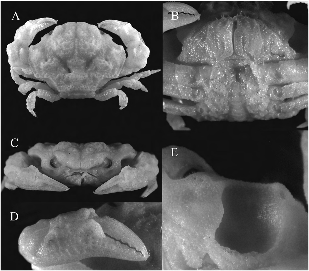
Description. – Carapace ( Fig. 7A View Fig ) broader than long, subpentagonal in its outline, with deep cavity at anterior portion of anterolateral margins characteristic for genus; regions more or less defined, grooves separating them distinct; epigastric, protogastric and cardiac regions raised; 3L in branchial region raised; entire dorsal surface finely granular, densely pitted with cavities, mostly visible only under magnification, larger pits found in mesogastric, cardiac and intestinal regions, surrounded by reticulations formed by small, fused granules especially near posterior and posterolateral margins; with a pair of depressions on either side of posterior part of 3M, and 1 smaller depression on central part of each branchial region. Front ( Fig. 7A, C View Fig ) projecting, broad, about 0.4 times carapace width, with a median thin notch that continues into epigastric groove; frontal margin divergent posteriorly toward supraorbital margin. Orbit small, completely occupied by eyestalk and cornea; eyestalk short, stout, distal end with cornea lined with granules; cornea well developed. Postorbital region raised, supraorbital margin with small granules, no distinct external orbital tooth. Infraorbital region granulose, pitted, margin with small granules. Subhepatic region and most of pterygostomial region occupied by deep cavity ( Fig. 7C, E View Fig ), visible from dorsal view as a concavity in the anterolateral margin; upper margin of cavity thick, ventral and posterior margins thin, crested; walls of cavity granulose, bottom pitted; rest of pterygostomial region granulose, pitted. Anterolateral margin lined with small granules, anterior one-third excavated, posterior two-thirds gently convex, feebly cristate; meeting almost at right angles with posterolateral margin. Posterolateral margin lined with partially fused granules, appearing raised; deeply concave, last ambulatory leg can be coapted against this concavity; junction with posterior carapace margin angulated. Posterior carapace margin slightly concave, with distinctly deeper concavities adjacent to junctions with posterolateral margins for accommodating coxae of last ambulatory legs.
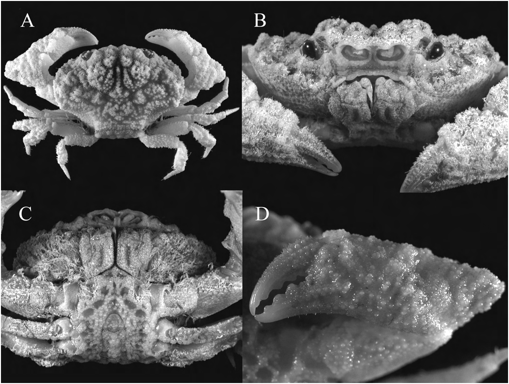
Antennules ( Fig. 8A View Fig ) folding almost transversely. Basal antennal segment broad, subrectangular, distal part granulose, remainder pitted; medial edge convex; occupying entire space between antennular fossa and internal orbital angle, orbital hiatus completely filled; antennal flagellum arising from distal end, short, not reaching external orbital angle. Ventral edge of epistome ( Fig. 7C View Fig ) out-turned, central region with pointed convexity, outer surface pitted. Posterolateral region of endostome with thin, oblique ridge.
Outer surface of third maxillipeds ( Figs. 7B View Fig , 8B View Fig ) eroded, pitted, with scattered granules, sparsely setose. Merus subquadrate, median length about half that of ischium; with submedian, granular hump bordered on either side by shallow depressions; anterior margin irregular and sparsely setose, anteroexternal margin rounded, external margin slightly concave, internal margin slightly convex. Ischium subrectangular, appearing more pitted than merus; with shallow, median, longitudinal sulcus; internal and external margins straight, granulose; separated from basis by a distinct suture. Exopod similarly pitted, proximal half expanded into a convexity, tapering to narrower distal end.
Surface of thoracic sternum ( Fig. 7B View Fig ) heavily pitted, rugose, uneven. Sternites 1 and 2 completely fused to form triangular plate. Sternites 3 and 4 completely fused, without any sutures visible; with shallow median depression just anterior to telson. Intersternal sutures in depressed, giving sternites 5, 6 and 7 a raised appearance. Abdominal cavity deep; with sternal condyle on sternite 5 adjacent to suture with sternite 4; abdomen almost reaching to imaginary line joining middle part of cheliped coxae in situ.
Chelipeds ( Fig. 7A, C, D View Fig ) similar, subequal. Fingers unpigmented, slightly shorter than palm, distal ends pointed, curved inward, cutting margins with at least 4 or 5 low triangular teeth. Dactylus about 0.8 times median length of palm (outer surface), slightly curved; with 4 longitudinal ridges—including upper margin—on distal half of outer surface, continuing proximally as rows of granules on proximal half. Fixed finger slightly deflexed, with 2 longitudinal ridges on distal half of outer surface, continuing as rows of fused granules in palm. Outer surface of palm with reticulated pattern of fused granules, rugose; inner surface granulose; distal portion of upper margin slightly keeled, granulose; proximal portion of upper margin, together with distal portion of carpus, forms an ovate aperture with effaced portion of carapace anterolateral margin when chela are coapted against carapace. Carpus large, outer surface irregular, rugose and covered in reticulate pattern of fused granules; inner surface granulose, distal portion with low, granular, triangular tooth. Merus mostly hidden from dorsal view, slightly longer than carpus, unarmed, surfaces and margins granulose.
Ambulatory legs ( Fig. 7A View Fig ) relatively short; first ambulatory leg longest, coxa to dactylus length about 0.8 times carapace width. Merus rectangular, subtriangular in cross-section; anterior and posterior edges granulose, with larger granules on posterior edge; dorsal and ventral surfaces finely granulose with scattered larger granules. Anterior edge of carpus serrated by conical granules, with short stiff setae; dorsal surface granulose or pitted, with traces of reticulate pattern of ridges; inner surface and lower posterior edge with granules. Propodus subrectangular; anterior and posterior edges serrated with conical granules, setose; dorsal surface with reticulate pattern of ridges, granulose or pitted. Dactylus straight; surface with conical granules, short stiff setae; terminating in simple chitinous claw.
Abdomen ( Fig. 8C View Fig ) appearing eroded, with pits and granules. Telson subtriangular, distal portion rounded. Penultimate segment subquadrate, median length as long as that of telson; distal one-third of lateral margins convex, proximal two-thirds straight. Segments 3–5 fused, with traces of sutures visible, lateral margins concave, surface heavily pitted. Segment 2 subtrapezoidal, granulose. Segment 1 divided into 2 parts by transverse ridge, subpentagonal, granulose.
Gonopod 1 ( Fig. 8D–F View Fig ) relatively long, tapering, with proximal third bent outwards prominently and a less substantial bend subterminally. Terminal end auriculiform, with about 4 subterminal, long, simple setae and subterminal flange. Distal half with scattered spines, especially on internal surface.
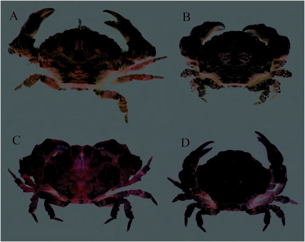
Live colouration. – Carapace and pereiopods ( Fig. 9D View Fig ) generally creamy white to very light pink; with a few small spots of brown or brownish-orange on carapace; similar spots and smudges seen on fingers, palms and carpus of chelipeds and on merus and carpus of ambulatory legs.
Etymology. – The specific epithet pumex is the Latin word for pumice, a light, porous volcanic rock, whose texture is similar to the carapace surface of this species. Used as a noun in apposition.
Remarks. – Comparing it to the excellent illustrations (cf. Sakai, 1935:77, Fig. 11, Pl. 7; 1939: 458, Fig. 28, Pl. 60; 1976: 416, Fig. 220a, Pl. 150; Takeda & Nagai, 1986: 548, Figs. 1c, 1d View Fig ) of the type species for the genus, Hepatoporus pumex , new species, is morphologically most similar to H. orientalis ( Sakai, 1935) in: 1) the general outline of the carapace; 2) the raised gastric, branchial and cardiac regions; and 3) the shape of the cavity formed by the carapace anterolateral margin and chela. It differs significantly from the type species by having: 1) a broader, more truncated front ( Fig. 7A, C View Fig ) (vs. triangular and acute); 2) a more deeply excavated anterolateral margin ( Fig. 7A, C View Fig ) (vs. shallow concavity); 3) the posterior two-thirds of the carapace anterolateral margin more even ( Fig. 7A View Fig ) (vs. irregular and jagged); 4) a large, distinct pit in the branchial region of the carapace ( Fig. 7A View Fig ) (vs. absent); and 5) reticulate patterns of fused granules and pits near the posterolateral and posterior margins of the carapace ( Fig. 7A View Fig ) (vs. simply granular).
Hepatoporus pumex was also compared to illustrations of H. guinotae ( Zarenkov, 1971) (see Serène, 1984; Davie & Turner, 1994). It is morphologically similar to H. guinotae in the general outline and surface texture of the carapace, and, to a certain extent, in the G1 morphology. It differs from H. guinotae in the following features: 1) the carapace dorsal surface, male thoracic sternum and third maxillipeds are not as pitted or reticulate in H. pumex ( Figs. 7A, B View Fig , 8B View Fig ) (vs. pronouncedly pitted and/or reticulate); 2) the concavity in the anterolateral margin is shallower ( Fig. 7A View Fig ) (vs. deeper); 3) the subhepatic cavity is more extensive, reaching further posteriad ( Fig. 7E View Fig ) (vs. not so posterior); 4) the lobes of the front are evenly convex ( Fig. 7A, C View Fig ) (vs. concave laterally); and 5) the fingers of the chelae are almost as long as the palm ( Fig. 7D View Fig ) (vs. distinctly shorter). Furthermore, the G1 of H. pumex differs distinctly from that of H. guinotae as figured by Serène (1984: Fig. 40): it is more slender and tapering ( Fig. 8D View Fig ) (vs. stouter and more uniform in thickness); the terminal flap/process is more circular ( Fig. 8E, F View Fig ) (vs. more elongate); there are fewer, about four, subterminal setae ( Fig. 8E, F View Fig ) (vs. more subterminal setae, about 7). Davie & Turner (1994) believe that H. distinctus (Takeda & Nagai, 1986) and H. guinotae may be conspecific. We find that H. pumex differs from H. distinctus , with the exception of G1 characters (G1 of H. distinctus not known), in the same features stated above for H. guinotae .
Hepatoporus pumex , new species, differs from H. asper Davie & Turner, 1994 in the general outline and surface features of the carapace, as well as the morphology of the G1. The carapace of H. pumex is relatively broader ( Fig. 7A View Fig ) (vs. narrower, almost as long as broad); the frontal and protogastric region is not eroded ( Fig. 7A View Fig ) (vs. eroded with large pits, some coalescing into a deep median groove); the branchial regions are finely granular and pitted, with some sections raised as tubercles ( Fig. 7A View Fig ) (vs. branchial regions with a deep crescent-shaped cavity lined with fungiform granules); the sternum is granular and rugose with shallow depressions ( Fig. 7B View Fig ) (vs. more extensively eroded with deep depressions); the G1 tapers distally, has an auriculiform terminal process and fewer but longer subterminal setae ( Fig. 8D–F View Fig ) (vs. inflated subterminally, without an auriculiform terminal process and with more but shorter subterminal setae) (cf. Davie and Turner, 1994).
Hepatoporus pumex is thus far known only from the type locality, Pamilacan Island , and nearby Panglao Island , both in the Bohol Sea, central Philippines. It has been collected from coral rubble, reef walls, and sandy/rocky seafloors associated with coral reefs, and from depths ranging between 2–32 m.

No known copyright restrictions apply. See Agosti, D., Egloff, W., 2009. Taxonomic information exchange and copyright: the Plazi approach. BMC Research Notes 2009, 2:53 for further explanation.
|
Kingdom |
|
|
Phylum |
|
|
Class |
|
|
Order |
|
|
Family |
|
|
Genus |